

The effect of scan parameters on cone beam CT trabecular bone microstructural measurements of the human mandible
- PMID: 24132024
- PMCID: PMC3853518
- DOI: 10.1259/dmfr.20130206
The effect of scan parameters on cone beam CT trabecular bone microstructural measurements of the human mandible
Abstract
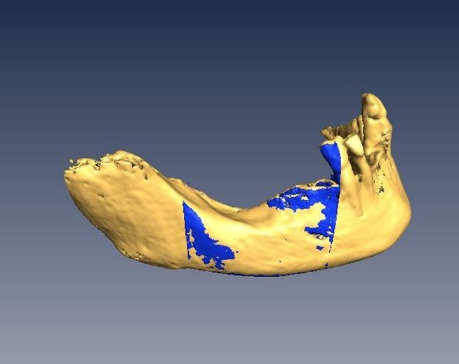
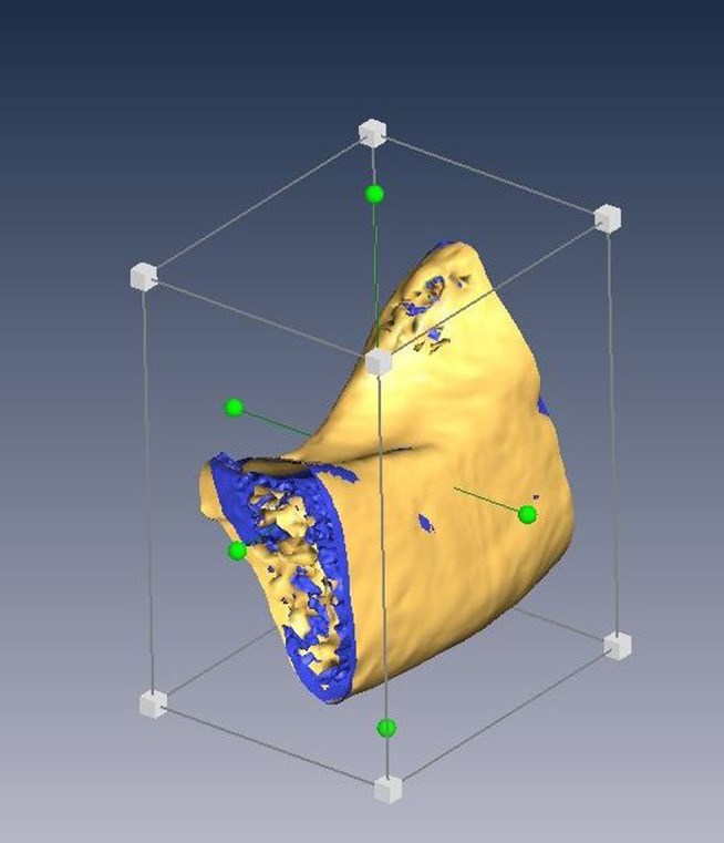
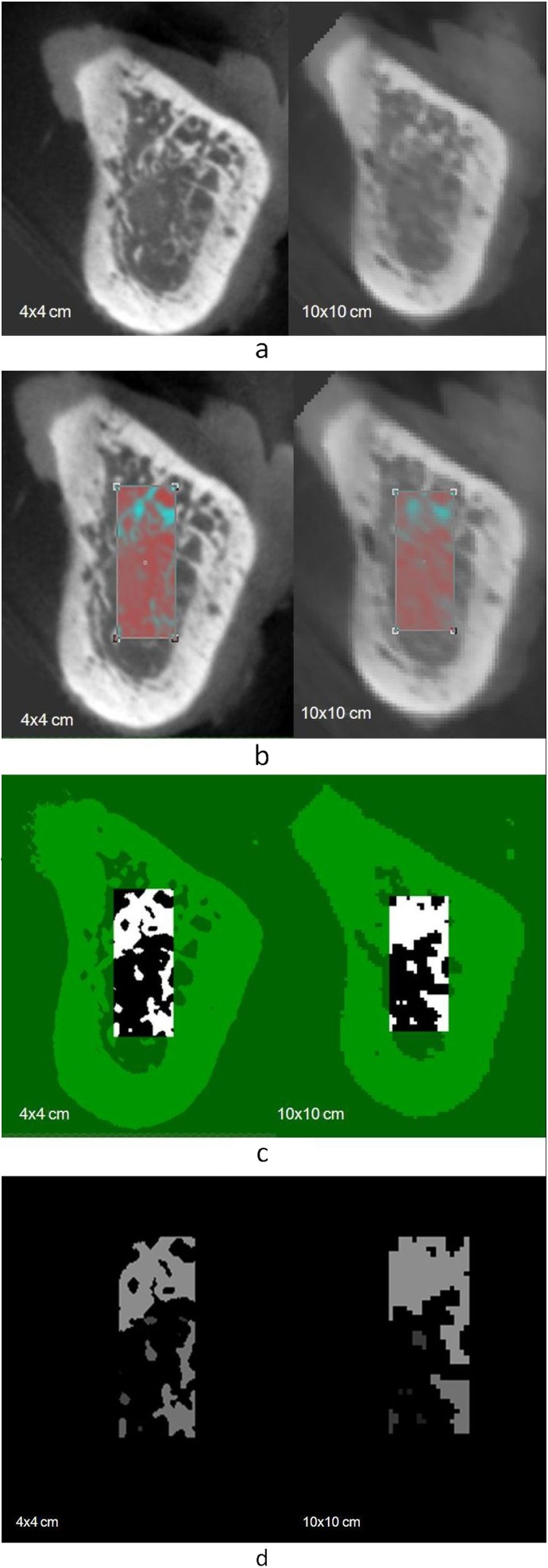
The objective of this study was to investigate the effect of different cone beam CT scan parameters on trabecular bone microstructure measurements. A human mandibular cadaver was scanned using a cone beam CT (3D Accuitomo 170; J.Morita, Kyota, Japan). 20 cone beam CT images were obtained using 5 different fields of view (4×4 cm, 6×6 cm, 8×8 cm, 10×10 cm and 10×5 cm), 2 types of rotation steps (180° and 360°) and 2 scanning resolutions (standard and high). Image analysis software was used to assess the trabecular bone microstructural parameters (number, thickness and spacing). All parameters were measured twice by one trained observer. Intraclass correlation coefficients showed high intraobserver repeatability (intraclass correlation coefficient, 0.95-0.97) in all parameters across all tested scan parameters. Trabecular bone microstructural measurements varied significantly, especially in smaller fields of view (p = 0.001). There was no significant difference in the trabecular parameters when using different resolutions (number, p = 0.988; thickness, p = 0.960; spacing, p = 0.831) and rotation steps (number, p = 1.000; thickness, p = 0.954; spacing, p = 0.759). The scan field of view significantly influences the trabecular bone microstructure measurements. Rotation steps (180° or 360°) and resolution (standard or high) selections are not relevant.
Keywords: cone beam CT; diagnostic imaging; scanning parameter; trabecular bone.
Figures
Similar articles
-
Influence of object location in different FOVs on trabecular bone microstructure measurements of human mandible: a cone beam CT study.Dentomaxillofac Radiol. 2014;43(2):20130329. doi: 10.1259/dmfr.20130329. Epub 2013 Nov 21. Dentomaxillofac Radiol. 2014. PMID: 24265395 Free PMC article.
-
Accuracy of trabecular bone microstructural measurement at planned dental implant sites using cone-beam CT datasets.Clin Oral Implants Res. 2014 Aug;25(8):941-5. doi: 10.1111/clr.12163. Epub 2013 Apr 15. Clin Oral Implants Res. 2014. PMID: 23581278
-
A comparative evaluation of cone beam CT and micro-CT on trabecular bone structures in the human mandible.Dentomaxillofac Radiol. 2013;42(8):20130145. doi: 10.1259/dmfr.20130145. Epub 2013 Jul 5. Dentomaxillofac Radiol. 2013. PMID: 23833320 Free PMC article.
-
Quantification of bone quality using different cone beam computed tomography devices: Accuracy assessment for edentulous human mandibles.Eur J Oral Implantol. 2016;9(4):411-424. Eur J Oral Implantol. 2016. PMID: 27990508
-
Influence of scan setting selections on root canal visibility with cone beam CT.Dentomaxillofac Radiol. 2012 Dec;41(8):645-8. doi: 10.1259/dmfr/27670911. Dentomaxillofac Radiol. 2012. PMID: 23166361 Free PMC article.
Cited by
-
Comparison of mandibular bone microarchitecture between micro-CT and CBCT images.Dentomaxillofac Radiol. 2015;44(5):20140322. doi: 10.1259/dmfr.20140322. Epub 2015 Jan 7. Dentomaxillofac Radiol. 2015. PMID: 25564887 Free PMC article.
-
Algorithms used in medical image segmentation for 3D printing and how to understand and quantify their performance.3D Print Med. 2022 Jun 24;8(1):18. doi: 10.1186/s41205-022-00145-9. 3D Print Med. 2022. PMID: 35748984 Free PMC article.
-
The effects of technical factors on the fractal dimension in different dental radiographic images.Eur Oral Res. 2023 May 4;57(2):68-74. doi: 10.26650/eor.2023984422. Eur Oral Res. 2023. PMID: 37525855 Free PMC article.
-
Influence of artefacts generated by titanium and zirconium implants in the study of trabecular bone architecture in cone-beam CT images.Dentomaxillofac Radiol. 2022 Sep 1;51(6):20220066. doi: 10.1259/dmfr.20220066. Epub 2022 May 4. Dentomaxillofac Radiol. 2022. PMID: 35466693 Free PMC article.
-
Quantifications of Mandibular Trabecular Bone Microstructure Using Cone Beam Computed Tomography for Age Estimation: A Preliminary Study.Biology (Basel). 2022 Oct 18;11(10):1521. doi: 10.3390/biology11101521. Biology (Basel). 2022. PMID: 36290424 Free PMC article.
References
-
- Song YD, Jun SH, Kwon JJ. Correlation between bone quality evaluated by cone-beam computerized tomography and implant primary stability. Int J Oral Maxillofac Implants 2009; 24: 59–64 - PubMed
-
- Kobayashi K, Shimoda S, Nakagawa Y, Yamamoto A. Accuracy in measurement of distance using limited cone-beam computerized tomography. Int J Oral Maxillofac Implants 2004; 19: 228–231 - PubMed
MeSH terms
LinkOut - more resources
Full Text Sources
Other Literature Sources
