. 2013 Nov 12;81(20):1732-40.
doi: 10.1212/01.wnl.0000435556.21319.e4. Epub 2013 Oct 16.

Amyloid-first and neurodegeneration-first profiles characterize incident amyloid PET positivity

Affiliations

Amyloid-first and neurodegeneration-first profiles characterize incident amyloid PET positivity

Clifford R Jack Jr et al. Neurology. .

Abstract

Objective: To estimate the incidence of and to characterize cognitive and imaging findings associated with incident amyloid PET positivity.

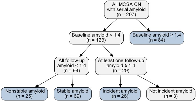
Methods: Cognitively normal (CN) participants in the Mayo Clinic Study of Aging who had 2 or more serial imaging assessments, which included amyloid PET, FDG-PET, and MRI at each time point, were eligible for analysis (n = 207). Twelve subjects with Alzheimer disease dementia were included for comparison.

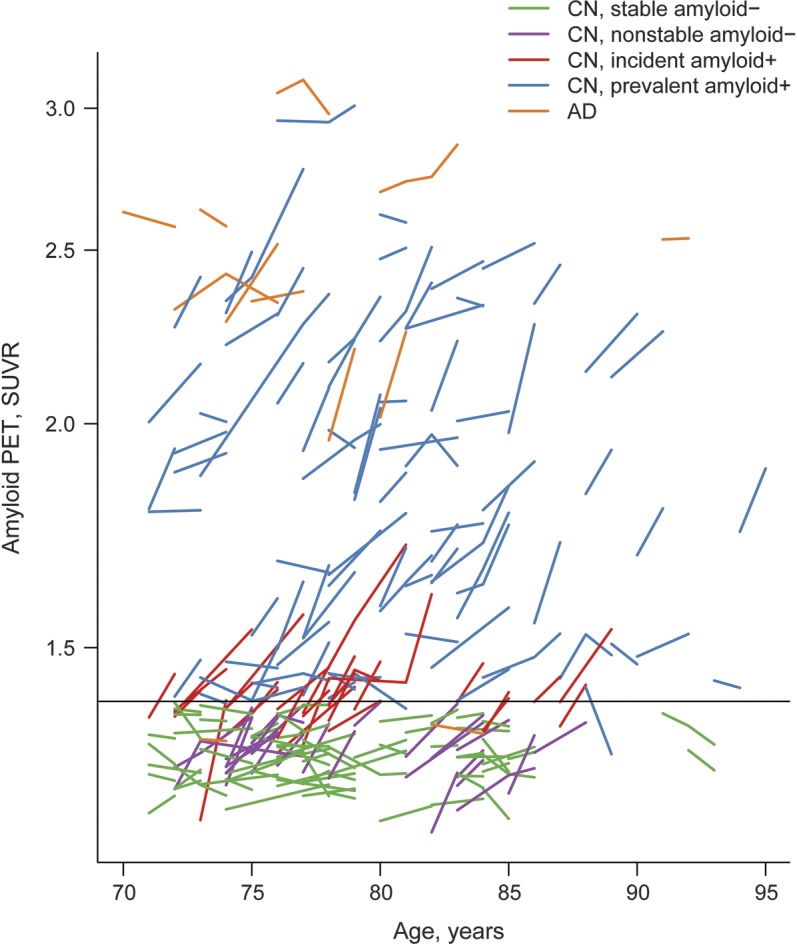
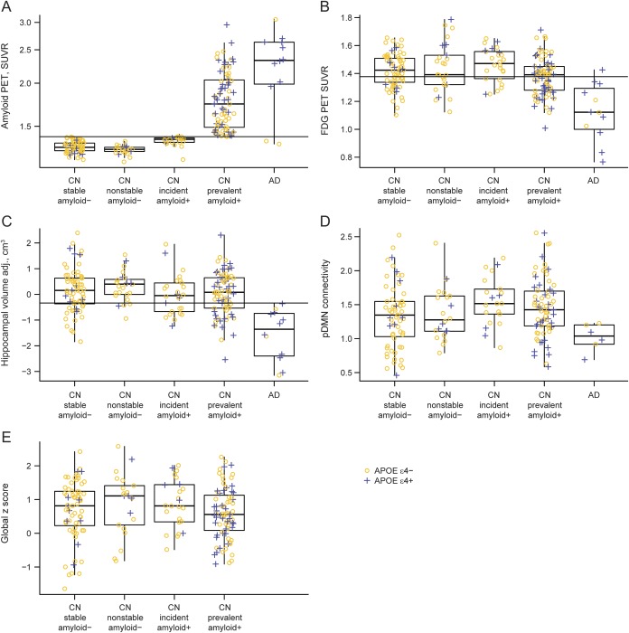
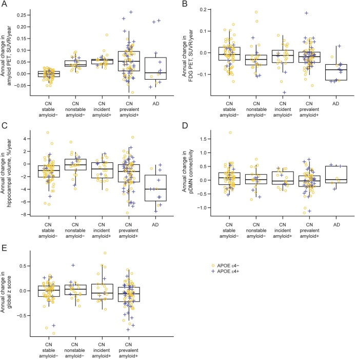
Results: Of the 123 CN participants who were amyloid-negative at baseline, 26 met criteria for incident amyloid PET positivity. Compared to the 69 subjects who remained stable amyloid-negative, on average these 26 did not differ on any imaging, demographic, or cognitive variables except amyloid PET (by definition) and task-free functional connectivity, which at baseline was greater in the incident amyloid-positive group. Eleven of the 26 incident amyloid-positive subjects had abnormal hippocampal volume, FDG-PET, or both at baseline.

Conclusions: The incidence of amyloid PET positivity is approximately 13% per year among CN participants over age 70 sampled from a population-based cohort. In 15/26 (58%), incident amyloid positivity occurred prior to abnormalities in FDG-PET and hippocampal volume. However, 11/26 (42%) incident amyloid-positive subjects had evidence of neurodegeneration prior to incident amyloid positivity. These 11 could be subjects with combinations of preexisting non-Alzheimer pathophysiologies and tau-mediated neurodegeneration who newly entered the amyloid pathway. Our findings suggest that both "amyloid-first" and "neurodegeneration-first" biomarker profile pathways to preclinical AD exist.


Figures

Figure 1
Figure 1. Flowchart
The cognitively normal (CN) groups in blue are the focus of this article. MCSA = Mayo Clinic Study of Aging.
Figure 2
Figure 2. Trajectory plots of amyloid by age
Stable amyloid PET-negative cognitively normal (CN) participants are represented with green lines, nonstable amyloid-negative CN with purple lines, incident amyloid-positive CN with red lines, prevalent amyloid-positive CN with blue lines, and Alzheimer disease (AD) with orange lines. Of the 84 amyloid-positive participants at baseline, 2 became amyloid-negative (<1.4) and decreased by >0.04 over time and thus could be considered to have reverted from amyloid-positive to -negative. We attribute this infrequent phenomenon (2/84 = 2%) to measurement noise in subjects who lie at the boundary of amyloid positivity. SUVR = standardized uptake value ratio.
Figure 3
Figure 3. Boxplot of baseline imaging and cognitive performance by diagnosis and amyloid group
APOE ε4-negative participants are represented with yellow circles and APOE ε4-positive participants are represented with blue plus signs. (A) Boxplot of baseline amyloid by diagnosis and amyloid group. The horizontal line represents a cutpoint of 1.40, which was used in the definition of incident amyloid. This cutpoint has a sensitivity of approximately 95% in 42 subjects with clinically diagnosed Alzheimer disease (AD). (B) Boxplot of baseline FDG-PET by diagnosis and amyloid group. The horizontal line represents a cutpoint of 1.38, which corresponds to a sensitivity of approximately 95% in 42 subjects with clinically diagnosed AD. (C) Boxplot of baseline adjusted hippocampal volume by diagnosis and amyloid group. The horizontal line represents a cutpoint of −0.34, which corresponds to a sensitivity of approximately 95% in 42 subjects with clinically diagnosed AD. (D) Boxplot of baseline posterior default mode network (pDMN) connectivity by diagnosis and amyloid group. (E) Boxplot of baseline global z score by amyloid group among cognitively normal (CN) participants. SUVR = standardized uptake value ratio.
Figure 4
Figure 4. Boxplot of annual change in imaging and cognitive performance by diagnosis and amyloid group
APOE ε4-negative participants are represented with yellow circles and APOE ε4-positive participants are represented with blue plus signs. (A) Boxplot of annual change in amyloid PET by diagnosis and amyloid group. (B) Boxplot of annual change in FDG-PET by diagnosis and amyloid group. (C) Boxplot of annual % change in hippocampal volume by diagnosis and amyloid group. (D) Boxplot of annual change in posterior default mode network (pDMN) connectivity by diagnosis and amyloid group. (E) Boxplot of annual change in global z score by amyloid group among cognitively normal (CN) participants. AD = Alzheimer disease; SUVR = standardized uptake value ratio.

Comment in

References

    1. Chetelat G, Villemagne VL, Villain N, et al. Accelerated cortical atrophy in cognitively normal elderly with high beta-amyloid deposition. Neurology 2012;78:477–484 - PubMed
    1. Jack CR, Jr, Lowe VJ, Weigand SD, et al. Serial PiB and MRI in normal, mild cognitive impairment and Alzheimer's disease: implications for sequence of pathological events in Alzheimer's disease. Brain 2009;132:1355–1365 - PMC - PubMed
    1. Villemagne VL, Pike KE, Chetelat G, et al. Longitudinal assessment of Abeta and cognition in aging and Alzheimer disease. Ann Neurol 2011;69:181–192 - PMC - PubMed
    1. Morris JC, Roe CM, Grant EA, et al. Pittsburgh compound B imaging and prediction of progression from cognitive normality to symptomatic Alzheimer disease. Arch Neurol 2009;66:1469–1475 - PMC - PubMed
    1. Sperling RA, Johnson KA, Doraiswamy PM, et al. Amyloid deposition detected with florbetapir F 18 ((18)F-AV-45) is related to lower episodic memory performance in clinically normal older individuals. Neurobiol Aging 2013;34:822–831 - PMC - PubMed

Publication types

MeSH terms