Review
. 2014 Jan 15;306(2):C98-C109.
doi: 10.1152/ajpcell.00289.2013. Epub 2013 Oct 16.

Physical biology in cancer. 4. Physical cues guide tumor cell adhesion and migration

Affiliations
Review

Physical biology in cancer. 4. Physical cues guide tumor cell adhesion and migration

Kimberly M Stroka et al. Am J Physiol Cell Physiol. .

Abstract

As tumor cells metastasize from the primary tumor location to a distant secondary site, they encounter an array of biologically and physically heterogeneous microenvironments. While it is well established that biochemical signals guide all stages of the metastatic cascade, mounting evidence indicates that physical cues also direct tumor cell behavior, including adhesion and migration phenotypes. Physical cues acting on tumor cells in vivo include extracellular matrix mechanical properties, dimensionality, and topography, as well as interstitial flow, hydrodynamic shear stresses, and local forces due to neighboring cells. State-of-the-art technologies have recently enabled us and other researchers to engineer cell microenvironments that mimic specific physical properties of the cellular milieu. Through integration of these engineering strategies, along with physics, molecular biology, and imaging techniques, we have acquired new insights into tumor cell adhesion and migration mechanisms. In this review, we focus on the extravasation and invasion stages of the metastatic cascade. We first discuss the physical role of the endothelium during tumor cell extravasation and invasion and how contractility of endothelial and tumor cells contributes to the ability of tumor cells to exit the vasculature. Next, we examine how matrix dimensionality and stiffness coregulate tumor cell adhesion and migration beyond the vasculature. Finally, we summarize how tumor cells translate and respond to physical cues through mechanotransduction. Because of the critical role of tumor cell mechanotransduction at various stages of the metastatic cascade, targeting signaling pathways involved in tumor cell mechanosensing of physical stimuli may prove to be an effective therapeutic strategy for cancer patients.

Keywords: cell adhesion; extravasation; matrix stiffness; three-dimensional migration; tumor metastasis.


Figures
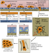
Fig. 1.
Fig. 1.
Physical cues influence the metastatic cascade. As circulating tumor cells collide with the vessel wall, transient adhesions form between various ligands on the tumor cell surface and E-, P-, and L-selectins on the vascular endothelium. The tumor cell arrests and firmly adheres to the endothelium via integrin binding with endothelial ICAM-1 and VCAM-1. Subendothelial matrix stiffness and Rho kinase/myosin light chain kinase signaling direct endothelial cell (EC) contractility (yellow arrows) and EC junction stability. The tumor cells may also release soluble factors that induce EC contractility and disruption, thus facilitating the extravasation process. Subsequently, tumor cells migrate through the extracellular matrix (ECM) of tissues and within longitudinal tracks between anatomic structures, where they are further influenced by physical cues, such as matrix stiffness and confinement. Finally, the tumor cells reach a destination where they form a secondary tumor. CEA, carcinoembryonic antigen; MUC16, mucin 16; PODXL, podocalyxin.
Fig. 2.
Fig. 2.
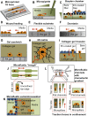
Diverse in vitro assays have been used to explore physical and biological cues affecting cell migration. Each of these assays mimics one or multiple cues presented to tumor cells (orange) and/or the endothelium (blue) during metastasis. Note that this is not an exhaustive list of migration assays. A: microphotopatterned or microcontact printed ECM protein lines (21, 33). B: micropipette chemotaxis assay (95). C: Boyden chamber chemotaxis assay, with bare filter containing nanometer- to micrometer-sized pores, or with EC-coated filter (56, 85). D: wound-healing assay. E: polyacrylamide (PA) gel flexible substrate assay (108). F: durotaxis assay using PA gel with a gradient of stiffness (87). G: collagen-PA gel sandwich assay (40). H: 3-dimensional (3D) collagen gel matrix assay (43). I: collagen gel invasion assay, with bare gel or EC-coated gel (93). J: microfluidic “trough” assay with 3 confining edges (106). K: microfluidic extra-/intravasation assay (70, 157). L: microfluidic chemotaxis assay with flat (5, 137) or micropillar-containing (114) channels. PDMS, polydimethylsiloxane. Red arrows indicate direction of cell migration.
Fig. 3.
Fig. 3.
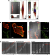
Physical confinement alters tumor cell adhesion and migration mechanisms. A: confocal reconstructions of the actin cytoskeleton (red; phalloidin staining) reveal significantly altered actin morphology in physically confined MDA-MB-231 metastatic breast tumor cells within narrow microchannels (3 μm wide × 10 μm high) compared with wide channels (50 μm wide × 10 μm high). B: confocal images of F-actin (red) and paxillin (green) on 2D planar surface (left) and in narrow and wide microchannels (right). Paxillin-positive punctate focal adhesions are much less pronounced in narrow than in wide channels. C: MDA-MB-231 cells migrate across narrow (3 μm wide × 10 μm high) channels, even in the absence of myosin II-mediated contractility (blebbistatin) or actin polymerization [latrunculin-A (LA)]. Time below each image series is in minutes. [From Balzer et al. (5).]

Similar articles

Cited by

References

    1. Al-Mehdi AB, Tozawa K, Fisher AB, Shientag L, Lee A, Muschel RJ. Intravascular origin of metastasis from the proliferation of endothelium-attached tumor cells: a new model for metastasis. Nat Med 6: 100–102, 2000 - PubMed
    1. Alcaide P, Newton G, Auerbach S, Sehrawat S, Mayadas TN, Golan DE, Yacono P, Vincent P, Kowalczyk A, Luscinskas FW. p120-Catenin regulates leukocyte transmigration through an effect on VE-cadherin phosphorylation. Blood 112: 2770–2779, 2008 - PMC - PubMed
    1. Baker EL, Srivastava J, Yu DH, Bonnecaze RT, Zaman MH. Cancer cell migration: integrated roles of matrix mechanics and transforming potential. PLos One 6: 2011 - PMC - PubMed
    1. Balgude AP, Yu X, Szymanski A, Bellamkonda RV. Agarose gel stiffness determines rate of DRG neurite extension in 3D cultures. Biomaterials 22: 1077–1084, 2001 - PubMed
    1. Balzer EM, Tong Z, Paul CD, Hung WC, Stroka KM, Boggs AE, Martin SS, Konstantopoulos K. Physical confinement alters tumor cell adhesion and migration phenotypes. FASEB J 26: 4045–4056, 2012 - PMC - PubMed

Publication types

LinkOut - more resources