. 2013 Dec 15;305(12):F1687-96.
doi: 10.1152/ajprenal.00211.2013. Epub 2013 Oct 16.

Regulation of NKCC2 activity by inhibitory SPAK isoforms: KS-SPAK is a more potent inhibitor than SPAK2

Affiliations

Regulation of NKCC2 activity by inhibitory SPAK isoforms: KS-SPAK is a more potent inhibitor than SPAK2

Hae J Park et al. Am J Physiol Renal Physiol. .

Abstract

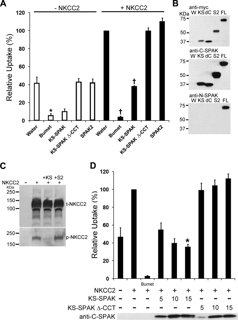
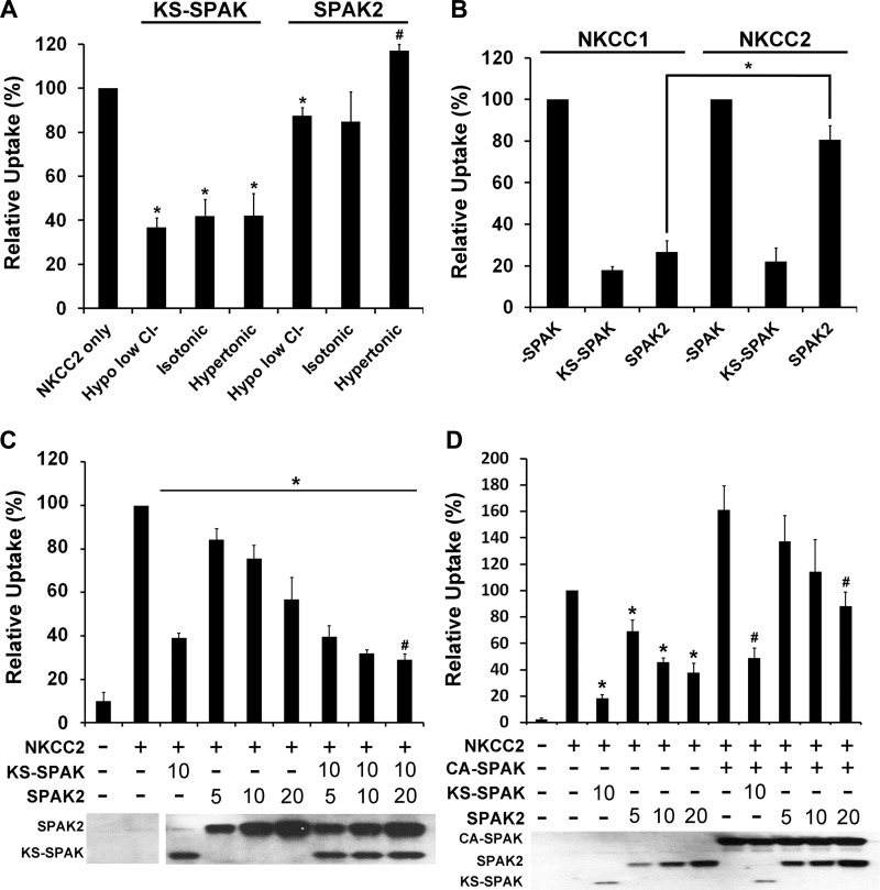
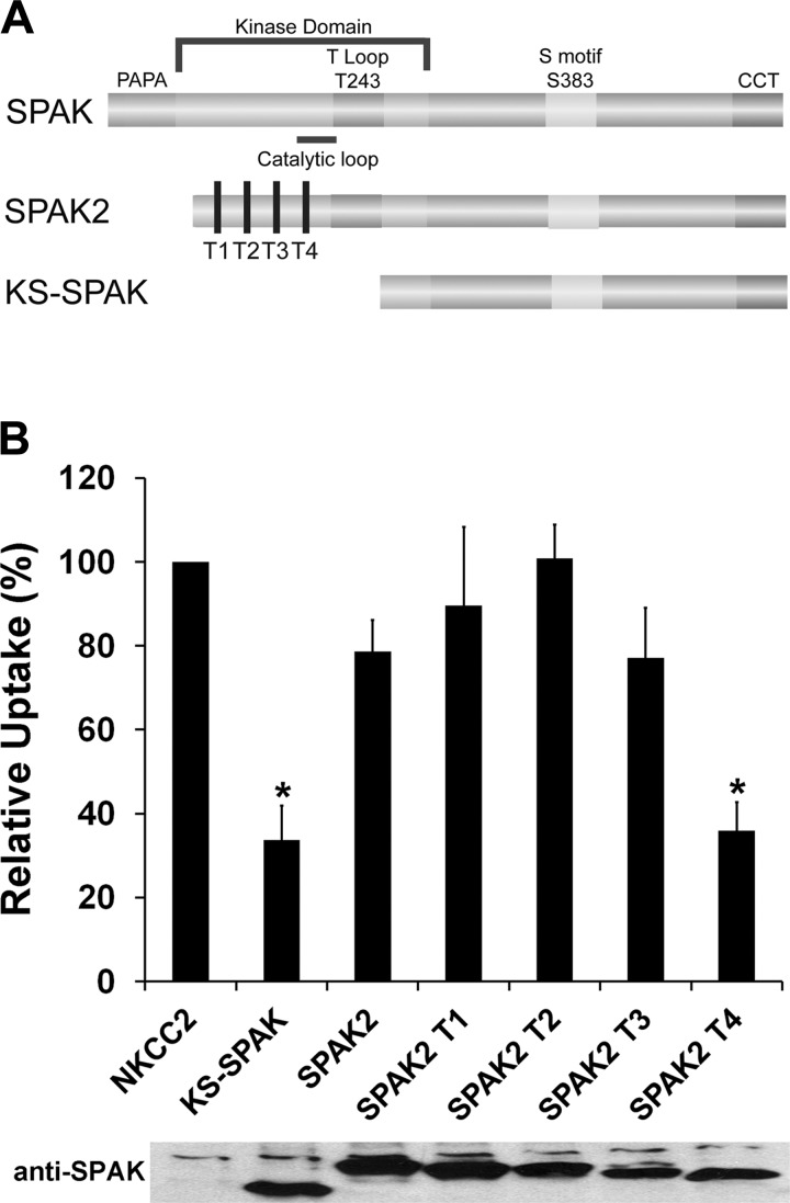
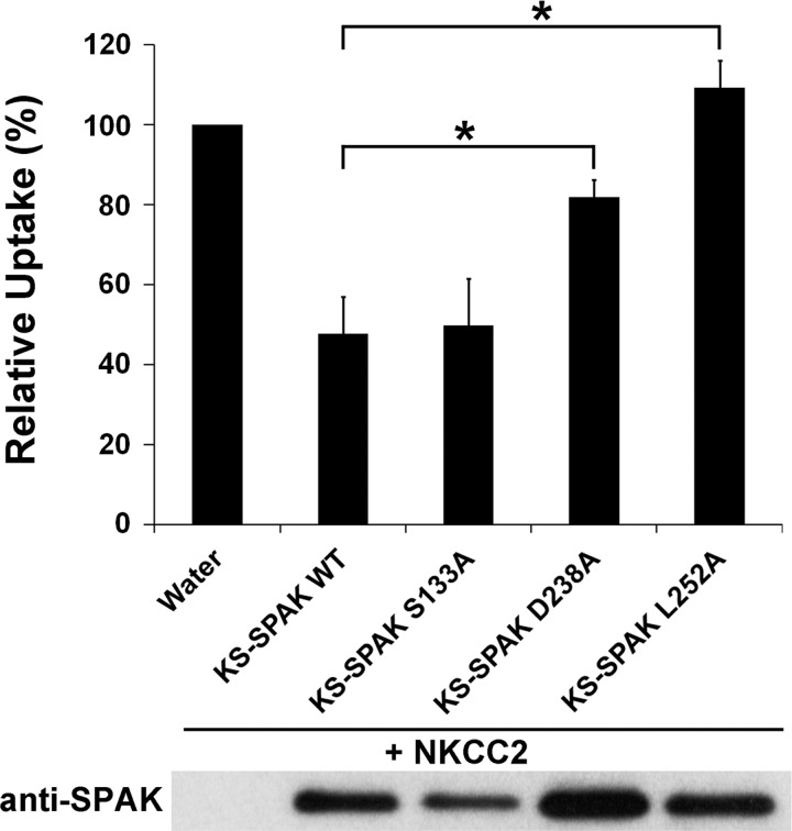
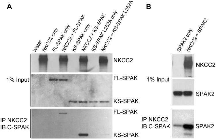
The cation cotransporters Na(+)-K(+)-2Cl(-) cotransporter 1 and 2 (NKCC1 and NKCC2) and Na(+)-Cl cotransporter (NCC) are phosphorylated and activated by the kinases Ste20-related proline alanine-rich kinase (SPAK) and oxidative stress-responsive kinase (OSR1), and their targeted disruption in mice causes phenotypes resembling the human disorders Bartter syndrome and Gitelman syndrome, reflecting reduced NKCC2 and NCC activity, respectively. We previously cloned a kinase-inactive kidney-specific SPAK isoform, kidney-specific (KS)-SPAK, which lacks the majority of the kinase domain present in full-length SPAK. Another putative inactive SPAK isoform, SPAK2, which only lacks the initial portion of the kinase domain, is also highly expressed in kidney. The functional relevance of inactive SPAK isoforms is unclear. Here, we tested whether KS-SPAK and SPAK2 differentially affect cation cotransporter activity. While KS-SPAK and SPAK2 both strongly inhibited NKCC1 activity, SPAK2 was a much weaker inhibitor of NKCC2 activity. Removal of the catalytic loop from SPAK2 resulted in an inhibitory effect on NKCC2 similar to that of KS-SPAK. Full-length SPAK is phosphorylated and activated by members of the with-no-lysine[K] (WNK) kinase family. Mutation of a WNK phosphorylation in KS-SPAK did not alter its ability to inhibit NKCC2 activity. In contrast, we found that residues involved in KS-SPAK interactions with cation cotransporters are required for it to inhibit cotransporter activity. Finally, both KS-SPAK and SPAK2 associated with NKCC2, as demonstrated by coimmunoprecipitation. Together, these data identify the structural basis for the differential effects of KS-SPAK and SPAK2 on cation cotransporter activity that may be physiologically important.

Keywords: cation cotransporter; hypertension; kinase.


Figures

Fig. 1.
Fig. 1.
Kidney-specific (KS)-Ste20-related proline alanine-rich kinase (SPAK) inhibits cation cotransporter activity in Xenopus oocytes. A: 13–14 oocytes per group were injected with water or 10 ng of each capped RNA (cRNA) as shown. After 3 days 86Rb uptake was measured for pooled groups of oocytes under hypertonic (300 mosM) conditions. Data are expressed relative to water-injected oocytes (100%) ± SE. Water-injected oocytes treated with 1 mM bumetanide (Bumet) confirmed that the uptake in the water-injected group was through endogenous Na+-K+-2Cl cotransporter 1 (NKCC1) or coinjected NKCC1 or NKCC2. Deletion of the conserved carboxy terminus (CCT), required for interactions with both cation cotransporters and with-no-lysine[K] (WNK) kinases, prevented inhibition of uptake by KS-SPAK (KS-SPAK Δ-CCT). *P < 0.001, compared with water injected; †P < 0.001, compared with NKCC2 only-injected; n = 4 (different frogs). B: Western blotting confirmed expression of the appropriate SPAK isoforms. W, water injected; KS, KS-SPAK; dC, KS-SPAK Δ-CCT; S2, SPAK2; FL, FL-SPAK. All isoforms have an NH2-terminal myc tag (top); each SPAK possesses the SPAK COOH terminus (middle). Anti-N-SPAK confirmed that only FL-SPAK has the SPAK NH2 terminus. C: immunoblotting showed that KS-SPAK, but not SPAK2, reduced phosphorylation of NKCC2 at the SPAK/OSR1 phosphorylation sites T95/T100 (human numbering), without an effect on total NKCC2 expression levels. D: titration of KS-SPAK revealed a dose-dependent inhibitory effect of KS-SPAK and confirmed the requirement for the Δ-CCT. *P = 0.05, compared with 5 ng KS-SPAK; n = 4 (different frogs).
Fig. 2.
Fig. 2.
SPAK2 is a strong inhibitor of NKCC1 but a relatively weak inhibitor of NKCC2. AD: data are expressed relative to uptake by NKCC2-injected oocytes, set to 100% for each condition ± SE; n = 4–5 (different frogs). Uptake by water-injected oocytes was only 3–17% of that of oocytes injected with NKCC2 alone; bumetanide inhibited uptake by oocytes injected with NKCC2 alone by 90–96% (data not shown), confirming that the majority of uptake is through injected NKCC2 (A) At least 30 oocytes were injected as indicated with 20 ng of NKCC2 and 10 ng of KS-SPAK or SPAK2 cRNAs used. In this experiment, the preincubation and uptake conditions differ from those used in Fig. 1 (see materials and methods). The night before the uptake experiment, each injection group was divided into groups of 10 and preincubated in the appropriate solution. 86Rb uptake was then measured for the pooled groups of oocytes in the appropriate uptake medium. *P ≤ 0.01, lower activity, and #P < 0.01, higher activity, compared with the NKCC2-injected group at the same tonicity. B: 10–20 oocytes were injected as indicated with 20 ng of NKCC1 or NKCC2 cRNA and 10 ng of KS-SPAK or SPAK2 cRNA. 86Rb uptake was performed after 3 days under hypotonic conditions, following an overnight preincubation in hypotonic low Cl solution. *P ≤ 0.001, lower degree of inhibition of uptake by SPAK2 in NKCC2- than in NKCC1-coinjected oocytes. C: 12–20 oocytes were injected with 20 ng of NKCC2 cRNA and KS-SPAK and/or SPAK2 cRNA (in ng) as indicated. 86Rb uptake was performed after 3 days under hypertonic conditions, following an overnight preincubation in hypertonic solution. *P < 0.01, lower uptake compared with oocytes injected with NKCC2 only; #P < 0.05, higher uptake by oocytes injected with NKCC2, KS-SPAK and SPAK2 (20 ng), compared with water-injected oocytes. D: 10–22 oocytes were injected with 20 ng of NKCC2 cRNA and constitutively active FL-SPAK (CA-SPAK), KS-SPAK and/or SPAK2 cRNA (in ng) as indicated. 86Rb uptake was performed after 3 days under hypotonic conditions, following an overnight preincubation in hypotonic low Cl solution. *P < 0.001, lower uptake compared with oocytes injected with NKCC2 only; #P < 0.05, lower uptake by oocytes injected with NKCC2 and CA-SPAK with either KS-SPAK (10 ng) or SPAK2 (20 ng), compared with oocytes injected with NKCC2 and CA-SPAK only. C and D, bottom: representative Western blots confirming expression of SPAK isoforms.
Fig. 3.
Fig. 3.
Deletion of the SPAK2 catalytic loop enhances its inhibitory effect on cation cotransporter activity. A: schematic of SPAK isoforms and SPAK2 truncations. The NH2 terminus of SPAK2 truncations used in the experiments presented in B are shown by vertical black bars (T1-T4); note that the truncation from T1 to T4 removes the catalytic loop, and that SPAK2 T4 is still 28 amino acids longer than KS-SPAK. B: 15–20 oocytes were injected with 20 ng of NKCC2 cRNA and with 10 ng of KS-SPAK, SPAK2, or SPAK2 truncations (T1-T4) cRNA, as indicated. 86Rb uptake was performed after 3 days under hypotonic low Cl conditions. A representative immunoblot using an antibody against the COOH terminus of SPAK is shown, confirming similar expression levels. Data are expressed relative to NKCC2-injected oocytes (100%) ± SE; n = 4–5 (different frogs); *P < 0.01, lower activity compared with the SPAK2-injected group. Uptake by water-injected oocytes was only 15% of that by NKCC2-injected oocytes, and bumetanide inhibited uptake by NKCC2-injected oocytes by 85% (data not shown).
Fig. 4.
Fig. 4.
KS-SPAK inhibition of cation cotransporter activity is independent of its phosphorylation, but requires specific residues required for protein-protein interactions. 10–20 oocytes were injected as indicated with 20 ng of NKCC2 cRNA and 10 ng of wild-type (WT) or mutant KS-SPAK cRNA. 86Rb uptake was performed after 3 days under hypotonic conditions, following an overnight preincubation in hypotonic low Cl solution. KS-SPAK S133A is mutated at a residue equivalent to S383 in full-length SPAK, which following phosphorylation by WNK kinase, leads to its activation. The residues in full-length SPAK equivalent to KS-SPAK residues D238 and L252 have been reported to play major roles its interactions with both NKCC1 and WNK kinase. Data are expressed relative to NKCC2-injected oocytes (100%) ± SE; n = 5 (different frogs); *P < 0.05, lower activity compared with the NKCC2-injected group. Uptake by water-injected oocytes was only 20% of that by NKCC2-injected oocytes, and bumetanide inhibited uptake by NKCC2-injected oocytes by 85% (data not shown). A representative immunoblot using an antibody against the COOH terminus of SPAK is shown to confirm expression.
Fig. 5.
Fig. 5.
KS-SPAK and SPAK2 interact with NKCC2. A: coimmunoprecipitations were performed in Xenopus laevis oocytes injected with 10 ng each of cRNA for NKCC2 (3× FLAG-NKCC2), FL-SPAK, KS-SPAK, or KS-SPAK L252A as indicated. 3 days after injection, lysate from 10 oocytes per group was precleared with mouse IgG-Agarose then incubated with anti-FLAG M2 Affinity Gel at 4°C overnight. After 5 washes with homogenization buffer, precipitated proteins were eluted and analyzed by immunoblotting to detect interactions, as were aliquots of the original lysate (1% of input), to confirm expression. NKCC2 was detected with an anti-Flag antibody, and SPAK isoforms were detected with an anti C-SPAK antibody. FL-SPAK and KS-SPAK only immunoprecipitated in the presence of NKCC2, and KS-SPAK L252A, which is mutated at a residue important for FL-SPAK interactions with NKCC1 (26), had greatly reduced binding compared with wild type KS-SPAK. B: same experiment was performed with SPAK2 instead of KS-SPAK L252A. Data obtained for KS-SPAK and FL-SPAK are not shown, but the results were similar to those shown in A.

Comment in

References

    1. Anselmo AN, Earnest S, Chen W, Juang YC, Kim SC, Zhao Y, Cobb MH. WNK1 and OSR1 regulate the Na+,K+,2Cl− cotransporter in HeLa cells. Proc Natl Acad Sci USA 103: 10883–10888, 2006 - PMC - PubMed
    1. Dowd BF, Forbush B. PASK (proline-alanine-rich STE20-related kinase), a regulatory kinase of the Na-K-Cl cotransporter (NKCC1). J Biol Chem 278: 27347–27353, 2003 - PubMed
    1. Flatman PW. Cotransporters, WNKs and hypertension: an update. Curr Opin Nephrol Hypertens 17: 186–192, 2008 - PubMed
    1. Gagnon KB, Delpire E. Molecular physiology of SPAK and OSR1: two Ste20-related protein kinases regulating ion transport. Physiol Rev 92: 1577–1617, 2012 - PMC - PubMed
    1. Gagnon KB, Delpire E. On the substrate recognition and negative regulation of SPAK, a kinase modulating Na+-K+-2Cl− cotransport activity. Am J Physiol Cell Physiol 299: C614–C620, 2010 - PMC - PubMed

Publication types

LinkOut - more resources