. 2013 Dec 6;288(49):35117-25.
doi: 10.1074/jbc.M113.521443. Epub 2013 Oct 16.

Identification of a dithiol-disulfide switch in collapsin response mediator protein 2 (CRMP2) that is toggled in a model of neuronal differentiation

Affiliations

Identification of a dithiol-disulfide switch in collapsin response mediator protein 2 (CRMP2) that is toggled in a model of neuronal differentiation

Manuela Gellert et al. J Biol Chem. .

Abstract

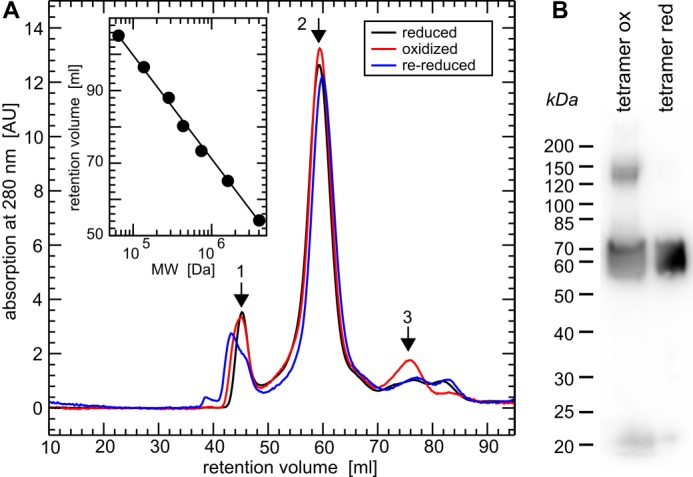
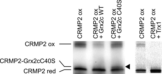
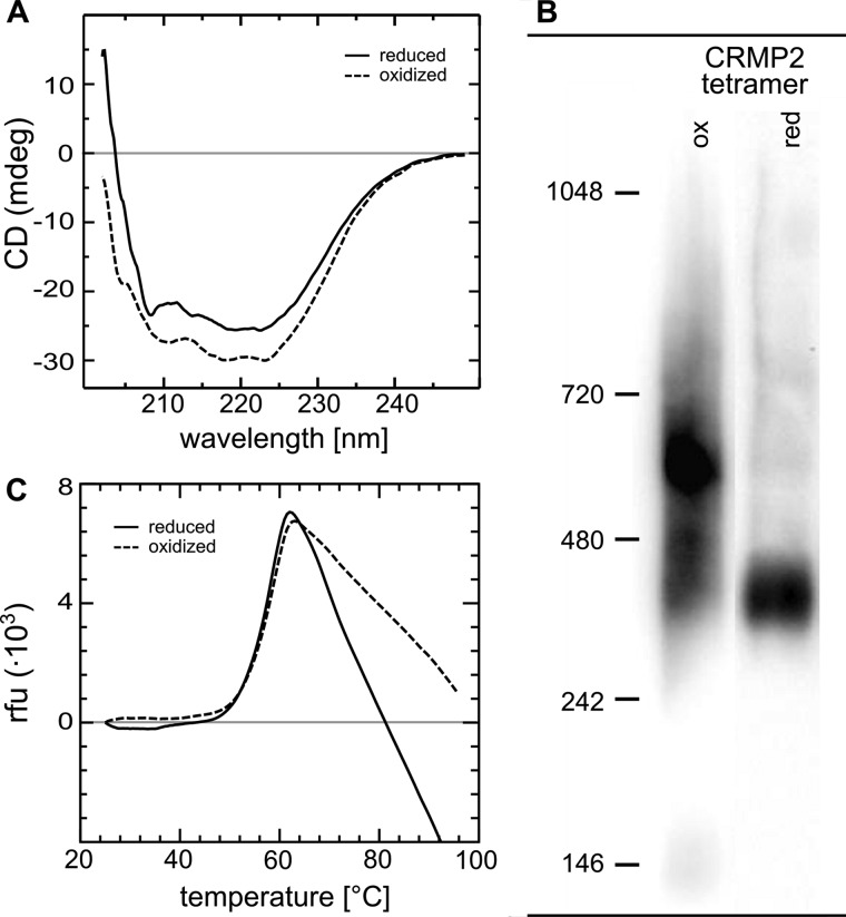
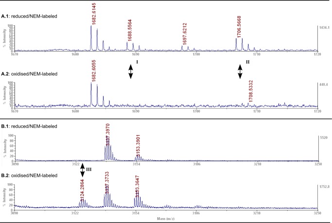
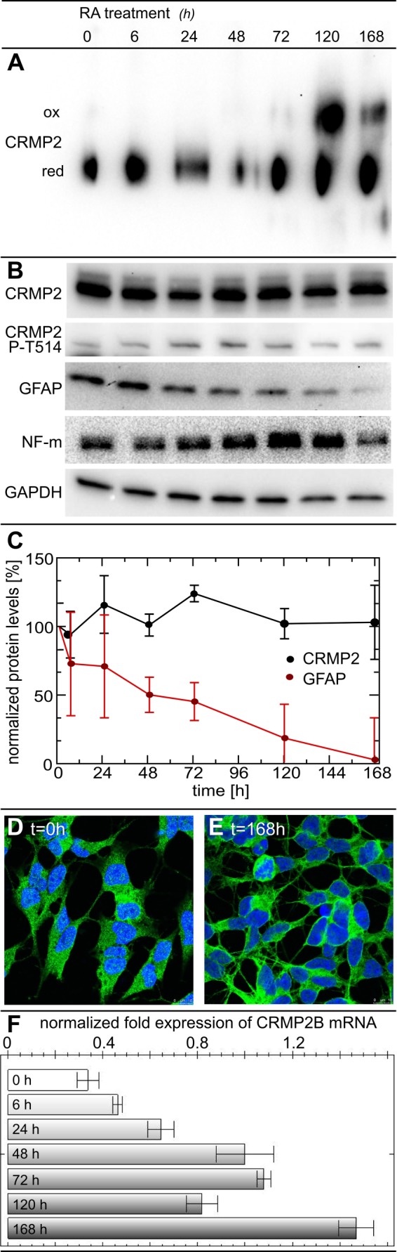
Vertebrate-specific glutaredoxin 2 (Grx2) is expressed in at least two isoforms, mitochondrial Grx2a and cytosolic Grx2c. We have previously shown that cytosolic Grx2 is essential for embryonic development of the brain. In particular, we identified collapsin response mediator protein 2 (CRMP2/DPYSL2), a mediator of the semaphorin-plexin signaling pathway, as redox-regulated target of Grx2c and demonstrated that this regulation is required for normal axonal outgrowth. In this study, we demonstrate the molecular mechanism of this regulation, a specific and reversible intermolecular Cys-504-Cys-504 dithiol-disulfide switch in homotetrameric CRMP2. This switch determines two conformations of the quaternary CRMP2 complex that controls axonal outgrowth and thus neuronal development.

Keywords: CRMP2; Disulfide; Glutaredoxin; Neurodifferentiation; Neuronal Differentiation; Redox Regulation; Redox Signaling; Redox Switch; Thiol.


Figures

FIGURE 1.
FIGURE 1.
Quaternary structure of reduced, oxidized, and re-reduced CRMP2. A, gel filtration chromatography (HiLoad 16/60 Superdex 200 column, GE Healthcare) of (Grx2c+DTT+TCEP) reduced, H2O2-oxidized, and re-reduced recombinant CRMP2 in their native, folded states. Calibration (inset) was done using the manufacturer's instructions: 1, ∼800 kDa, i.e. dodecamer (theoretical mass, 770.4 kDa); 2, 256 kDa, i.e. (homo)tetramer (theoretical mass, 256.8 kDa); 3, 65 kDa, i.e. monomeric CRMP2 (theoretical mass, 64.2 kDa). B, non-reducing SDS-PAGE and Western blot of the tetrameric fractions of oxidized and reduced CRMP2 following the gel-filtration chromatography depicted in A and denaturation of the protein complexes.
FIGURE 2.
FIGURE 2.
Grx2c reduces CRMP2 in a dithiol reaction mechanism. Incubation of reduced (red) Grx2c with oxidized (ox) CRMP2 did not lead to a complex between the proteins when analyzed by non-reducing SDS-PAGE. Only incubation with a Grx2c mutant lacking the second (resolving) cysteinyl residue in the CXXC (C40S) active site led to the formation of a mixed disulfide (see arrowhead). In addition, wild-type Trx1 was able to reduce the protein in vitro and did not form a stable complex with CRMP2.
FIGURE 3.
FIGURE 3.
Conformational changes of CRMP2 upon reduction. A, CD spectroscopy of oxidized and subsequently reduced CRMP2. The decrease in ellipticity between 210–220 nm indicates a decrease in α-helical content of reduced CRMP2. B, blue native gel electrophoresis of reduced and oxidized tetrameric CRMP2. 1 μg of the protein was reduced by a catalytic amount of Grx2c (1:100, +DTT+TCEP) or oxidized by H2O2 treatment. Both fractions were analyzed by blue native gel electrophoresis. The higher migration velocity of reduced CRMP2 indicated binding of more Coomassie dye molecules and thus the exposure of more hydrophobic surfaces compared with the oxidized CRMP2 tetramer. The marks depicted were introduced only for orientation purposes and do not reflect the actual molecular weight of the complexes (see Fig. 1). C, differential scanning fluorimetry of reduced and oxidized CRMP2. No significant differences were observed in the thermal stability of reduced and oxidized CRMP2.
FIGURE 4.
FIGURE 4.
Identification of a specific dithiol-disulfide switch in CRMP2 by MALDI-TOF mass spectrometry. Mass spectra details of reduced (A.1/B.1) or oxidized (A.2/B.2) NEM-labeled CRMP2. A.1/A.2 peptide I, GLYDGPVCEVSVTPK+NEM; peptide II, GLYDGPVCEVSVTPK + NEM/H2O at Cys-504 (A.1, I and II, theoretical masses, 1688.89/1706.81; observed, 1688.56/1706.57); B.1/B.2 peptide III, 2× GLYDGPVCEVSVTPK in form of disulfide-linked peptides (B.2, III, theoretical mass, 3124.52; observed, 3124.28) not present in the reduced protein (B.1).
FIGURE 5.
FIGURE 5.
The CRMP2 conformational switch in a cellular model of neuronal differentiation. For differentiation into a neuron-like phenotype, SH-SY5Y cells were treated with 10 μm retinoic acid (RA). At 6, 24, 48, 72, 120, and 168 h, cells were detached by trypsin treatment, washed with PBS, lysed, separated by blue native gel electrophoresis, and analyzed by anti-CRMP2 Western blotting (A). Western blots of CRMP2, CRMP2 phospho-Thr-514, glial fibrillary acidic protein (GFAP), neurofilament m (NF-m), and the GAPDH loading control following SDS-PAGE (B). The presence of the neuronal marker neurofilament m confirmed the neuronal character of the cells. Quantification of CRMP2 and glial fibrillary acidic protein levels by densitometry of Western blots from three independent experiments, normalized to GAPDH levels and the untreated controls (C). The phenotype and localization of CRMP2 were analyzed by immunofluorescence and confocal microscopy (D and E). CRMP2B mRNA was quantified by quantitative PCR and normalized to GAPDH mRNA levels (F). ox, oxidized; red, reduced.

References

    1. Holmgren A. (1979) Glutathione-dependent synthesis of deoxyribonucleotides. Purification and characterization of glutaredoxin from Escherichia coli. J. Biol. Chem. 254, 3664–3671 - PubMed
    1. Gravina S. A., Mieyal J. J. (1993) Thioltransferase is a specific glutathionyl mixed disulfide oxidoreductase. Biochemistry 32, 3368–3376 - PubMed
    1. Mieyal J. J., Gallogly M. M., Qanungo S., Sabens E. A., Shelton M. D. (2008) Molecular mechanisms and clinical implications of reversible protein S-glutathionylation. Antioxid. Redox Signal. 10, 1941–1988 - PMC - PubMed
    1. Lillig C. H., Berndt C., Holmgren A. (2008) Glutaredoxin systems. Biochim. Biophys. Acta 1780, 1304–1317 - PubMed
    1. Lillig C. H., Berndt C. (2013) Glutaredoxins in thiol/disulfide exchange. Antioxid. Redox Signal. 18, 1654–1665 - PubMed

Publication types

MeSH terms

LinkOut - more resources