Review
. 2013 Oct 14:4:303.
doi: 10.3389/fmicb.2013.00303.

Proteinaceous determinants of surface colonization in bacteria: bacterial adhesion and biofilm formation from a protein secretion perspective

Affiliations
Review

Proteinaceous determinants of surface colonization in bacteria: bacterial adhesion and biofilm formation from a protein secretion perspective

Caroline Chagnot et al. Front Microbiol. .

Abstract

Bacterial colonization of biotic or abiotic surfaces results from two quite distinct physiological processes, namely bacterial adhesion and biofilm formation. Broadly speaking, a biofilm is defined as the sessile development of microbial cells. Biofilm formation arises following bacterial adhesion but not all single bacterial cells adhering reversibly or irreversibly engage inexorably into a sessile mode of growth. Among molecular determinants promoting bacterial colonization, surface proteins are the most functionally diverse active components. To be present on the bacterial cell surface, though, a protein must be secreted in the first place. Considering the close association of secreted proteins with their cognate secretion systems, the secretome (which refers both to the secretion systems and their protein substrates) is a key concept to apprehend the protein secretion and related physiological functions. The protein secretion systems are here considered in light of the differences in the cell-envelope architecture between diderm-LPS (archetypal Gram-negative), monoderm (archetypal Gram-positive) and diderm-mycolate (archetypal acid-fast) bacteria. Besides, their cognate secreted proteins engaged in the bacterial colonization process are regarded from single protein to supramolecular protein structure as well as the non-classical protein secretion. This state-of-the-art on the complement of the secretome (the secretion systems and their cognate effectors) involved in the surface colonization process in diderm-LPS and monoderm bacteria paves the way for future research directions in the field.

Keywords: MSCRAMM; adhesin; aggregation; cell surface; pili/fimbriae/curli; protein secretion system; secreted protein; secretome.


Figures
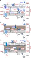
Figure 1
Figure 1
Gene Ontology (GO) for cellular components and protein subcellular location in bacteria. (A) In diderm-LPS bacteria, five clearly defined compartments are considered (i) the cytoplasm (CP; GO:0005737), (ii) the cytoplasmic membrane (CM; GO:0005886), (iii) the periplasm (PP; GO:0042597), (iv) the outer membrane (OM; GO:0019867), and (v) the extracellular milieu (EM; GO:0005576). The cell envelope (GO:0010339) is constituted of the OM and CM, also called inner membrane (IM), as well as a thin cell wall (CW) located in-between. The situation at the CM can be discriminated even further between locations either intrinsic (GO:0031226) or extrinsic (GO:0019897) to the CM. The former refers to gene products with covalently attached moieties embedded in the CM, which splits into locations (i) integral to CM (GO:0005887) where some part of the peptide sequence spans all or part of the CM, i.e., the integral membrane proteins (IMPs), and (ii) anchored to the external side of CM (GO: 0031362) corresponding to proteins tethered to the CM by non-polypeptidic covalently attached anchor, i.e., the lipoproteins. The latter refers to proteins extrinsic to the CM, i.e., neither anchored by covalent bonds to any moiety nor directly embedded in the CM. Peripheral proteins are loosely bound to the surface components of the CM on the internal (GO:0031234) or external side (GO:0031232). Some proteins localized at the OM can also be subunits of supramolecular protein complexes (GO:0043234). The situation at the OM can be discriminated even further between locations intrinsic (GO:003230) or extrinsic (GO:0031242) to the OM. The former refers to gene products with a covalently attached moiety embedded in the OM, which splits into locations (i) integral to OM (GO:00045203) where some part of the peptide sequence spans all or part of the OM, i.e., the outer membrane proteins (OMPs), and (ii) anchored to internal side of OM (GO:0036406) corresponding to proteins tethered to the OM by non-polypeptidic covalently attached anchor, i.e., some lipoproteins. (B) In monoderm bacteria, four clearly defined compartments are considered (i) the cytoplasm (CP; GO:0005737), (ii) the cytoplasmic membrane (CM; GO:0005886), (iii) the cell wall (CW; GO:0009275), and (iv) the extracellular milieu (EM; GO:0005576). An inner wall zone (IWZ) (Matias and Beveridge, 2005) has been identified (GO:0030287); importantly, it should not be considered sensu stricto as a periplasm since the CW is porous and therefore it is not bordered (bounded) contrary to the situation in diderm-LPS bacteria where the periplasmic space is strictly delimited by two biological membranes. The CM and CW constitute the cell envelope (GO:0010339). The situation at the CM is similar to what is described for diderm-LPS bacteria. Some proteins localized at the CM or CW can also be subunits of protein complex (GO:0043234) or be extrinsic to the CW (GO:0010339). (C) In diderm-mycolate bacteria, five clearly defined compartments are considered (i) the cytoplasm (CP; GO:0005737), (ii) the cytoplasmic membrane (CM; GO:0005886), (iii) the cell wall (CW; GO:0009275), (iv) the mycolate outer membrane (MOM) or mycomembrane (GO:0036407), and (v) the extracellular milieu (EM; GO:0005576). The cell envelope (GO:0010339) is constituted of the MOM, CW, and CM; a pseudo-periplasm might exist but remains to be evidenced. The situation at the CM is similar to what is described for diderm-LPS or diderm bacteria. The situation at the MOM (GO:0036407) can be discriminated further between location integral to the mycomembrane (GO:0036419), i.e., the MOM proteins (MOMPs), or extrinsic to the MOM (GO:0036420). Location at the cell surface (GO:0009986) refers (i) in diderm-LPS, to the OM and/or external side of the OM and is intended to proteins exposed externally (GO:0031242) or intrinsic to the OM (GO:0031230) (similar rermarks apply to diderm-mycolate bacteria and the MOM), and (ii) in monoderm bacteria, to the CW and/or external side of the CM and is intended to proteins exposed externally or attached to the CW (GO:0009275) or the CM, i.e., integrated (GO:0005887), anchored (GO:0046658) or loosely bound (GO:0031232). As the CW is not a permeability barrier in monoderm bacteria but porous, the surface proteins do not necessarily have domains protruding from the confine of the cell envelope to interact with the external environment. Altogether with the subset of proteins localized extracellularly (GO:0005576), i.e., the exoproteome, these gene products (GO numbers in red) correspond to the extracytoplasmic proteins, i.e., the extracytoproteome (proteins depicted in blue).
Figure 2
Figure 2
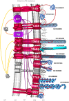
The complement of the secretome involved in colonization process in diderm-LPS bacteria. Among the 9 distinct secretion systems through which a secreted protein can be translocated across the OM in diderm-LPS bacteria, the T1SS, T2SS, T3SS, T4SS, T5SS, T7SS, T8SS, and T9SS can be involved in colonization process (depicted in red). Translocation machineries depicted in violet are protein export pathway participating to the protein transport of effectors involved in colonization process. The T1SS can secrete adhesins, which the release into the extracellular milieu (GO:0005576) and/or the association to the external side of the OM (GO:0031244) remain to be clarified. Besides the secretion of adhesion factors, the T2SS is involved in the formation of Type 4 pilus (GO:0044096), i.e., the T2SS subfamily c (T2cSS). The subfamily a of the T3SS (T3aSS) can be involved in the formation of pilus structure (GO:0009289), i.e., either the injectisome or the Hrp (hypersensitive response and pathogenicity) pilus, whereas the subfamily b of the T3SS (T3bSS) is involved in flagellum assembly (GO:009288). The T4SS is involved in the formation of pili (GO:0009289), either pilus T (T4aSS) or pilus F (T4bSS). The T5SS is involved in the secretion of adhesion either integral (GO:0045203) or extrinsic (GO:0031244) to the OM. The T7SS (CUP; chaperone-usher pathway) is involved in the formation of Type 1 pilus, and the T8SS (ENP; extracellular nucleation-precipitation pathway) in the formation of pilus of the type curli. The T9SS (Por secretion system) can secrete adhesins involved in gliding motility. In addition, some surface proteins could use systems as yet uncovered, the so-called non-classical (NC) secretion. Only branches corresponding to the complement of the secretome involved in bacterial colonization are colored. Extracytoplasmic proteins, i.e., single proteins and supramolecular protein structures, potentially involved in surface colonization are depicted in blue. Orange and yellow arrows indicate the routes for proteins targeted to the CM possessing or lacking an N-terminal SP, respectively. Violet arrows indicate the routes for exported proteins and red arrows for secreted proteins. CP, cytoplasm; IM, inner membrane; PP, periplasm; OM, outer membrane; EC, extracellular milieu; SP, signal peptide.
Figure 3
Figure 3
The complement of the secretome involved in colonization process in monoderm bacteria. Among the 8 distinct secretion systems through which a secreted protein can be translocated across the CM, the Sec, FPE, Tra, and FEA pathways can potentially be involved in colonization process in monoderm bacteria (depicted in red). The Sec pathway covers (i) integration of membrane protein (GO:0005887) via YidC, (ii) the anchoring to CM (GO:0046658) via the lipoprotein maturation pathway, (iii) the anchoring to the CW (GO:0044426) in a covalent or non-covalent manner, (iv) the association on the external side of the CW (GO:0010339), (v) the formation of cell surface supramolecular structure, namely pilus (GO:0009289) and cellulosome (GO:0043263), and (vi) protein secretion in the extracellular milieu (GO:0005576). It is worth noting that some proteins with no N-terminal SP can be translocated via Sec in a SecA2-dependent manner in monodermata (Rigel and Braunstein, ; Renier et al., 2013). The FPE is involved the formation of Type 4 pilus (GO:0044096). The Tra system (misleadingly called “Type IV-like secretion system” in monoderm bacteria) is involved in the formation of conjugative pili (GO:0009289). The FEA is involved in the secretion and assembly of the flagellum protein subunits (GO:009288). In addition, some surface proteins could use systems as yet uncovered, the so-called non-classical (NC) secretion. Extracytoplasmic proteins, i.e., single proteins and supramolecular protein structures, potentially involved in surface colonization are depicted in blue. Only branches corresponding to the complement of the secretome involved in bacterial colonization are colored. Orange and yellow arrows indicate the routes for proteins targeted to the CM possessing or lacking an N-terminal SP. Violet arrows indicate the routes for exported/secreted proteins (export and secretion are synonymous in monoderm bacteria). Green arrow indicates proteins integrated into the CM. CP: cytoplasm; CM: cytoplasmic membrane; CW: cell wall; EC, extracellular milieu; SP: signal peptide.
Figure 4
Figure 4
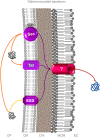
Protein secretion in diderm-mycolate bacteria. In diderm-mycolate bacteria, three protein export systems are currently recognized, the Sec, Tat and ESX (ESAT-6 system) (Digiuseppe Champion and Cox, ; Feltcher et al., ; Ligon et al., 2012). So far, none have been clearly reported and characterized as involved in surface colonization process. As in monodermata, some proteins with no N-terminal SP can be translocated via Sec in a SecA2-dependent manner (Feltcher and Braunstein, 2012). While it is clear some exported proteins (i.e., first translocated across the CM by these export systems) are further secreted into the extracellular milieu (i.e., translocated across the MOM), no mycomembrane machinery allowing the translocation across the MOM have been identified to date (Niederweis et al., ; Houben et al., ; Ligon et al., ; Freudl, ; Van Der Woude et al., 2013). In other no words (Table 1), no protein secretion system has been sensu stricto identified as yet in diderm-mycolate bacteria. It is still an enigma whether one MOM translocon or specific translocons for each of the three protein export systems are present or if the protein secretion is completed in a one-step or two-steps process (Desvaux et al., 2009a). This (or those) MOM translocon would truly correspond to a protein secretion system. For these different reasons and others, numbering the ESX (as “Type VII secretion system”) in diderm-mycolate bacteria is clearly premature and misleading (Desvaux et al., 2009a,b). Orange and yellow arrows indicate the routes of proteins targeted to the CM possessing or lacking an N-terminal SP. Violet arrows indicate the routes for exported proteins and red arrows for secreted proteins. CP, cytoplasm; CM, cytoplasmic membrane; CW, cell wall; MOM, mycolate outer membrane (or mycomembrane); EC, extracellular milieu; SP, signal peptide.

References

    1. Abdallah A. M., Gey Van Pittius N. C., Champion P. A., Cox J., Luirink J., Vandenbroucke-Grauls C. M., et al. (2007). Type VII secretion–mycobacteria show the way. Nat. Rev. Microbiol. 5, 883–891 10.1038/nrmicro1773 - DOI - PubMed
    1. Alvarez-Martinez C. E., Christie P. J. (2009). Biological diversity of prokaryotic Type IV secretion systems. Microbiol. Mol. Biol. Rev. 73, 775–808 10.1128/MMBR.00023-09 - DOI - PMC - PubMed
    1. An Y. H., Friedman R. J. (1998). Concise review of mechanisms of bacterial adhesion to biomaterial surfaces. J. Biomed. Mater. Res. 43, 338–348 - PubMed
    1. Antelmann H., Tjalsma H., Voigt B., Ohlmeier S., Bron S., Van Dijl J. M., et al. (2001). A proteomic view on genome-based signal peptide predictions. Genome Res. 11, 1484–1502 10.1101/gr.182801 - DOI - PubMed
    1. Antelmann H., Van Dijl J. M., Bron S., Hecker M. (2006). Proteomic survey through secretome of Bacillus subtilis. Methods Biochem. Anal. 49, 179–208 - PubMed

LinkOut - more resources