

Primary bone carcinosarcoma of the fibula with chondrosarcoma and squamous cell carcinoma components
- PMID: 24133601
- PMCID: PMC3796245
Primary bone carcinosarcoma of the fibula with chondrosarcoma and squamous cell carcinoma components
Abstract
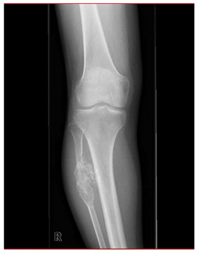
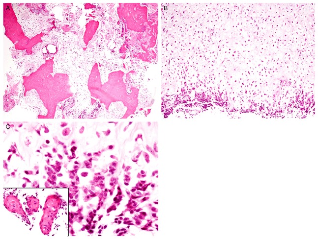
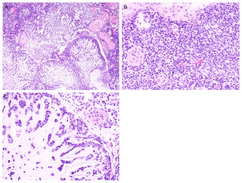
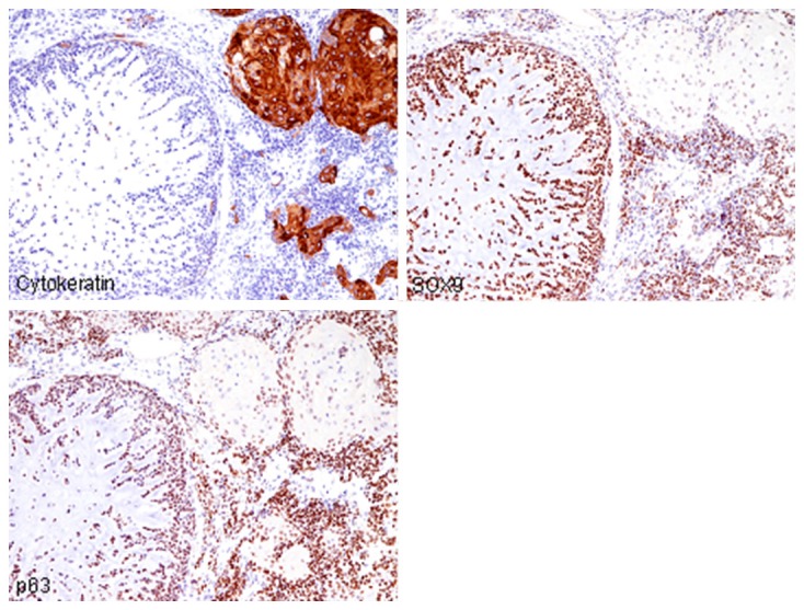
Carcinosarcoma is defined as a malignant neoplasm that is composed of both carcinomatous and sarcomatous components. The occurrence of carcinosarcoma in the bone is extremely rare. In this report, we describe the third documented de novo case of carcinosarcoma of the bone. A 59-year-old Japanese female presented with a painful tumor in her right lower leg. Plane radiography revealed an osteolytic destructive lesion with periosteal reaction and mineralization in the right fibula. Resection of the fibula tumor was performed under a clinical diagnosis of chondrosarcoma. Histopathological study revealed that the tumor was comprised of three components. The main component was proliferation of small round to short spindle cells (approximately 50%), and the remaining components were chondrosarcoma (30%) and squamous cell carcinoma (20%). Immunohistochemically, SOX9 was expressed in the small round to spindle cells and chondrosarcoma component, and p63 and p40 were expressed in all three components. Accordingly, an ultimate diagnosis of carcinosarcoma of the bone was made. The clinicopathological analysis of carcinosarcoma of the bone revealed that this type of tumor affects the middle-aged to elderly persons and occurs in the long bone. All three de novo cases had chondrosarcoma and squamous cell carcinoma components. One of the 3 patients died of the disease. The histogenesis of carcinosarcoma of the bone remains a matter of controversy, although a multpotential stem cell theory has been proposed. Additional studies are required to clarify the clinical behavior and histogenesis of carcinosarcoma of the bone.
Keywords: Carcinosarcoma; bone; chondrosarcoma; squamous cell carcinoma.
Figures
References
-
- Kondi-Pafiti A, Grapsa D, Hasiakos D, Gennatas K, Fotiou S. Carcinosarcomas of the uterus and ovary: a clinicopathologic and immunohistochemical study of 11 cases. Eur J Gynaecol Oncol. 2009;30:93–7. - PubMed
-
- Synder MJ, Robboy SJ, Vollmer RT, Dodd LG. An abnormal cervicovaginal cytology smear in uterine carcinosarcoma is an adverse prognostic sign. Analysis of 25 cases. Am J Clin Pathol. 2004;122:434–9. - PubMed
-
- Ikegami H, Iwasaki H, Ohjimi Y, Takeuchi T, Ariyoshi A, Kikuchi M. Sarcomatoid carcinoma of the urinary bladder: A clinicopathologic and immunohistochemical analysis of 14 patients. Hum Pathol. 2000;31:332–40. - PubMed
Publication types
MeSH terms
LinkOut - more resources
Full Text Sources
Medical
Research Materials