

Case Reports
doi: 10.3350/cmh.2013.19.3.315.
Epithelioid hemangioendothelioma of the liver
Affiliations
- PMID: 24133671
- PMCID: PMC3796683
- DOI: 10.3350/cmh.2013.19.3.315
Item in Clipboard
Case Reports
Epithelioid hemangioendothelioma of the liver
Clin Mol Hepatol.
2013 Sep.
No abstract available
Keywords: Hemangioendothelioma, Epithelioid; Liver.
Conflict of interest statement
The authors have no conflicts to disclose.
Figures

Magnetic resonance imaging findings. (A) Axial T2-weighted image shows high signal intensity in the central zone (arrow) and concentric alteration in signal intensity, corresponding to regions of different histology. (B) Axial T1-weighted image shows low signal intensity in the central sclerotic zone (arrow).
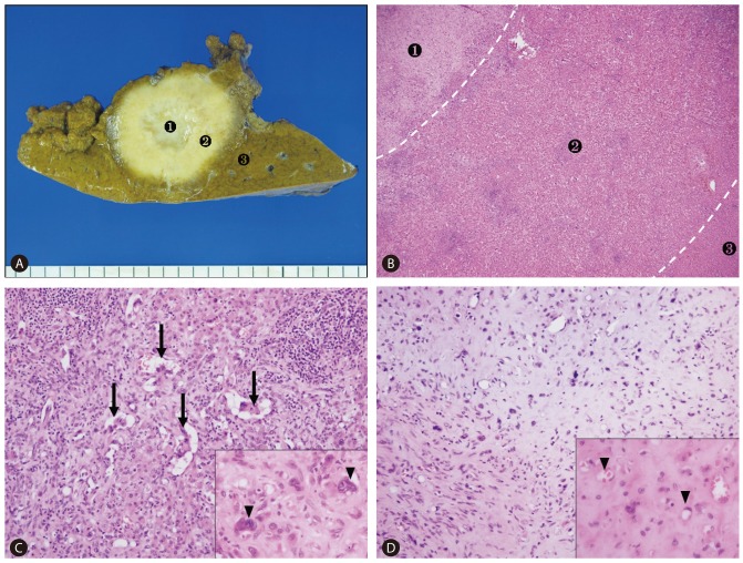
Macroscopic and histological examination. (A) The cut surface reveals a firm, yellowish-white mass with irregular margins and central fibrosis (No.1: central zone, No.2: peripheral zone, No.3: normal liver). (B) Histological findings correspond to the gross appearance of the tumor (No.1: central zone, No.2: peripheral zone, No.3: normal liver) (H&E, ×40). (C) At the peripheral zone, epithelioid tumor cells form polypoid projections (arrows) in dilated sinusoids, and epithelioid cells show atypical nuclei with prominent nucleoli (inlet, arrowheads) (H&E, ×200). (D) At the central zone, the dendritic tumor cells are embedded in myxochondroid and sclerotic stroma. Characteristically, the tumor cells have intracytoplasmic vacuoles containing red blood cells, which resemble signet ring-like structures (inlet, arrowhead) (H&E, ×200).
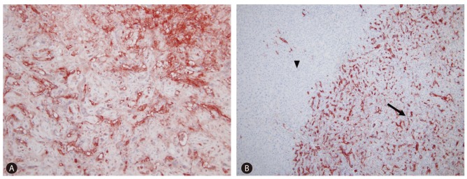
Immunohistochemical results. (A) The tumor cells synthesize factor VIII-related antigen (von Willebrand factor) in the cytoplasm and neoplastic vascular lumens. (B) The tumor cells with CD34 positivity (right arrow), in contrast to the neighboring normal hepatocytes, which show no immunoreactivity for CD34 (left arrowhead) and the immunoreactivity pattern for CD31 is identical to CD34 (figure not shown).
Similar articles
-
[Hepatic epithelioid hemangioendothelioma: a case report].Zhonghua Bing Li Xue Za Zhi. 2005 Jun;34(6):383. Zhonghua Bing Li Xue Za Zhi. 2005. PMID: 16185519 Chinese. No abstract available.
-
Epithelioid hemangioendothelioma of the liver.Clin Gastroenterol Hepatol. 2005 Jul;3(7):xxxii. doi: 10.1016/s1542-3565(05)00361-7. Clin Gastroenterol Hepatol. 2005. PMID: 16206490 No abstract available.
-
Hepatic epitheloid hemangioendothelioma: multiphase CT appearance and correlation with pathology.Crit Rev Comput Tomogr. 2004;45(5-6):343-54. Crit Rev Comput Tomogr. 2004. PMID: 15747575 Review.
-
[Characteristics of ERG, Fli-1, CD34, CD31 and FⅧRAg expression in hepatic malignant vascular tumors].Zhonghua Bing Li Xue Za Zhi. 2017 Nov 8;46(11):760-763. doi: 10.3760/cma.j.issn.0529-5807.2017.11.005. Zhonghua Bing Li Xue Za Zhi. 2017. PMID: 29136688 Chinese.
-
[Hepatic epithelioid hemangioendothelioma in needle biopsy specimens: report of 5 cases with review of literature].Zhonghua Bing Li Xue Za Zhi. 2011 Jan;40(1):23-6. Zhonghua Bing Li Xue Za Zhi. 2011. PMID: 21429354 Review. Chinese.
Cited by
-
Spectrum of appearances on CT and MRI of hepatic epithelioid hemangioendothelioma.BMC Gastroenterol. 2015 Jun 19;15:69. doi: 10.1186/s12876-015-0299-x. BMC Gastroenterol. 2015. PMID: 26088585 Free PMC article.
-
Hepatic epithelioid hemangioendothelioma: Update on diagnosis and therapy.World J Clin Cases. 2020 Sep 26;8(18):3978-3987. doi: 10.12998/wjcc.v8.i18.3978. World J Clin Cases. 2020. PMID: 33024754 Free PMC article. Review.
-
Imaging findings of hepatic epithelioid hemangioendothelioma and fibrolamellar hepatocellular carcinoma: a critical appraisal of current literature about imaging features of two rare liver cancers.Transl Cancer Res. 2019 Apr;8(Suppl 3):S297-S310. doi: 10.21037/tcr.2018.11.33. Transl Cancer Res. 2019. PMID: 35117109 Free PMC article. Review.
-
Hepatic epithelioid hemangioendothelioma metastasized to the peritoneum, omentum and mesentery: a case report.Int J Clin Exp Pathol. 2015 May 1;8(5):5883-9. eCollection 2015. Int J Clin Exp Pathol. 2015. PMID: 26191313 Free PMC article. Review.
-
Updated information regarding management of hepatic epithelioid hemangioendothelioma.Intractable Rare Dis Res. 2022 Nov;11(4):211-214. doi: 10.5582/irdr.2022.01113. Intractable Rare Dis Res. 2022. PMID: 36457586 Free PMC article.
References
-
- Ishak KG, Sesterhenn IA, Goodman ZD, Rabin L, Stromeyer FW. Epithelioid hemangioendothelioma of the liver: a clinicopathologic and follow-up study of 32 cases. Hum Pathol. 1984;15:839–852. - PubMed
-
- Makhlouf HR, Ishak KG, Goodman ZD. Epithelioid hemangioendothelioma of the liver: a clinicopathologic study of 137 cases. Cancer. 1999;85:562–582. - PubMed
-
- Mehrabi A, Kashfi A, Fonouni H, Schemmer P, Schmied BM, Hallscheidt P, et al. Primary malignant hepatic epithelioid hemangioendothelioma: a comprehensive review of the literature with emphasis on the surgical therapy. Cancer. 2006;107:2108–2121. - PubMed
-
- Weiss SW, Enzinger FM. Epithelioid hemangioendothelioma: a vascular tumor often mistaken for a carcinoma. Cancer. 1982;50:970–981. - PubMed
-
- Weiss SW, Goldblum JR. Hemangioendothelioma: vascular tumors of intermediate malignancy. In: Weiss SW, Goldblum JR, editors. Enzinger and Weiss's Soft Tissue Tumors. 5th edition. PA: Mosby; 2008. pp. 681–702.
Publication types
MeSH terms
Substances
LinkOut - more resources
Full Text Sources
Other Literature Sources
Medical
