. 2013 Oct 17:14:125.
doi: 10.1186/1471-2202-14-125.

670 nm light mitigates oxygen-induced degeneration in C57BL/6J mouse retina

Affiliations

670 nm light mitigates oxygen-induced degeneration in C57BL/6J mouse retina

Rizalyn Albarracin et al. BMC Neurosci. .

Abstract

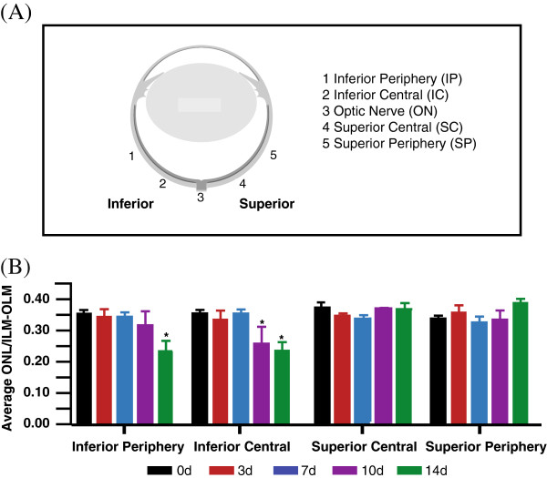
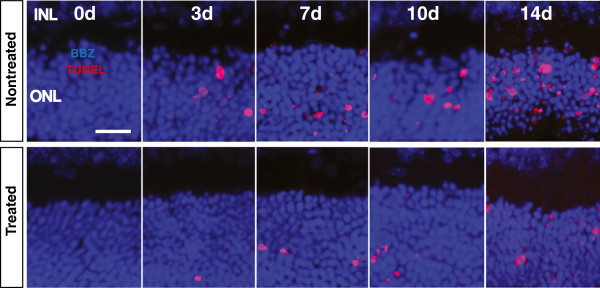
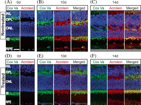
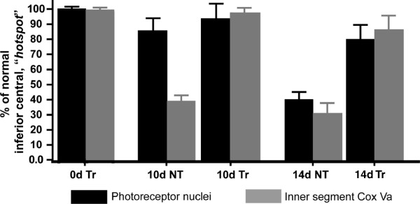
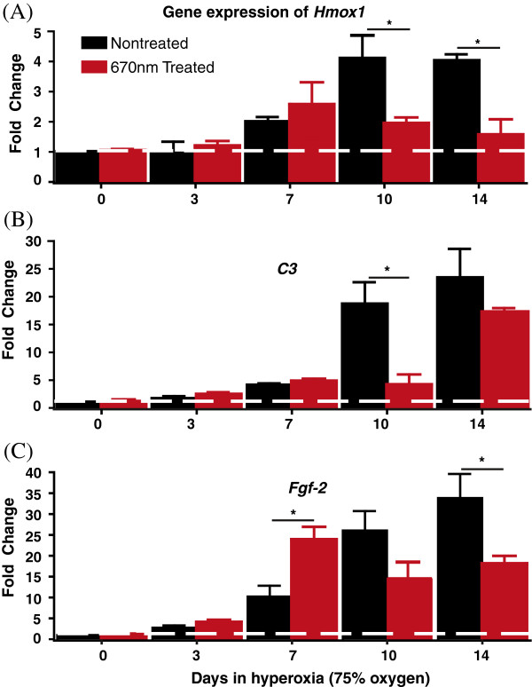
Background: Irradiation with light wavelengths from the far red (FR) to the near infrared (NIR) spectrum (600 nm -1000 nm) has been shown to have beneficial effects in several disease models. In this study, we aim to examine whether 670 nm red light pretreatment can provide protection against hyperoxia-induced damage in the C57BL/6J mouse retina. Adult mice (90-110 days) were pretreated with 9 J/cm2 of 670 nm light once daily for 5 consecutive days prior to being placed in hyperoxic environment (75% oxygen). Control groups were exposed to hyperoxia, but received no 670 nm light pretreatment. Retinas were collected after 0, 3, 7, 10 or 14 days of hyperoxia exposure (n = 12/group) and prepared either for histological analysis, or RNA extraction and quantitative polymerase chain reaction (qPCR). Photoreceptor damage and loss were quantified by counting photoreceptors undergoing cell death and measuring photoreceptor layer thickness. Localization of acrolein, and cytochrome c oxidase subunit Va (Cox Va) were identified through immunohistochemistry. Expression of heme oxygenase-1 (Hmox-1), complement component 3 (C3) and fibroblast growth factor 2 (Fgf-2) genes were quantified using qPCR.

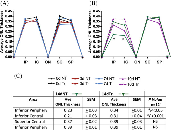
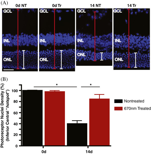
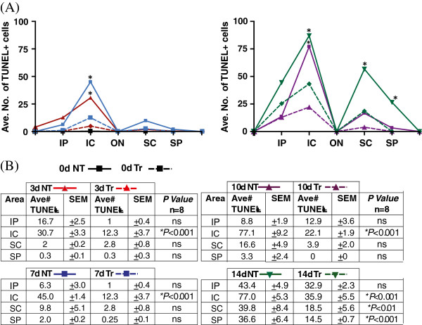
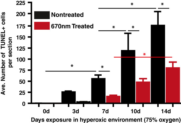
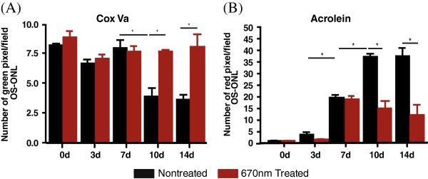
Results: The hyperoxia-induced photoreceptor loss was accompanied by reduction of metabolic marker, Cox Va, and increased expression of oxidative stress indicator, acrolein and Hmox-1. Pretreatment with 670 nm red light reduced expression of markers of oxidative stress and C3, and slowed, but did not prevent, photoreceptor loss over the time course of hyperoxia exposure.

Conclusion: The damaging effects of hyperoxia on photoreceptors were ameliorated following pretreatment with 670 nm light in hyperoxic mouse retinas. These results suggest that pretreatment with 670 nm light may provide stability to photoreceptors in conditions of oxidative stress.


Figures

Figure 1
Figure 1
Change in ONL thickness with exposure to hyperoxia. Quantitative analysis of the impact of hyperoxia exposure (75% oxygen) to the photoreceptor population was performed in retinas of adult C57BL/6J mice (n = 12). (A) The average thickness of the outer nuclear layer (ONL) was sampled from four main areas; the inferior periphery, inferior central, superior central and superior periphery of the retinas from 0d control (black), 3d (red), 7d (blue), 10d (purple), and 14d (green). (B) Significant thinning of the ONL is evident by 10d of hyperoxic exposure, specifically in the inferior central area of the retina. At 14d in hyperoxia, depletion of the ONL has spread from the inferior central to the inferior periphery. The error bars representing the ± SEM. *Statistically significant thinning of the ONL (p < 0.05) compared to control animals.
Figure 2
Figure 2
Quantitative assessment of the effects of 670 nm light on the ONL thickness of hyperoxic mice. The average thickness of the ONL was measured along the retinas of the nontreated (NT), hyperoxia-exposed (solid colored lines) and 670 nm-treated (Tr), hyperoxia-exposed mice (broken colored lines) from (A) 0d, 3d, 7d and (B) 10d and 14d groups. The retinas from 0d groups (NT, solid black line and Tr, broken black line) served as controls. (C) At 14d, the ONL of the Tr group (broken green line) in the inferior region was significantly thicker than the NT retinas (solid green line). *Statistically significant difference (p < 0.001) compared to the 670 nm light-treated retinas of the same time point.
Figure 3
Figure 3
Effects of 670 nm light treatment on the population of photoreceptors hyperoxic mice. (A) Representative micrographs of retinal sections showing the nuclear layers stained with Bisbenzimide (blue). To account for obliquely cut retina and possible artefacts, the ratio of the ONL (white line) to the OLM-ILM (red line) was used for analyses. (B) Quantitative analysis of photoreceptor nuclei density in the inferior central region of NT group (black bar) and Tr mice (red bar). Compared with the nontreated retinas at 14d hyperoxia (14dNT), treatment with 670 nm red light induced a significant level of photoreceptor nuclei density preservation in the 14dTr group (*p < 0.001). Scale bar 50 μm in A.
Figure 4
Figure 4
Effects of 670 nm light pretreatment on photoreceptor cell death following hyperoxia. Representative images of TUNEL-stained sections sampled from the inferior region of the NT and Tr retinas exposed to hyperoxia at different timepoints. TUNEL + labelling (red) was undetectable in the NT and Tr retinas from 0d (controls). A small population of TUNEL + photoreceptors was present in 3dNT retinas and continued to increase in number from 7, 10 and 14 days of exposure to hyperoxia. TUNEL + cells are also present in the 670 nm light-treated groups with significantly reduced number. The blue staining is Bisbenzimide, a DNA-specific stain, identifying the nuclear layers of the retina. Scale bar 25 μm.
Figure 5
Figure 5
Quantitative analyses of photoreceptors death. (A) TUNEL + photoreceptor cells were counted across the retina (from the inferior to superior) in 0d, 3d, 7d, 10d and 14d animals and data from Nontreated (solid colored lines) and 670 nm Treated (broken colored lines) groups were compared. TUNEL + cells were present in the inferior central area (hotspot) of the nontreated retinas from 3d (solid red line) and the number of cell death increased as a function of hyperoxia-exposure time. (B) In the 14dNT retinas (solid green line), hyperoxia exposure caused widespread cell death that also affected the photoreceptors in the superior retina. By contrast, these numbers were significantly reduced by 670 nm light pretreatment. *Statistically significant difference (p < 0.001) compared to NT groups.
Figure 6
Figure 6
Quantitative analysis of the average total number of TUNEL + cells. Total counts of TUNEL + photoreceptors across the entire section of NT (black bar) and Tr (red bar) retinas from 0d, 3d, 7d, 10d and 14d groups. *Statistically significant difference (p < 0.05) between groups marked with black or red lines.
Figure 7
Figure 7
Immunohistochemical analysis of Cox Va and acrolein. (A-F) Effects of 670 nm light treatment on the metabolic status and oxidative stress profiles of the mouse retinas following exposure to 10d and 14d of hyperoxic exposure were examined by immunohistochemical staining. Cryosectioned retinas from the inferior region of the NT and Tr groups were labelled with marker for mitochondrial metabolism, Cox Va (green) and indicator of oxidative stress, acrolein (red). Compared to 0d control retina (A), Cox Va labelling patterns (green) in the 10dNT and 14dNT were disrupted (B-C, green) but not in the 10dTr and 14dTr groups (E-F, green). Exposure to hyperoxia caused a significant increase in the acrolein expression in 10dNT and 14dNT retinas (B-C, red) compared to 0d control (D). The acrolein labelling was also present in the 10dTr and 14dTr groups but the intensity was much lower than the NT retinas (E-F, red). Scale bar 25 μm.
Figure 8
Figure 8
670 nm light pretreatment regulates the expression of acrolein and Cox Va. (A-B) Quantitative measurement of the fluorescent labelling intensity of retinal section from the inferior region labelled with mitochondrial Cox Va (A) and acrolein (B) from the nontreated (black) and 670 nm light-treated (red) hyperoxia-exposed retinas for 0, 3, 7, 10 and 14 days. *Statistically significant difference (p < 0.05) between groups highlighted by the solid black vertical line.
Figure 9
Figure 9
Correlation between the population of photoreceptor nuclei (black bar) and the mitochondrial Cox Va immunostaining (gray bar) in the inner segments. The level of Cox Va expressed by the mitochondria in the inner segments is proportional to the number of photoreceptor nuclei in all groups, except for retinas in 10dNT group. Animals that from 0dNT (nontreated, non-hyperoxic) group were used as baseline controls.
Figure 10
Figure 10
Quantitative gene expression analyses. (A-C) Differential expression profiles of oxidative stress-inducible (A)Hmox-1, (B) proinflammatory C3 and (C) neuroprotectant Fgf-2 mRNA transcripts were measured using real time quantitative PCR. cDNAs from NT (black bar) and 670 nm-Tr (red) C57BL/6J retinas exposed to 0d, 3d, 7d, 10d 14d hyperoxia were used for quantitative analysis. The results were averaged from three independent experiments. The broken horizontal white line represents the fold change (FC) of 1 which means no change in the gene expression. The fold change of >1 represents gene upregulation while FC < 1 indicates downregulation. *Statistically significant difference (p < 0.05) between groups highlighted by the solid black horizontal line.

References

    1. Whelan HT, Smits RL, Buchman EV, Whelan NT, Turner SG, Margolis DA, Cevenini V, Stinson H, Ignatius R, Martin T. et al.Effect of NASA light-emitting diode irradiation on wound healing. Lasers Surg Med. 2001;14(6):305–314. doi: 10.1089/104454701753342758. - DOI - PubMed
    1. Whelan HT. DARPA soldier self care: rapid healing of laser eye injuries with light emitting diode technology. USA: St Pete Beach; 2004. (RTO HFM symposium on combat casualty care in ground based tactical situations: trauma technology and emergency medical procedures. vol. 109).
    1. Zhang Y, Song S, Fong CC, Tsang CH, Yang Z, Yang M. cDNA microarray analysis of gene expression profiles in human fibroblast cells irradiated with red light. J Invest Dermatol. 2003;14(5):849–857. doi: 10.1046/j.1523-1747.2003.12133.x. - DOI - PubMed
    1. Mester E, Spiry T, Szende B, Tota JG. Effect of laser rays on wound healing. Am J Surg. 1971;14(4):532–535. doi: 10.1016/0002-9610(71)90482-X. - DOI - PubMed
    1. Liang H, Whelan H, Eells J, Wong-Riley M. Near-infrared light via light-emitting diode treated is therapeutic against rotenone- and MPP + −induced neurotoxicity. Neuroscience. 2008;14(4):963–974. doi: 10.1016/j.neuroscience.2008.03.042. - DOI - PMC - PubMed

Publication types

MeSH terms

LinkOut - more resources