. 2014 Mar;50(3):637-46.
doi: 10.1165/rcmb.2013-0199OC.

Chemosensory functions for pulmonary neuroendocrine cells

Affiliations

Chemosensory functions for pulmonary neuroendocrine cells

Xiaoling Gu et al. Am J Respir Cell Mol Biol. 2014 Mar.

Abstract

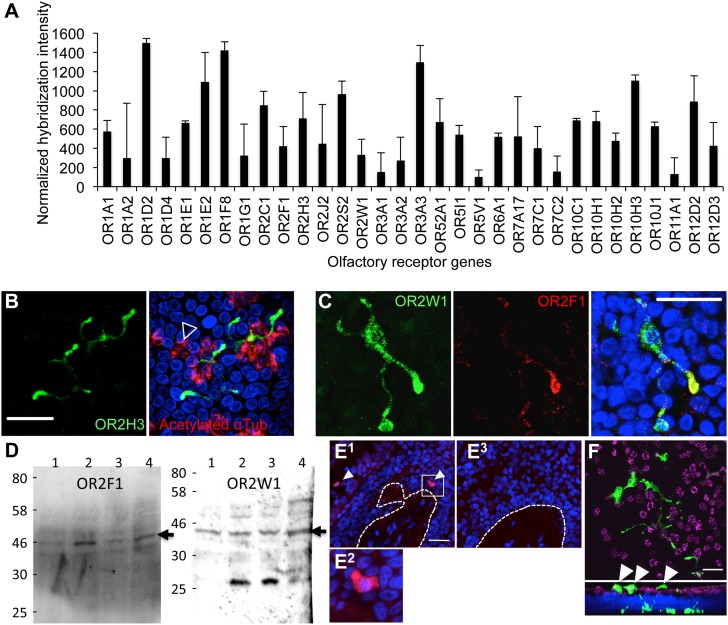
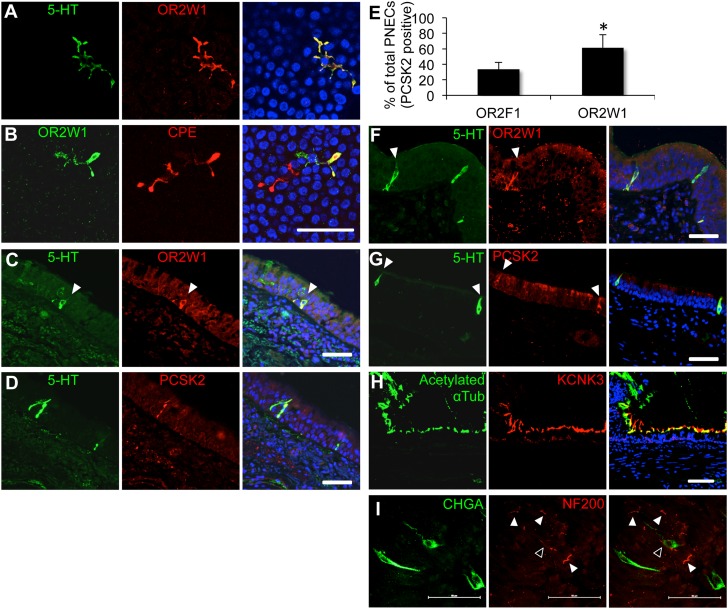
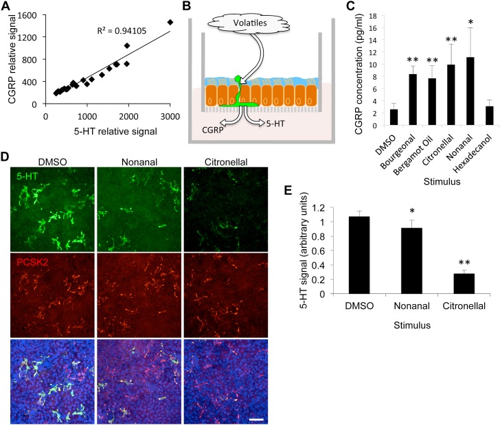
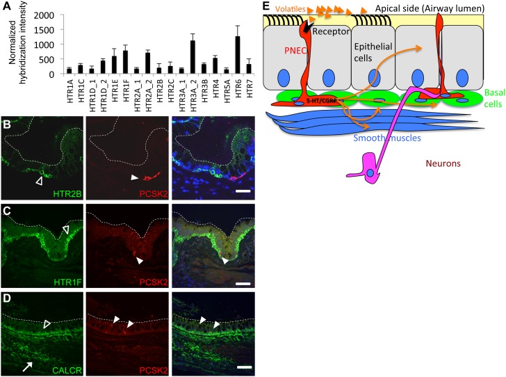
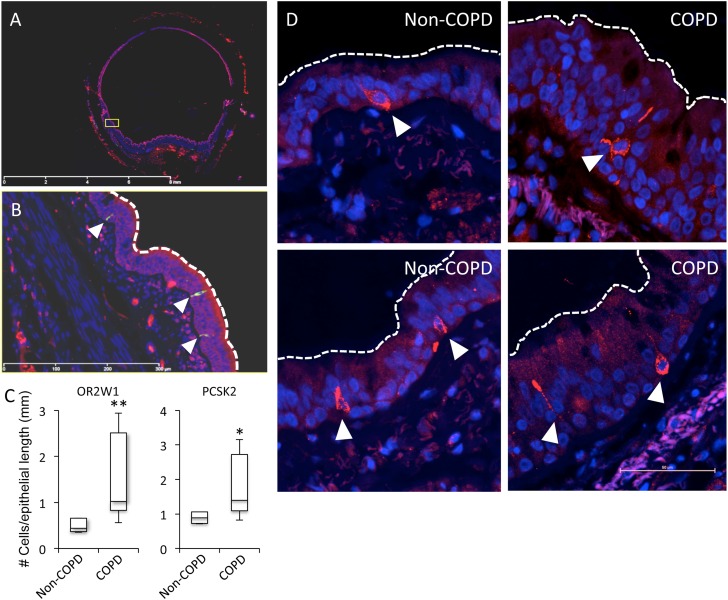
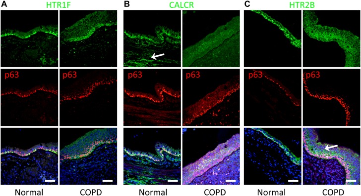
The mammalian airways are sensitive to inhaled stimuli, and airway diseases are characterized by hypersensitivity to volatile stimuli, such as perfumes, industrial solvents, and others. However, the identity and function of the cells in the airway that can sense volatile chemicals remain uncertain, particularly in humans. Here, we show that solitary pulmonary neuroendocrine cells (PNECs), which are morphologically distinct and physiologically undefined, might serve as chemosensory cells in human airways. This conclusion is based on our finding that some human PNECs expressed members of the olfactory receptor (OR) family in vivo and in primary cell culture, and are anatomically positioned in the airway epithelium to respond to inhaled volatile chemicals. Furthermore, apical exposure of primary-culture human airway epithelial cells to volatile chemicals decreased levels of serotonin in PNECs, and the led to the release of the neuropeptide calcitonin gene-related peptide (CGRP) to the basal medium. These data suggest that volatile stimulation of PNECs can lead to the secretion of factors that are capable of stimulating the corresponding receptors in the lung epithelium. We also found that the distribution of serotonin and neuropeptide receptors may change in chronic obstructive pulmonary disease, suggesting that increased PNEC-dependent chemoresponsiveness might contribute to the altered sensitivity to volatile stimuli in this disease. Together, these data indicate that human airway epithelia harbor specialized cells that respond to volatile chemical stimuli, and may help to explain clinical observations of odorant-induced airway reactions.


Figures

Figure 1.
Figure 1.
Olfactory receptors (ORs) expressed in cultured preparations of human airway epithelial cells. (A) OR gene expression by microarray analysis of mRNAs isolated from primary airway cell preparations. Shown is the mean (± SEM) from 10 independent donors. (B) Representative photomicrograph of immunostaining for the OR, OR2H3 (green), in primary-culture human airway cells demonstrating an interdigitating morphology. Cilia are labeled in red (acetylated α-tubulin, open arrowhead). Images are confocal z-stacks. (C) Colocalization of odor receptors, OR2W1 (green) and OR2F1 (red), in human airway preparations. Images are confocal z-stacks. (D) Protein blot analyses. OR2F1 and OR2W1 are expressed in human primary-culture airway cells. Each lane represents a sample from a single donor. The expected size of the corresponding ORs is indicated (∼ 45 kD; closed arrows). (E) In situ hybridization of human airway tissue sections using an OR2W1-specific riboprobe. (E1) antisense probe; (E2) enlargement of box in (E1); (E3) sense control riboprobe. Dotted lines represent the basal aspect of the stratified epithelium. Both sense and antisense probes were hybridized to sequential sections of the same tissue. (F) Cultured cells immunostained for 5-HT show extensive interdigitating morphology. Upper panel is a confocal x,y section. Lower panel is a z,y section (“stack”) of the upper panel. Note that some of the cellular extensions show projections to the apical side (white arrowheads). Nuclei are labeled with 4′,6-diamidino-2-phenylindole (blue). Scale bars, 50 μm.
Figure 2.
Figure 2.
Human pulmonary olfactory cells are solitary pulmonary neuroendocrine cells (PNECs). The OR, OR2W1, is coexpressed with 5-HT (serotonin) (A) and the enzyme, carboxipeptidase E (CPE) (B), in human primary airway epithelial cell preparations. The OR, OR2W1, is coexpressed with the neuroendocrine markers, 5-HT (C) and proprotein convertase subtilisin/kexin type 2 (PCSK2) (D), in human lung tissue sections. (E) OR abundance in preparations from a single donor. Single culture membranes were cut into two pieces and costained for the specified receptor and the PNEC marker, PCSK2. Mean receptor abundance (% cells) is shown for the receptor and PCSK2 out of the total PCSK2-positive cells. Error bars denote SEM. *P < 0.05 (one-tailed t test; n = 3 independent donors). (F and G) PNECs in lung sections from the rhesus macaque. (H) Photomicrographs of the two-pore potassium channel subfamily K member 3 (KCNK3) and the cilia marker, acetylated α-tubulin. (I) Some PNECs might be innervated. Whole-mount immunohistochemistry in the imaged region shows that at least one cell might interact with a neuronal fiber (open arrowhead). Other cells in the imaged area do not seem to interact with neuronal afferents (white arrowheads). PNECs were labeled with anti–chromogranin A (CHGA) antibody (green), and neuronal fibers were labeled with anti–neurofilament H antibody (NF200; red). Scale bars, 50 μm. See Figure E6 for larger images, and Movie E1 for a three-dimensional reconstruction.
Figure 3.
Figure 3.
PNECs release neuroendocrine factors in response to volatile apical stimuli. (A) Levels of the biogenic amine, 5-HT, and the peptide calcitonin gene-related peptide (CGRP) in individual PNECs (R2 = 0.94; n = 27 cells from three independent donors, 7–10 cells per donor). Signals represent average pixel intensity of cell bodies that were coimmunostained for 5-HT and CGRP. (B) An illustration of the supported membrane insert system used for culture of primary airway epithelial cells (20, 50). Treatment experiments were accomplished by applying the various ligands apically, while measuring the basal release of neuroendocrine factors. (C) CGRP release in basal medium after apical treatment with indicated volatile. Mean (± SEM) of CGRP levels measured by ELISA (P < 0.05, Kruskal-Wallis ANOVA; n = 4 inserts per treatment). (D and E) Treatments of primary cell preparations with nonanal or citronellal followed by immunostaining for 5-HT levels relative to controls. Shown are means (± SEM) (one-way ANOVA; P < 0.001; n = 4 inserts from a single donor per treatment). *P < 0.05; **P < 0.01. Scale bars, 50 μm. DMSO, dimethyl sulfoxide.
Figure 4.
Figure 4.
(A) 5-HT receptor (HTR) gene expression by microarray analysis of mRNAs isolated from primary airway cell preparations from 10 independent donors (as in Figure 1A). (B and C) Serotonin receptors, 5-HTR 2B and HTR1F, in basal cells (open arrowheads) detected by immunostaining. White arrowheads point to solitary PNECs. (D) The calcitonin/CGRP receptor (CALCR) is enriched in basal cells (open arrowheads) and in airway smooth muscles or possibly the neurons that innervate them (white arrow). White arrowheads in C and D mark PNECs. Dashed white lines show the apical side of the stratified epithelium. Scale bars, 50 μm. (E) Model for the possible local impact of olfactory stimulation of PNECs followed by neuroendocrine release of 5-HT and CGRP on various cell types in the airways (orange arrows).
Figure 5.
Figure 5.
PNECs are more abundant in airways from patients with chronic obstructive pulmonary disease (COPD) relative to healthy donors. (A) Representative confocal scan of a human bronchial tissue section used to quantify PNEC frequencies. (B) Magnification of yellow box in (A); dotted line indicates an example of the region used to normalize number of cells to epithelial length. Cells in the images shown were positive for both OR2W1 and PCSK2. (C) Box plots represent PNEC frequencies in tissue sections from lungs of subjects with and without COPD (*P < 0.05; **P < 0.01; Mann-Whitney U Test; n = 6–7 individuals per group). (D) The morphology and size of OR-expressing PNECs in COPD and non-COPD lungs are similar. Left panels, non-COPD; right panels, COPD. Cells were labeled with an anti-OR2W1 antibody. White arrowheads mark PNECs.
Figure 6.
Figure 6.
Spatial expression patterns of 5-HT and CGRP receptors are different in normal versus COPD lungs. (A) Representative photomicrographs of immunostaining for HTR1F (green). Staining is enriched in basal cells in normal and COPD lungs (basal cells marked with the p63 marker, red nuclei). Note that the morphology of COPD basal cells is elongated, and the distribution of the receptor uniform, compared with those in normal lungs. (B) Representative photomicrographs of immunostaining for CALCR (green). Staining is highly enriched in basal cells (marked by p63, red nuclei) and smooth muscle or neuronal fibers (white arrow) in normal donors. However, expression is lower and more diffused in COPD lungs. (C) Representative photomicrographs of immunostaining for HTR2B (green). Receptor is enriched basal cells of lungs from non-COPD subjects when compared with normal lungs. Note that in lungs of subjects with COPD, staining for HTR2B is more diffused and higher in nonbasal epithelial cells relative to basal cells (white arrow). Scale bars, 50 μm.

References

    1. Massaro GD, Massaro D. Formation of pulmonary alveoli and gas-exchange surface area: quantitation and regulation. Annu Rev Physiol. 1996;58:73–92. - PubMed
    1. Patton JS, Byron PR. Inhaling medicines: delivering drugs to the body through the lungs. Nat Rev Drug Discov. 2007;6:67–74. - PubMed
    1. Millqvist E, Bende M, Löwhagen O. Sensory hyperreactivity—a possible mechanism underlying cough and asthma-like symptoms. Allergy. 1998;53:1208–1212. - PubMed
    1. Deshpande DA, Wang WC, McIlmoyle EL, Robinett KS, Schillinger RM, An SS, Sham JS, Liggett SB. Bitter taste receptors on airway smooth muscle bronchodilate by localized calcium signaling and reverse obstruction. Nat Med. 2010;16:1299–1304. - PMC - PubMed
    1. Shah AS, Ben-Shahar Y, Moninger TO, Kline JN, Welsh MJ. Motile cilia of human airway epithelia are chemosensory. Science. 2009;325:1131–1134. - PMC - PubMed

Publication types

MeSH terms