

The immature human ovary shows loss of abnormal follicles and increasing follicle developmental competence through childhood and adolescence
- PMID: 24135076
- PMCID: PMC3860895
- DOI: 10.1093/humrep/det388
The immature human ovary shows loss of abnormal follicles and increasing follicle developmental competence through childhood and adolescence
Abstract
Study question: Do the ovarian follicles of children and adolescents differ in their morphology and in vitro growth potential from those of adults?
Summary answer: Pre-pubertal ovaries contained a high proportion of morphologically abnormal non-growing follicles, and follicles showed reduced capacity for in vitro growth.
What is known already: The pre-pubertal ovary is known to contain follicles at the early growing stages. How this changes over childhood and through puberty is unknown, and there are no previous data on the in vitro growth potential of follicles from pre-pubertal and pubertal girls.
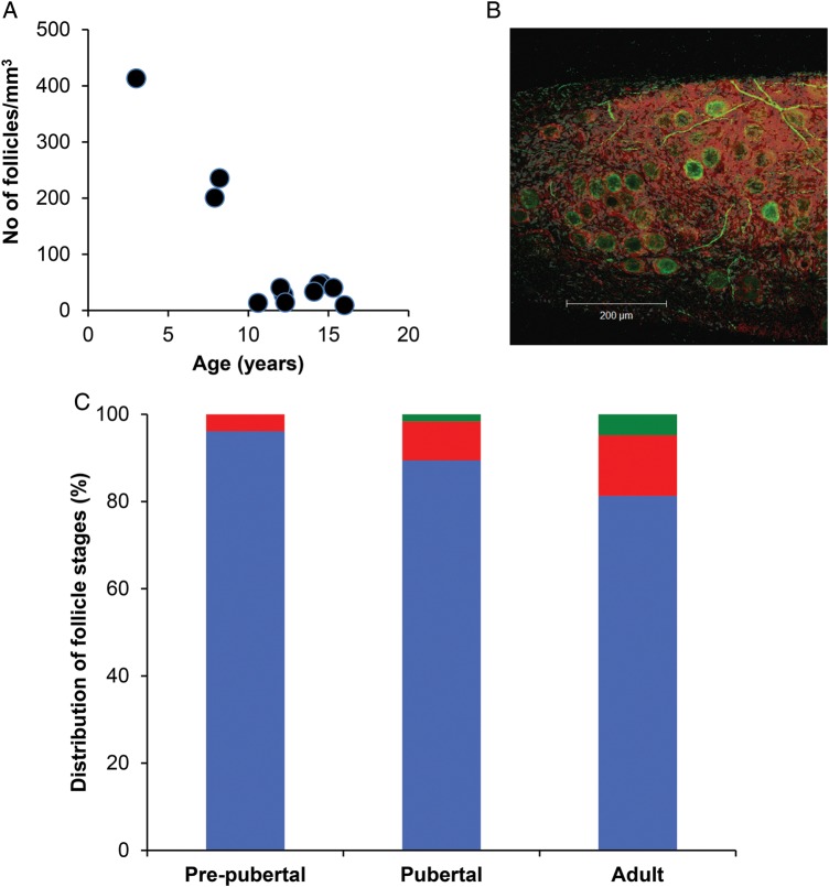
Study design, size, duration: Ovarian biopsies from five pre-pubertal and seven pubertal girls and 19 adult women were analysed histologically, cultured in vitro for 6 days, with growing follicles then isolated and cultured for a further 6 days.
Participants/materials, setting, methods: Ovarian biopsies were obtained from girls undergoing ovarian tissue cryopreservation for fertility preservation, and compared with biopsies from adult women. Follicle stage and morphology were classified. After 6 days in culture, follicle growth initiation was assessed. The growth of isolated secondary follicles was assessed over a further 6 days, including analysis of oocyte growth.
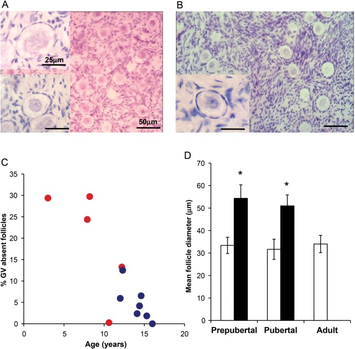
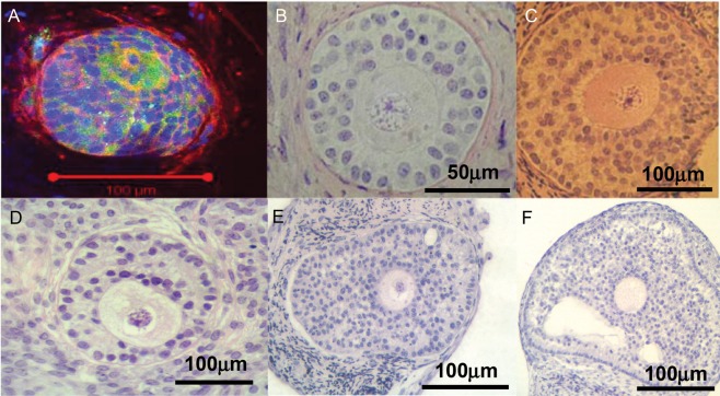
Main results and the role of chance: Pre-pubertal ovaries contained a high proportion of abnormal non-growing follicles (19.4 versus 4.85% in pubertal ovaries; 4004 follicles analysed; P = 0.02) characterized by indistinct germinal vesicle membrane and absent nucleolus. Follicles with this abnormal morphology were not seen in the adult ovary. During 6 days culture, follicle growth initiation was observed at all ages; in pre-pubertal samples there was very little development to secondary stages, while pubertal samples showed similar growth activation to that seen in adult tissue (pubertal group: P = 0.02 versus pre-pubertal, ns versus adult). Isolated secondary follicles were cultured for a further 6 days. Those from pre-pubertal ovary showed limited growth (P < 0.05 versus both pubertal and adult follicles) and no change in oocyte diameter over that period. Follicles from pubertal ovaries showed increased growth; this was still reduced compared with follicles from adult women (P < 0.05) but oocyte growth was proportionate to follicle size.
Limitations, reasons for caution: These data derive from only a small number of ovarian biopsies, although large numbers of follicles were analysed. It is unclear whether the differences between groups are related to puberty, or just age.
Wider implications of the findings: These findings show that follicles from girls of all ages can be induced to grow in vitro, which has important implications for some patients who are at high risk of malignant contamination of their ovarian tissue. The reduced growth of isolated follicles indicates that there are true intrafollicular differences in addition to potential differences in their local environment, and that there are maturational processes occurring in the ovary through childhood and adolescence, which involve the loss of abnormal follicles, and increasing follicle developmental competence.
Study funding/competing interest(s): Funded by MRC grants G0901839 and G1100357. No competing interests.
Keywords: adolescence; childhood; follicle; ovary; puberty.
Figures

References
-
- Abir R, Feinmesser M, Yaniv I, Fisch B, Cohen IJ, Ben-Haroush A, Meirow D, Felz C, Avigad S. Occasional involvement of the ovary in Ewing sarcoma. Hum Reprod. 2010;25:1708–1712. - PubMed
-
- Albamonte MI, Albamonte MS, Stella I, Zuccardi L, Vitullo AD. The infant and pubertal human ovary: Balbiani's body-associated VASA expression, immunohistochemical detection of apoptosis-related BCL2 and BAX proteins, and DNA fragmentation. Hum Reprod. 2013;28:698–706. - PubMed
-
- Albertini DF, Barrett SL. Oocyte-somatic cell communication. Reprod Suppl. 2003;61:49–54. - PubMed
-
- Anderson RA, Wallace WH. Fertility preservation in girls and young women. Clin Endocrinol (Oxf) 2011;75:409–419. - PubMed
-
- Anderson RA, Wallace WHB, Baird DT. Ovarian cryopreservation for fertility preservation: indications and outcomes. Reproduction. 2008;136:681–689. - PubMed
Publication types
MeSH terms
Grants and funding
LinkOut - more resources
Full Text Sources
Other Literature Sources
Research Materials
Miscellaneous
