. 2013 Nov;184(2):203-11.
doi: 10.1016/j.jsb.2013.10.003. Epub 2013 Oct 14.

3-D analysis of dictyosomes and multivesicular bodies in the green alga Micrasterias denticulata by FIB/SEM tomography

Affiliations

3-D analysis of dictyosomes and multivesicular bodies in the green alga Micrasterias denticulata by FIB/SEM tomography

Gerhard Wanner et al. J Struct Biol. 2013 Nov.

Abstract

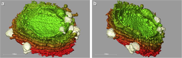
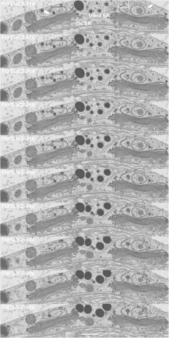
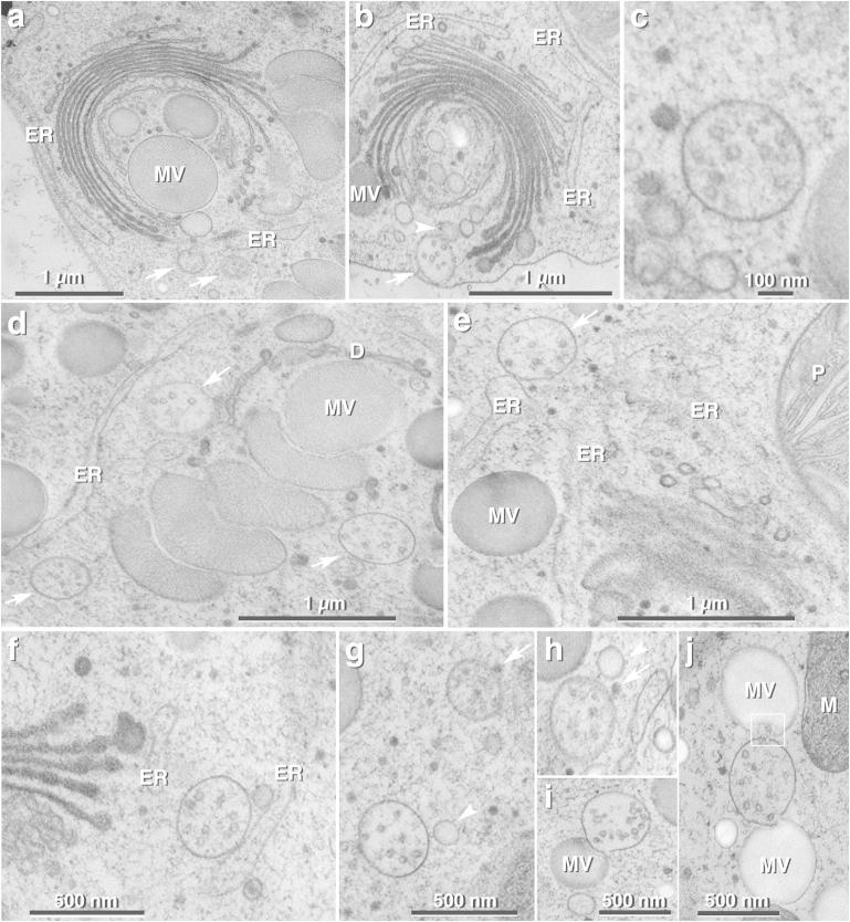
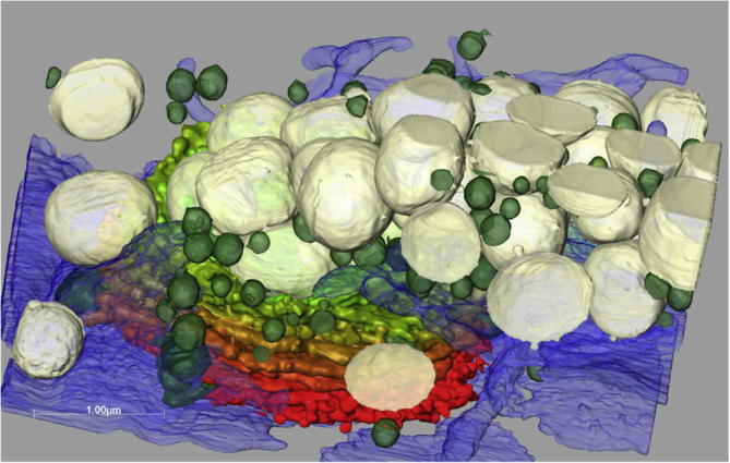
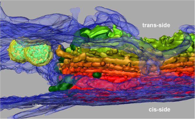
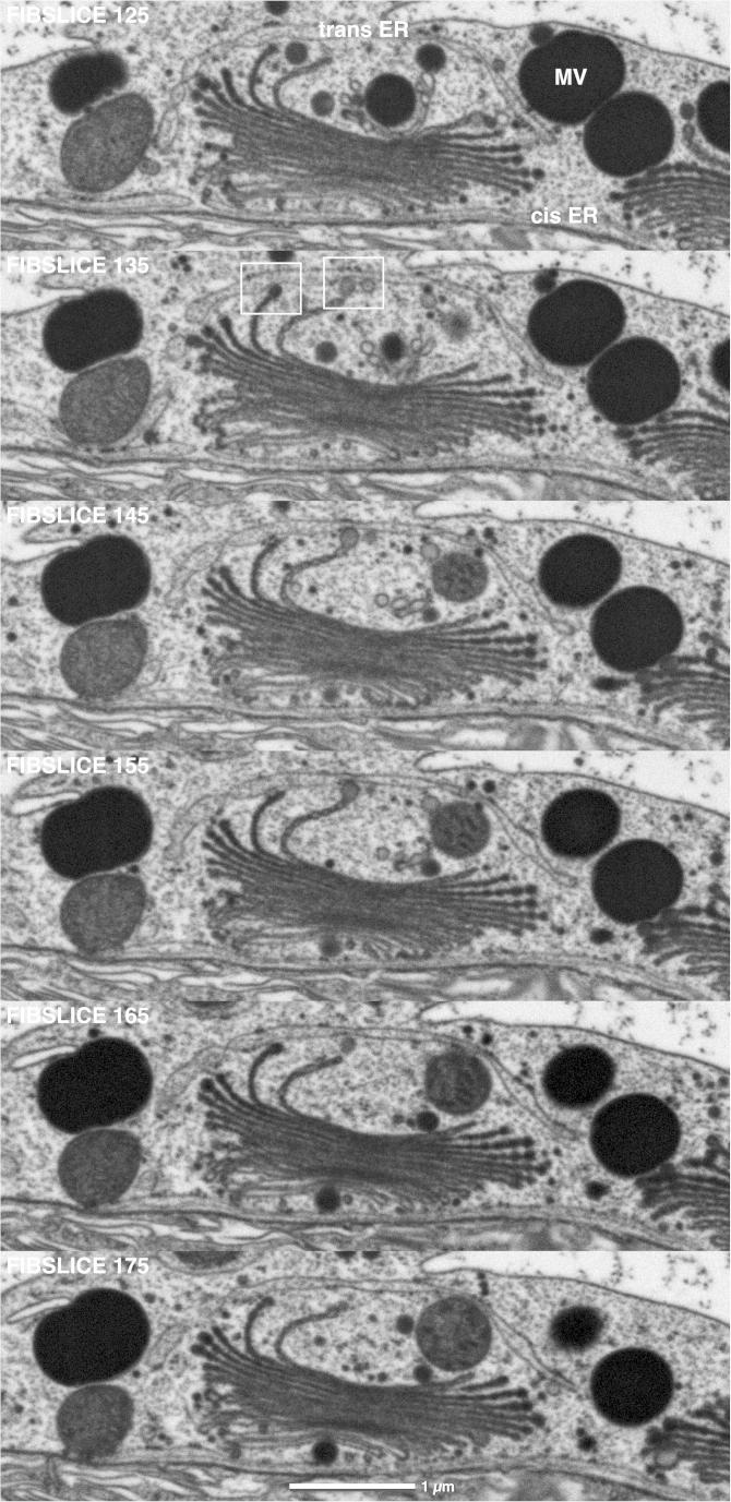
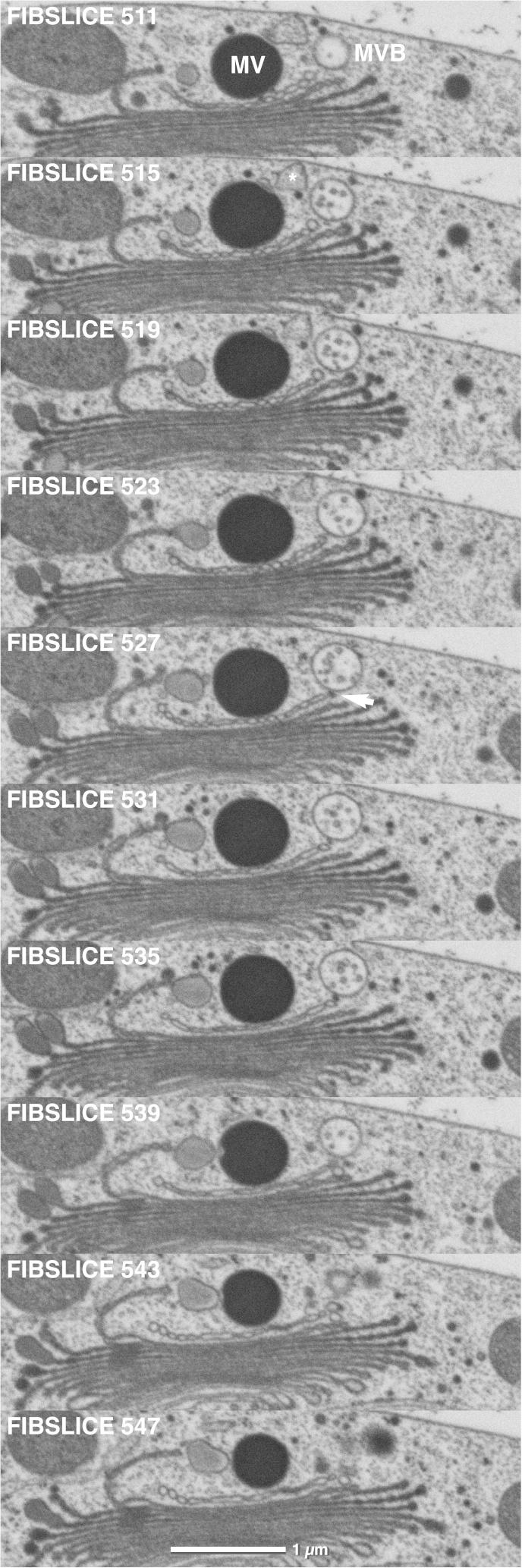
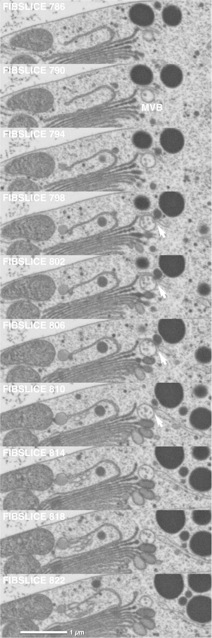
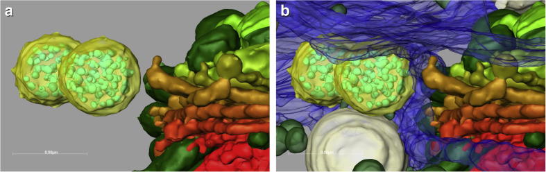
In the present study we employ FIB/SEM tomography for analyzing 3-D architecture of dictyosomes and formation of multivesicular bodies (MVB) in high pressure frozen and cryo-substituted interphase cells of the green algal model system Micrasterias denticulata. The ability of FIB/SEM of milling very thin 'slices' (5-10 nm), viewing the block face and of capturing cytoplasmic volumes of several hundred μm(3) provides new insight into the close spatial connection of the ER-Golgi machinery in an algal cell particularly in z-direction, complementary to informations obtained by TEM serial sectioning or electron tomography. Our FIB/SEM series and 3-D reconstructions show that interphase dictyosomes of Micrasterias are not only closely associated to an ER system at their cis-side which is common in various plant cells, but are surrounded by a huge "trans-ER" sheath leading to an almost complete enwrapping of dictyosomes by the ER. This is particularly interesting as the presence of a trans-dictyosomal ER system is well known from mammalian secretory cells but not from cells of higher plants to which the alga Micrasterias is closely related. In contrast to findings in plant storage tissue indicating that MVBs originate from the trans-Golgi network or its derivatives our investigations show that MVBs in Micrasterias are in direct spatial contact with both, trans-Golgi cisternae and the trans-ER sheath which provides evidence that both endomembrane compartments are involved in their formation.

Keywords: Dictyosomes; ER; FIB/SEM tomography; Micrasterias denticulata; Multivesicular bodies; TEM.


Figures

Fig.1
Fig.1
(a), and (b) Different 3-D views of the trans-side of a dictyosome of M. denticulata reconstructed from FIB/SEM series. Cisternal rims are lacerated, outermost-trans-cisterna are shorter than others and undulated. Secretory vesicles (grey) are connected to edges of trans-cisternae. From red to green: cis- to trans-cisternae.
Fig.2
Fig.2
FIB/SEM series of two dictyosomes of an interphase Micrasterias cell. Trans-most Golgi cisternae are shorter and involute respectively circular (arrows). ER cisternae present at both cis- and trans-side of the right dictyosome. Framed area indicates vesicular contact between a trans-Golgi cisterna and the trans-ER. Small secretory vesicles and large mucilage vesicles are visible around the left dictyosome. 10 nm slices, every second image shown.
Fig.3
Fig.3
TEM micrographs of ultrathin sections of high-pressure frozen and freeze substituted interphase cells of M. denticulata. (a) Dictyosomes with adjacent ER cisterna at cis- and trans-side. Trans-most cisterna is chain-like constricted. Large mucilage vesicles (MV) are in contact with rims of trans-cisternae. Two MVBs (arrows) are located in close proximity to the trans-Golgi side and to a trans-ER cisterna. (b) MVB (arrow) is in contact with vacuolar membrane. translucent vesicles (arrow head) are in contact with trans-dictyosomal cisterna and MVB. ER = endoplasmic reticulum. (c) Representative MVB of Micrasterias with intraluminal vesicles. (d) Trans-side of dictyosome (D) with stack of mucilage vesicles (MV). Three MVBs (arrows) are in close proximity to the dictyosome and to a trans-ER cisterna. ER = endoplasmic reticulum. (e) Dictyosome and parts of the enwrapping ER. Contact between MVB (arrow) and part of a trans-ER cisterna visible. MV = mucilage vesicle; ER = endoplasmic reticulum; P = Plastid. (f) Part of a dictyosome and MVB attached to an ER cisterna. ER = endoplasmic reticulum. (g,h) MVBs in spatial contact with small electron-dense vesicle (arrow) and with larger translucent vesicle (arrow head). (i,j) MVBs attached to mucilage vesicles (MV). (j) Intraluminal vesicle of MVB discharged into upper mucilage vesicle (framed area). M = mitochondrion.
Fig.4
Fig.4
3-D reconstruction of interphase dictyosome of Micrasterias with enwrapping ER system (blue) and different vesicles populations. Small secretory vesicles are shown in green, mucilage vesicles in grey. Dictyosomal cis-side in red, trans-side in green.
Fig.5
Fig.5
3-D reconstruction of dictyosome and ER envelope (blue) at cis- and trans-side of the dictyosome. Two MVBs (yellow) are visible in proximity of the dictyosomal trans-cisternae.
Fig.6
Fig.6
FIB/SEM series of dictyosome in contact to a cis- and a trans-ER cisterna. Trans-ER covers the trans-side of the dictyosome almost entirely. At least two of the trans-cisternae (framed areas) are in contact with the trans-ER either directly or via vesicular connections. The trans-most cisterna is vesicular and disconnected from Golgi stack appearing as TGN. Large mucilage vesicles (MV) are visible in close vicinity to dictyosome. 10 nm slices, every 10th image shown.
Fig.7
Fig.7
FIB/SEM series of MVB in contact with trans-most dictyosomal cisterna via small vesicles (arrow). Maturating and ripe mucilage vesicle in contact to each other and close to trans-Golgi cisternae. 10 nm slices, every 4th image shown.
Fig.8
Fig.8
FIB/SEM series of MVB in direct spatial contact to both a trans-dictyosomal cisterna and a trans-ER cisterna (arrow). The MVB also contacts one of the maturating mucilage vesicles.
Fig.9
Fig.9
(a,b). 3-D reconstruction of spatial relationship between trans-Golgi cisternae and MVBs (yellow) without (a) and with (b) enwrapping ER system (blue). The reconstructions show that the membranes of the MVBs are in direct contact with the membranes of the ER but not with the Golgi cisternae.

References

    1. Affenzeller M.J., Darehshouri A., Andosch A., Lütz C., Lütz-Meindl U. Salt stress-induced cell death in the unicellular green alga Micrasterias denticulata. J. Exp. Bot. 2009;60:939–954. - PMC - PubMed
    1. Affenzeller M.J., Darehshouri A., Andosch A., Lütz C., Lütz-Meindl U. PCD and autophagy in the unicellular green alga Micrasterias denticulata. Autophagy. 2009;5:854–855. - PMC - PubMed
    1. Aichinger N., Lütz-Meindl U. Organelle interactions and possible degradation pathways visualized in high-pressure frozen algal cells. J. Microsc. 2005;219:86–94. - PubMed
    1. Andosch A., Affenzeller M.J., Lütz C., Lütz-Meindl U. A freshwater green alga under cadmium stress: ameliorating calcium effects on ultrastructure and photosynthesis in the unicellular model Micrasterias. J. Plant Physiol. 2012;169:1489–1500. - PubMed
    1. Bassham D.C., Laporte M., Marty F., Moriyasu Y., Ohsumi Y., Olsen L.J., Yoshimoto K. Autophagy in development and stress responses of plants. Autophagy. 2006;2:2–11. - PubMed

Publication types

MeSH terms

LinkOut - more resources