. 2013 Nov;123(11):4714-30.
doi: 10.1172/JCI67333. Epub 2013 Oct 25.

Developmental and tumoral vascularization is regulated by G protein-coupled receptor kinase 2

Developmental and tumoral vascularization is regulated by G protein-coupled receptor kinase 2

Verónica Rivas et al. J Clin Invest. 2013 Nov.

Abstract

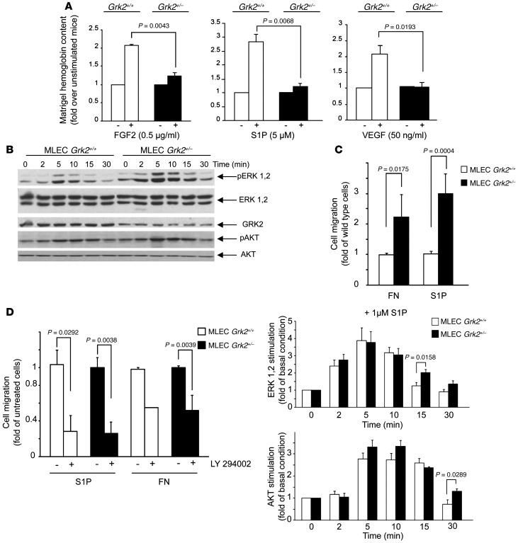
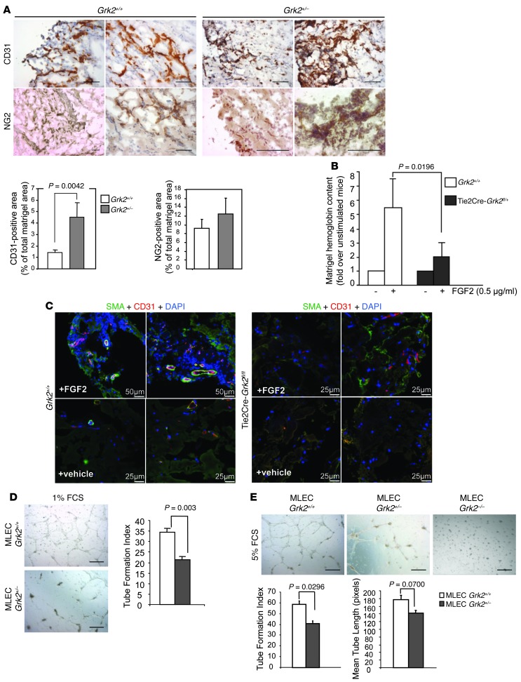
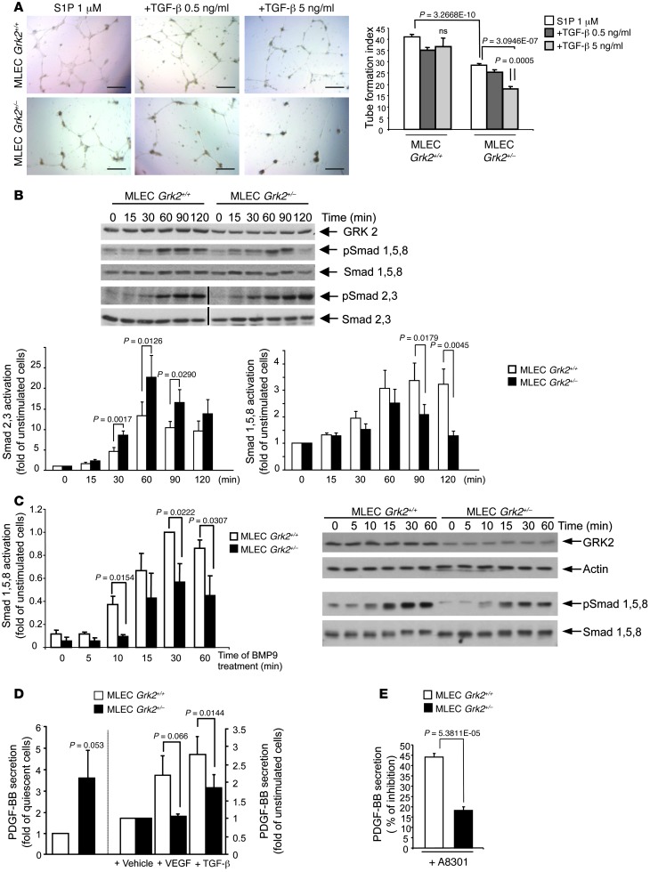
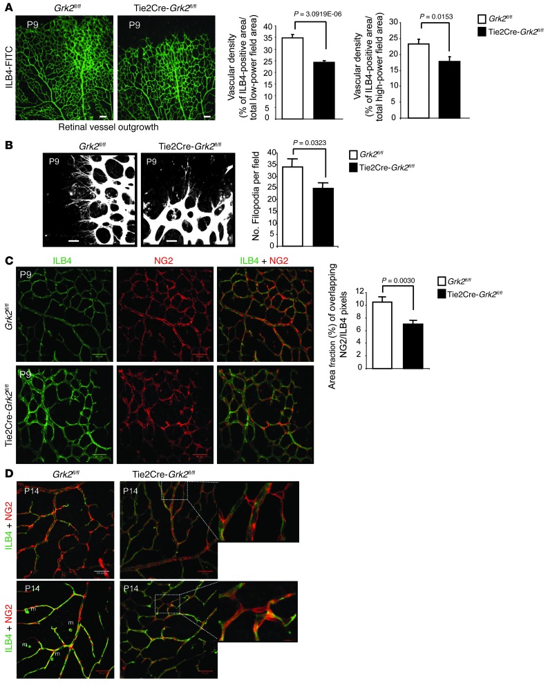
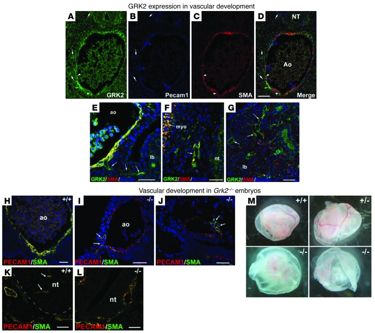
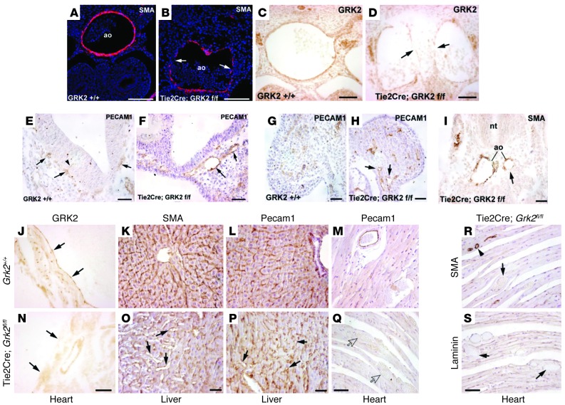
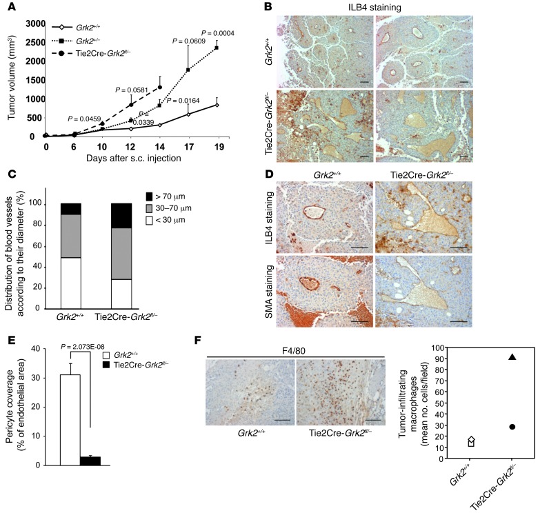
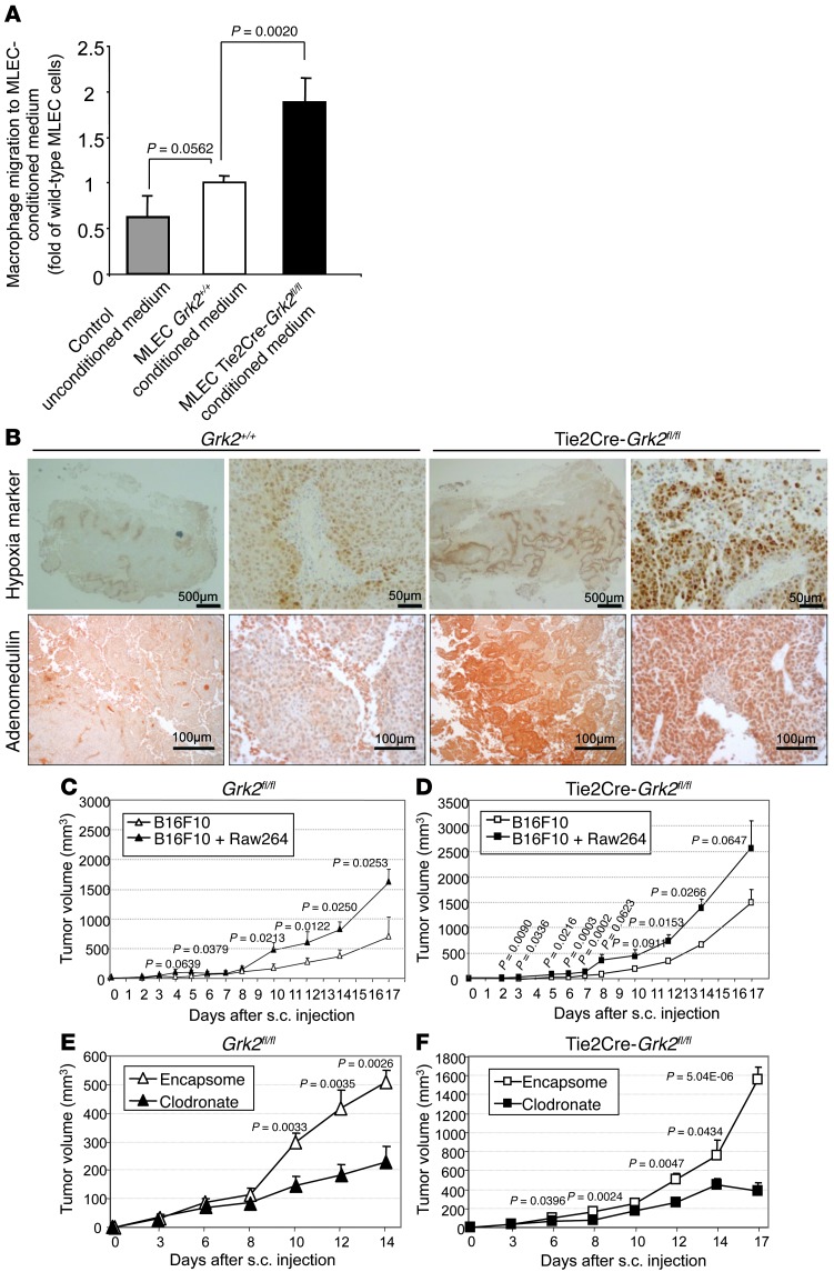
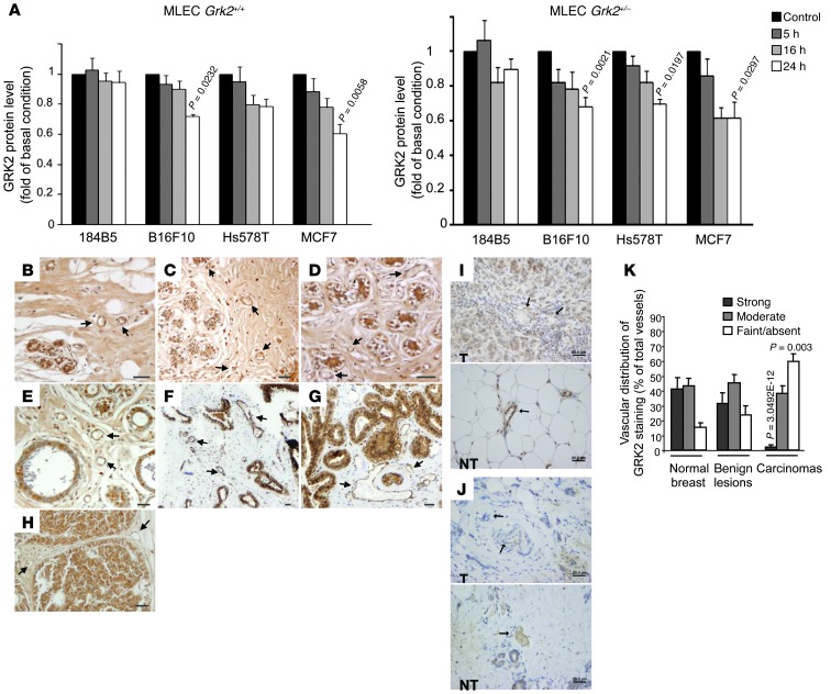
Tumor vessel dysfunction is a pivotal event in cancer progression. Using an in vivo neovascularization model, we identified G protein-coupled receptor kinase 2 (GRK2) as a key angiogenesis regulator. An impaired angiogenic response involving immature vessels was observed in mice hemizygous for Grk2 or in animals with endothelium-specific Grk2 silencing. ECs isolated from these animals displayed intrinsic alterations in migration, TGF-β signaling, and formation of tubular networks. Remarkably, an altered pattern of vessel growth and maturation was detected in postnatal retinas from endothelium-specific Grk2 knockout animals. Mouse embryos with systemic or endothelium-selective Grk2 ablation had marked vascular malformations involving impaired recruitment of mural cells. Moreover, decreased endothelial Grk2 dosage accelerated tumor growth in mice, along with reduced pericyte vessel coverage and enhanced macrophage infiltration, and this transformed environment promoted decreased GRK2 in ECs and human breast cancer vessels. Our study suggests that GRK2 downregulation is a relevant event in the tumoral angiogenic switch.


Figures

Figure 1
Figure 1. GRK2 deficiency results in an impaired in vivo angiogenesis, despite effective EC activation.
(A) Matrigel implants mixed with the indicated stimuli or vehicle were injected into WT (Grk2+/+) or global hemizygous (Grk2+/–) mice, and hemoglobin content was quantified as described in the Methods. The angiogenic response data (calculated as fold over control conditions) were obtained from 9 to 12 animals for each condition in 3 to 4 independent experiments. (B) GRK2 downregulation does not compromise the S1P-induced activation of ERK1/2 and AKT pathways in MLECs. Data from 3 to 4 independent experiments are shown. Representative blots are shown. (C and D) GRK2 downmodulation enhances the PI3K-dependent directed cell migration of ECs in response to fibronectin (FN) and S1P. Data of 4 to 6 independent experiments performed in duplicate are shown. ERK1/2 and AKT activation and cell migration assays are detailed in Methods.
Figure 2
Figure 2. Endothelial GRK2 protein levels regulate vascular morphogenesis.
(AC) Angiogenesis in mice with global or endothelial-specific reduction of GRK2 expression is characterized by a lack of functional vessels, concomitant with the occurrence of aberrant vessel-like structures with a poor mural coverage. Sections of FGF2-embeded Matrigel plugs excised (A) from WT Grk2+/+ and global Grk2+/– or (C) from Tie2Cre-Grk2fl/fl mice were analyzed by immunohistochemistry, and the density of invasion by either endothelial or mural cells was determined with (A) anti-CD31 or anti-NG2 or (C) SMA antibodies, respectively, as detailed in Methods. Ratios of CD31-positive cells to total DAPI-stained cells in FGF2-stimulated WT Grk2+/+ and Tie2Cre-Grk2fl/fl mice were 0.22 ± 0.06 and 0.58 ± 0.28, respectively. (B) Endothelial GRK2 downmodulation is sufficient to impair the functionality of angiogenic vessels. The angiogenic response in WT or Tie2Cre-GRK2 f/+ mice was determined as in Figure 1A. Data from 12 animals for each condition in 4 independent experiments are shown. (D and E) ECs with deficient GRK2 expression are less competent to form capillary-like networks in vitro. MLECs from WT (Grk2+/+), global hemizygous (Grk2+/–), or endothelial-specific (Tie2Cre-Grk2fl/fl) GRK2 knockout mice were seeded on Matrigel-coated wells in the presence of (D) 1% FCS or (E) 5% FCS. Photographs were taken after 16 to 18 hours in culture (original magnification, ×5), and formation of tubular networks was quantified as described in Methods. Data were obtained from 2 to 3 independent assays performed in duplicate. Scale bar: 100 μm (A); 25 μm (C, Tie2Cre-Grk2fl/fl and vehicle-treated Grk2+/+); 50 μm (C, FGF2-treated Grk2+/+); 500 μm (D and E).
Figure 3
Figure 3. Effect of GRK2 downregulation on TGF-β1 endothelial signaling.
(A) ECs with reduced expression of GRK2 are more sensitive to dose-dependent inhibitory effects of TGF-β1 on tube network formation. In vitro tube formation was monitored in the presence of 1 μM S1P supplemented with or without 0.5 or 5 ng/ml TGF-β1 and quantified as in Figure 2, D and E. Data from 3 independent assays performed in duplicate are shown. Scale bar: 500 μm. (B and C) Proper signaling of Smads downstream of ALK1 and ALK5 receptor activation depends on GRK2 expression. (B) Grk2+/– MLECs display enhanced TGF-β1–triggered activation of Smad2/3 paralleled by a decrease in Smad1/5/8 stimulation. The lanes were run on the same gel but, where indicated by the black vertical line, were noncontiguous. (C) BMP9-induced activation of Smad1/5/8 via ALK1 is reduced in Grk2+/– MLECs. (D) Both basal and angiogenic factor–induced PDGF-BB secretion are altered in Grk2+/– MLECs. Serum-starved cells were incubated for 48 hours in 1% FCS supplemented with or without 50 ng/ml VEGF or 5 ng/ml TGF-β1, and PDGF-BB was determined in the cell-conditioned media as described in Methods. (E) The production of PDGF-BB in Grk2+/– MLECs is less sensitive to ALK5 signaling inhibitors. Media of cells treated with 5 ng/ml TGF-β1 for 48 hours in the presence or absence of the ALK5 inhibitor A8301 (5 μM) was processed for PDGF-BB quantification. In BE data from 3–4 independent experiments are shown.
Figure 4
Figure 4. Loss of endothelial GRK2 impairs developmental outgrowth and maturation of the retinal vasculature.
(A and B) Expansion of the primary vascular plexus and endothelial filopodia was reduced in the absence of GRK2. (A) Whole-mount P9 retinas from WT (Grk2fl/fl; n = 5) or Tie2Cre-Grk2fl/fl (n = 5) pups were stained with endothelial FITC-ILB4 marker. The ILB4-positive area was quantified in the entire retinal surface (low magnification) or in proximal regions of the vascular plexus (high magnification) (WT, n = 15; mutant, n = 12) as detailed in Methods. (B) Filopodia of tip cells were counted in high-power fields of 5 WT retinas (n = 17) and 6 Tie2Cre-Grk2fl/fl retinas (n = 18). (C and D) Delayed recruitment of pericytes to retinal ECs and perturbed cell-cell interactions in mutant mice. Whole-mount (C) P9 and (D) P14 retinas of WT (n = 5) and Tie2Cre-Grk2fl/fl (n = 5) mice were double stained with ILB4-FITC and anti-NG2 antibodies, and the corresponding positive areas were quantified (n = 14 and n = 11 images, respectively) as detailed in Methods. (C) Pericyte coverage was expressed as the percentage fraction of colocalizing pericyte- and endothelial-positive areas. (D) Abnormal association of pericytes with endothelial capillaries in the primary vascular plexus of P14 Tie2Cre-Grk2fl/fl retinas. Some pericytes remain dissociated from ECs and make irregular connections between endothelial capillaries (zoomed images). High-magnification fields (n = 12 from retinas of 5 Grk2fl/fl; n = 20 from retinas of 5 Tie2Cre-Grk2fl/fl) were inspected. m, macrophages. Scale bar: 100 μm (A, C, and D); 25 μm (B).
Figure 5
Figure 5. Abnormal vascular network formation and morphology in global GRK2 knockout embryos.
(AD) Vascular expression pattern of GRK2 in WT embryos. Triple immunostaining of (A) GRK2 and (B) endothelial and (C) smooth muscle markers in an E10.5 Grk2+/+ embryo. GRK2 is expressed in cells around the aorta (ao), colocalizing with SMA (arrowheads). Most aortic endothelium is GRK2 negative (blue arrows), while GRK2 colocalizes with PECAM1 in some small vessels (white arrows). (E) A small GRK2-positive vessel emerging from the aorta toward the lung bud (lb) in an E11.5 embryo (arrows). By this stage, colocalization of GRK2 with SMA is more prominent in the aortic media. (F and G) GRK2-positive ECs in small vessels of the perineural plexus around the (F) neural tube (nt) and (G) lung bud from an E11.5 embryo. Note GRK2/SMA colocalization in the myotome (myo). (HJ) Vascular phenotype of E10.5 GRK2-null (Grk2–/–) embryos. (H) In contrast to normal aortic wall architecture in the WT embryo, (I and J) the wall of large- and medium-sized vessels of the GRK2-null embryos was constituted of only PECAM-positive ECs, with few SMA-positive cells loosely connected to the endothelium (arrows). (K and L) Lack of angiogenic growth of vessels (arrows) within the neural tube of (L) GRK2-null compared with (K) WT embryos. Note the disorganized vessel wall of the perineural plexus in the mutant embryo. (M) Whole-mount view of E10.5 yolk sacs isolated from Grk2+/+, Grk2+/–, and Grk2–/– embryos. Large blood vessels are completely absent in Grk2–/– mice. Scale bar: 25 μm (AJ); 50 μm (K and L).
Figure 6
Figure 6. Specific downregulation of endothelial GRK2 expression causes vascular malformations during embryonic development and at adulthood.
(AD) Endothelial-specific ablation of GRK2 leads to a delayed formation of the aortic media. GRK2 immunoreactivity is reduced in endothelium of E11.5 embryos (arrows in D), while GRK2 staining of nervous ganglia adjacent to the aorta is unaffected. The aortic endothelium shows defective covering of SMA-positive cells (arrows in B) in mutant animals. (EI) Vascular defects in E10.5 embryos upon specific ablation of endothelial GRK2. Angiogenesis of the hindbrain is delayed in Tie2Cre-Grk2fl/fl embryos. PECAM1-positive vessels can be seen within the neuroectoderm of control embryos (arrowhead in E), but they are scarce or absent in Tie2Cre-Grk2fl/fl embryos (arrows in EI indicate representative vessels). Vessels of the perineural plexus (arrows in E) and of the limb bud (arrows in H) are dilated in the mutant embryo as compared with the WT (F and G, respectively). The paired aorta from this Tie2Cre-Grk2fl/fl embryo also shows a reduced left branch (arrow in I), similar to that of the systemic mutant (see Supplemental Figure 5). (JS) Adult mice with endothelial-specific ablation of GRK2 show disorganization and dilatation of small vessels and capillaries in the liver (arrows in O and P). GRK2 staining is strongly reduced in the cardiac endocardium (compare to arrows in J and N) and coronary endothelium of Tie2Cre-Grk2fl/fl mice. Extravascular blood accumulations without PECAM1-positive endothelium covering (arrows in Q) and lacking SMA and laminin immunoreactivity (arrows in R and S) suggests microhemorrhages. A normal coronary vessel is shown (arrowhead in R). Scale bar: 25 μm (CF); 50 μm (A, B, GI, K, L, O, and P); 100 μm (J, M, N, and QS).
Figure 7
Figure 7. Endothelial GRK2 levels influence tumor growth by altering pericyte recruitment and tumor microvasculature.
(A) Tumors implanted in mice with reduced endothelial expression of GRK2 exhibit higher growth rates. B16F10 melanoma cells were subcutaneously injected in mice and tumor size was monitored as described in Methods (n = 3–4 animals for each condition in 3 independent experiments). P values indicated in comparisons between WT and hemizygous mice and between hemizygous and Tie2Cre-Grk2fl/– mice. (B and C) Higher occurrence of large and dilated vessels in tumors grown in Tie2Cre-Grk2fl/– mice. Tumor sections were stained with the endothelial biotinylated ILB4 marker and hematoxylin counterstained. Vessel size was calculated as detailed in Methods. Distribution of vessel diameter in each condition is shown in C. (D and E) Reduced pericyte recruitment in tumors implanted in Tie2Cre-Grk2fl/– mice. Sections of tumors were (D) stained with biotinylated ILB4 and anti-SMA antibodies or (E) double stained with fluorescein-ILB4 and anti-SMA antibodies to quantify the mural coverage as described in Methods. Data were expressed as a percentage of the total ILB4-positive area. (F) Tumors grown in Tie2Cre-Grk2fl/– mice show increased macrophage density compared to those implanted in WT mice. The number of F4/80-positive cells in different sections covering the whole tumor mass is shown. Two distinct specimens were analyzed for each condition. Scale bar: 100 μm.
Figure 8
Figure 8. Absence of endothelial GRK2 expression enhances monocyte migration in vitro and promotes tumor growth in a macrophage-dependent manner.
(A) Enhanced RAW267.4 macrophage recruitment in response to conditioned media from ECs lacking GRK2. 48-hour-conditioned media of MLECs from WT Grk2+/+ or Tie2Cre-Grk2fl/fl mice were used as chemoattractant in Transwell migration assays (data from 3 to 4 independent experiments performed in duplicate). (B) Lack of endothelial GRK2 contributes to enhance intratumoral hypoxia and adrenomedullin expression. B16F10 melanoma cells were subcutaneously injected in mice, and tumors were removed 17 days after and stained with the anti-pimonidazole–based adducts FITC-Mab1 antibody for detection of hypoxia or anti-adrenomedullin antibody, as detailed in Methods, and hematoxylin counterstained. Scale bar: 500 μm (hypoxia, first and third images); 50 μm (hypoxia, second and fourth images); 100 μm (adrenomedullin). (C and D) M2-polarized macrophages enhance tumor growth. B16F10 melanoma cells were subcutaneously injected in WT or Tie2Cre-Grk2fl/fl mice in the presence or absence of IL4-pretreated RAW267.4 cells as described in Methods. Sizes of tumors implanted in WT mice equal those in Tie2Cre-Grk2fl/fl mice in control conditions when tumor cells in the formers are implemented with macrophages. (E and F) Effects of clodronate-promoted depletion of macrophages on tumor growth. Tumor-bearing mice were treated with control liposomes (encapsome) or clodronate-encapsulated liposomes by intraperitoneal injection starting 24 hours before tumor cell inoculation, followed by treatments every 4 days. Macrophages are involved in the higher growth of tumor cells implanted in Tie2Cre-Grk2fl/fl mice compared to WT animals, as such differential growth is abrogated in clodronate-treated Tie2Cre-Grk2fl/fl mice. Five to eight animals were analyzed for each condition.
Figure 9
Figure 9. Tumor-derived factors reduce the expression of GRK2 in the endothelium of tumor microvasculature.
(A) GRK2 downregulation in MLECs exposed to factors secreted by tumoral but not by nontransformed cell lines. WT or GRK2+/– derived MLECs were cocultured with the different malignant or normal mammary (184B5) cell lines as described in Methods (n = 3–4 independent experiments). (BJ) GRK2 expression is markedly decreased in tumor EC cells of breast cancer patients. Immunohistochemical staining of normal and different benign and malignant lesions of the mammary gland reveals that vessels from nontumoral tissues (BG) show a noticeable signal for GRK2 at the endothelial layer, while tumoral vessels of breast carcinomas (HJ) display lower or even undetectable GRK2 levels. Normal vessels at the tumor margin retain GRK2 staining. Blood vessels were identified by the presence of blood cells and by anatomic features. Representative vessels are indicated with arrows. (BD) Normal breast, (E) mild hyperplasia, (F) fibroadenoma, (G) adenosis, and (HJ) breast carcinomas samples were analyzed. T, tumoral area; NT, nontumoral area. (K) Relative vessel immunoreactivity was evaluated by two investigators in a blinded fashion and classified as strong, moderate, or faint/absent GRK2-expressing vessels. The percentage ratio of each vessel group to total vessels analyzed in each breast tissue condition was represented. The number of vessels analyzed was 73 in 7 cases of normal breast tissue, 139 in 14 cases of benign lesions, and 138 in 35 patients of breast carcinoma. P values compare normal cells or normal breast tissues. Scale bar: 20 μm.

References

    1. Carmeliet P, Jain RK. Molecular mechanisms and clinical applications of angiogenesis. Nature. 2011;473(7347):298–307. doi: 10.1038/nature10144. - DOI - PMC - PubMed
    1. Weis SM, Cheresh DA. Tumor angiogenesis: molecular pathways and therapeutic targets. Nat Med. 2011;17(11):1359–1370. doi: 10.1038/nm.2537. - DOI - PubMed
    1. Kiefer F, Siekmann AF. The role of chemokines and their receptors in angiogenesis. Cell Mol Life Sci. 2011;68(17):2811–2830. doi: 10.1007/s00018-011-0677-7. - DOI - PMC - PubMed
    1. van Meeteren LA, Goumans MJ, ten Dijke P. TGF-β receptor signaling pathways in angiogenesis; emerging targets for anti-angiogenesis therapy. Curr Pharm Biotechnol. 2011;12(12):2108–2120. doi: 10.2174/138920111798808338. - DOI - PubMed
    1. Holderfield MT, Hughes CC. Crosstalk between vascular endothelial growth factor, notch, and transforming growth factor-beta in vascular morphogenesis. Circ Res. 2008;102(6):637–652. doi: 10.1161/CIRCRESAHA.107.167171. - DOI - PubMed

Publication types

MeSH terms

Substances