. 2013 Nov;123(11):4950-62.
doi: 10.1172/JCI68793. Epub 2013 Oct 25.

Human iPS cell-derived alveolar epithelium repopulates lung extracellular matrix

Human iPS cell-derived alveolar epithelium repopulates lung extracellular matrix

Mahboobe Ghaedi et al. J Clin Invest. 2013 Nov.

Abstract

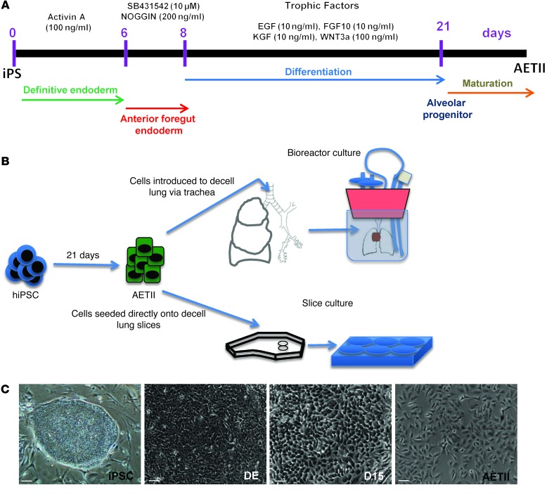
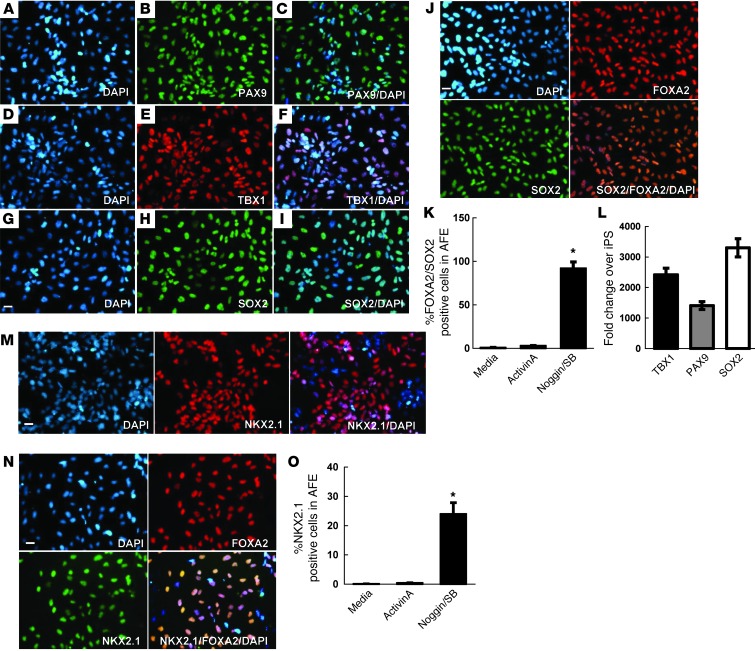
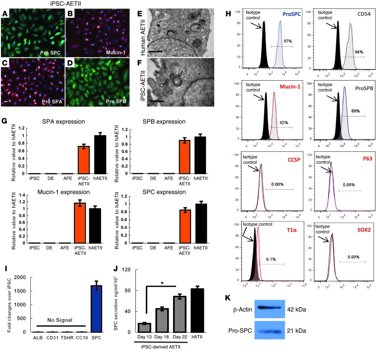
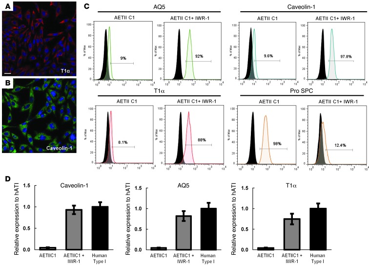
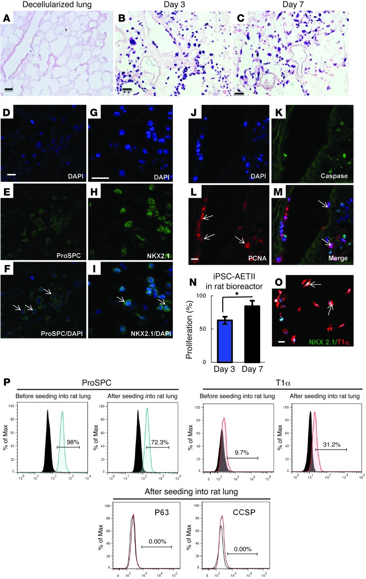
The use of induced pluripotent stem cells (iPSCs) has been postulated to be the most effective strategy for developing patient-specific respiratory epithelial cells, which may be valuable for lung-related cell therapy and lung tissue engineering. We generated a relatively homogeneous population of alveolar epithelial type II (AETII) and type I (AETI) cells from human iPSCs that had phenotypic properties similar to those of mature human AETII and AETI cells. We used these cells to explore whether lung tissue can be regenerated in vitro. Consistent with an AETII phenotype, we found that up to 97% of cells were positive for surfactant protein C, 95% for mucin-1, 93% for surfactant protein B, and 89% for the epithelial marker CD54. Additionally, exposing induced AETII to a Wnt/β-catenin inhibitor (IWR-1) changed the iPSC-AETII-like phenotype to a predominantly AETI-like phenotype. We found that of induced AET1 cells, more than 90% were positive for type I markers, T1α, and caveolin-1. Acellular lung matrices were prepared from whole rat or human adult lungs treated with decellularization reagents, followed by seeding these matrices with alveolar cells derived from human iPSCs. Under appropriate culture conditions, these progenitor cells adhered to and proliferated within the 3D lung tissue scaffold and displayed markers of differentiated pulmonary epithelium.


Figures

Figure 1
Figure 1. Schematic summarizing the experiment.
(A) Schematic protocol for directed differentiation of iPSCs to AETII in vitro in 22 days. Cytokines were added at different steps indicated on top of panel. (B) Schematic summarizing the iPSC differentiation and decellularization-recellularization of both rat and human lung with iPSC-derived AETII cells. decell, decellularized. (C) Phase-contrast images of iPSCs at day 0, DE cells at day 6, and differentiated cells at day 15 and 22, which are termed AETII cells. Scale bars: 63 μm.
Figure 2
Figure 2. Characterization of cells at day 8 of differentiation to produce AFE.
(AI) Immunofluorescence analysis of AFE markers. (A, D, and G) DAPI staining for nuclei; (B, E, and H) PAX9-, TBX1- and SOX2-positive cells. (C, F, and I) Merge at day 8. (J) Immunofluorescence staining showing AFE cells are positive for both SOX2 and FOXA2. (K) Flow cytometric analysis of double-positive cells for SOX2 and FOXA2 in AFE cells at day 8 compared with cells cultured in activin A and RPMI medium only (y axis: percentage of positive cells for FOXA2/SOX2 ). (L) mRNA expression of SOX2, TBX1 and PAX9 in AFE generated from DE cells in vitro at day 8 (data expressed as quantification of mRNA normalized to GAPDH and average fold change in gene expression over iPSCs; y axis, fold changes in gene expression compared with iPSC). (M) Expression of NKX2.1 on day 13 after induction AFE quantified by DAPI staining for nuclei, NKX2.1 positive cells, and merge. (N) Immunofluorescence staining showing NKX2.1 cells in AFE stained positive for FOXA2, indicating these cells are more lung progenitor rather than thyroid progenitor. (O) Flow cytometric analysis of positive cells for NKX2.1. Up to 24% of AFE cells were positive for NKX2.1 at day 13. (y axis, percentages of cells positive for NKX2.1). Bars indicate mean ± SEM of n = 3 independent experiments for qRT-PCR and flow cytometry. *P < 0.05. Scale bars: 31 μm.
Figure 3
Figure 3. Functional characterization of AETII cells derived from iPSCs, day 22 of differentiation (C1 clone).
(AD) Immunostaining of AETII marker: (A) pro-SPC, (B) mucin-1, (C) pro-SPA, (D) pro-SPB. Scale bar: 63 μm. (E and F) Transmission electron microscopy represents (E) human AETII and (F) iPSC-derived AETII containing characteristic cytoplasmic laminar bodies. Scale bars: 0.5 μm. (G) qRT-PCR analysis in undifferentiated iPSC, DE, AFE, and differentiated AETII cells compared with human AETII from 3 independent experiments. Values from the triplicate PCR reactions for a GOI (SPA, SPB, SPC, and mucin-1) were normalized against average GAPDH Ct values from the same cDNA sample. Fold change of GOI tra-script levels between iPS-derived AETII and human type II cells equals 2–ΔΔCt, where ΔCt = Ct(GOI) – Ct(GAPDH), and ΔΔCt = ΔCt(AETII) – ΔCt(ATII). (H) Flow cytometry analysis for the percentage of positive cells for AETII and AETI markers at day 22. Cells were negative for p63 and SOX2. (I) Expression of albumin, CD31, TSHR, and CC10 (CCSP) in iPSC-AETII. Cells were negative for genes indicative of other lineages at day 22. (J) Amount of secreted SPC in the iPSC-derived AETII supernatants collected during the time course of differentiation compared with human type II cells determined by ELISA. (K) Western blot for pro-SPC in iPSC-AETII at day 22 and β-actin as an internal control. Bars indicate ± SEM and n = 3 independent experiments for qRT-PCR, ELISA, and flow cytometry. *P < 0.05.
Figure 4
Figure 4. Functional characterization of AETI cells derived from iPSCs, day 29 of differentiation (C1 clone).
(A and B) Immunofluorescent staining of alveolar type I marker (A) T1α (B) caveolin-1. Scale bar: 63 μm. (C) Flow cytometry analysis for the percentage of positive cells for alveolar type I marker at day 29 in the presence and absence of IWR-1. (y axis, percentage of positive cells). (D) qRT-PCR analysis in AETI cells as compared with native human type I (AETI) cells, from 3 independent experiments. Values from the triplicate PCR reactions for a GOI (AQ5, T1α, caveolin-1) were normalized against average GAPDH Ct values from the same cDNA sample. Fold change of GOI transcript levels between iPS-derived AETI and human type I cells equals 2–ΔΔCt, where ΔCt = Ct(GOI) – Ct(GAPDH) and ΔΔCt = ΔCt(AETI) – ΔCt(hAETI). (y axis, relative gene expression compared with human type I cells). Bars indicate ± SEM and n = 3 independent experiments for qRT-PCR, ELISA, and flow cytometry.
Figure 5
Figure 5. iPSC-derived AETII recellularized 3D rat lung tissue scaffolds in a bioreactor.
(A) H&E staining of decellularized rat lung; (B and C) H&E staining of 3- and 7-day seeded rat lung with iPSC-derived AETII cells cultured in a bioreactor. Scale bars: 25 μm. (DF) Immunofluorescent staining for pro-SPC in AETII seeded cells at day 3. (D) DAPI staining; (E) pro-SPC; (F) merge (arrows in F indicate cells positive for pro-SPC). (GI) Immunostaining for NKX2.1 at day 7. (G) DAPI, (H) NKX2.1, (I) merge (arrows in 5I indicate cells positive for NKX2.1) (JM) Caspase and PCNA immunostaining at day 7 (arrows indicates cells positive for PCNA in L and caspase in M). Scale bar: 25 μm. (N) Proliferation at day 7 compared with day 3. iPSC-AETII displayed a significantly increased fractional proliferation (P < 0.05) after 7 days when they were stained for PCNA (y axis, percentage proliferation based on the number of positive nuclei stained for PCNA) (O) Immunostaining of the few engrafted epithelial cells that acquired flattened morphology, positive for T1α, and negative for NKX2.1 at day 7. Scale bar: 63 μm. Arrows in O indicate cells positive for T1α. (P) Flow cytometry for SPC, T1α, CCSP, p63, and SOX2 before and after seeding into rat lung scaffold in bioreactor. The number of SPC-positive cells decreased during 7-day culture, while the number of cells positive for T1α increased from 9% to 31.2%. All differentiated cells from iPSCs were negative for CCSP, p63, and SOX2 before and after cell seeding.
Figure 6
Figure 6. iPSC-derived AETII (C1 clone) adhere to sections of acellular rat and human lung matrix.
(AG) iPSC-AETII on human lung sections at day 7. (A) H&E. Scale bar: 200 μm (B) Immunostaining for SPC and CCSP. (C) Immunostaining for NKX2.1 and T1α (arrows indicates cells positive for SPC in B and T1α in C. Scale bar: 63 μm. (DF) Immunostaining for NKX2.1. (D), DAPI, (E) NKX2.1, (F) merging. Scale bar: 50 μm. (G) Caspase and PCNA immunostaining. Scale bar: 49 μm (HO) iPSC-derived AETII cultured on rat lung sections for 7 days. (H) H&E. Scale bar: 200 μm. (I) Immunostaining for SPC and CCSP. (J, K) Immunostaining for NKX2.1 and T1α (arrows indicate cells positive for SPC in I, T1α in J, and NKX2.1 in K). Scale bars: 63 μm. (L) DAPI staining, (M) immunostaining for PCNA, and (N) caspase, (O) merge. Scale bar: 50 μm. (PV) Native human AETII cells, isolated from fresh adult human lung, cultured on human lung sections for 7 days. (P) H&E. Scale bar: 200 μm. (Q) Immunostaining for SPC and CCSP. Scale bar: 63 μm (R) Immunostaining for NKX2.1 and T1α. Scale bar: 63 μm (arrows indicate cells positive for SPC in Q and T1α in R). (SU) Immunostaining for NKX2.1. (S) DAPI, (T) NKX2.1, (U) merge. Scale bar: 50 μm. (V) Caspase and PCNA immunostaining. Scale bar: 49 μm.

References

    1. Longmire TA, et al. Efficient derivation of purified lung and thyroid progenitors from embryonic stem cells. Cell Stem Cell. 2012;10(4):398–411. doi: 10.1016/j.stem.2012.01.019. - DOI - PMC - PubMed
    1. Petersen TH, Calle EA, Niklason LE. Strategies for lung regeneration. Mater Today. 2011;14(5):196–201. doi: 10.1016/S1369-7021(11)70114-6. - DOI
    1. Nichols JE, Niles JA, Cortiella J. Production utilization of acellular lung scaffolds in tissue engineering. J Cell Biochem. 2012;113(7):2185–2192. doi: 10.1002/jcb.24112. - DOI - PubMed
    1. McCurry KR, et al. Lung transplantation in the United States, 1998–2007. Am J Transplant. 2009;9(part 2):942–958. doi: 10.1111/j.1600-6143.2009.02569.x. - DOI - PubMed
    1. Wang D, Haviland DL, Burns AR, Zsigmond E, Wetsel RA. A pure population of lung alveolar epithelial type II cells derived from human embryonic stem cells. Proc Natl Acad Sci U S A. 2007;104(11):4449–4454. doi: 10.1073/pnas.0700052104. - DOI - PMC - PubMed

Publication types

MeSH terms