. 2014 Jan-Feb;6(1):273-85.
doi: 10.4161/mabs.26773.

Antibody-drug conjugate model fast characterization by LC-MS following IdeS proteolytic digestion

Antibody-drug conjugate model fast characterization by LC-MS following IdeS proteolytic digestion

Elsa Wagner-Rousset et al. MAbs. 2014 Jan-Feb.

Abstract

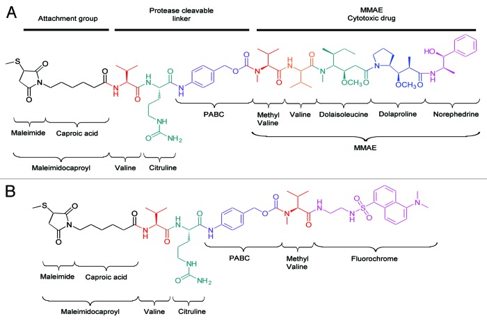
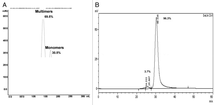
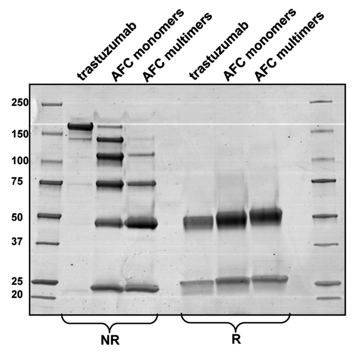
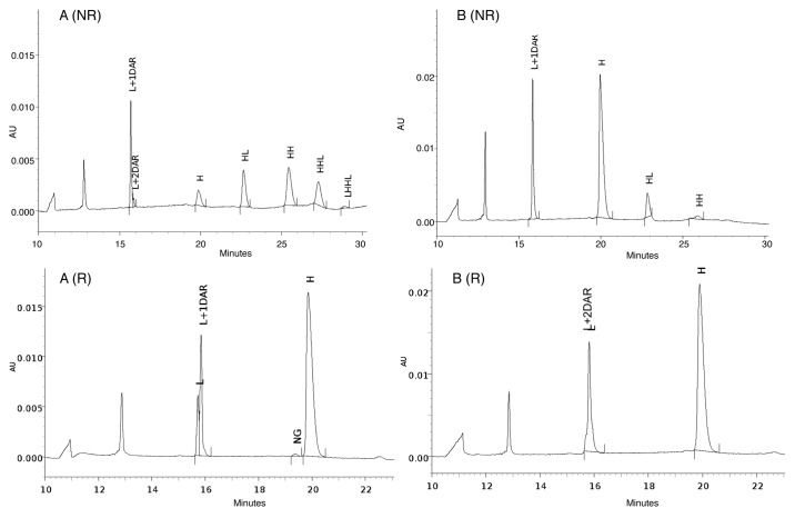
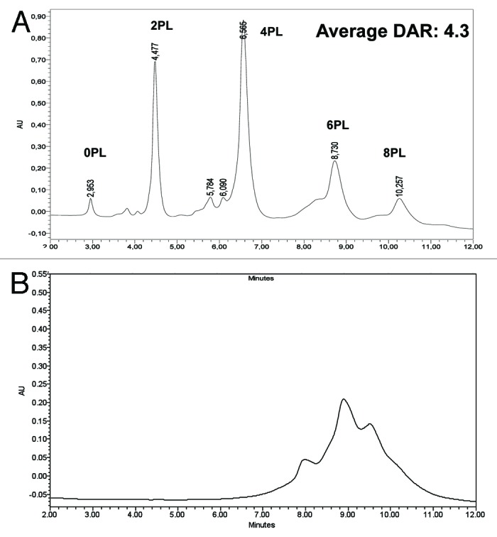
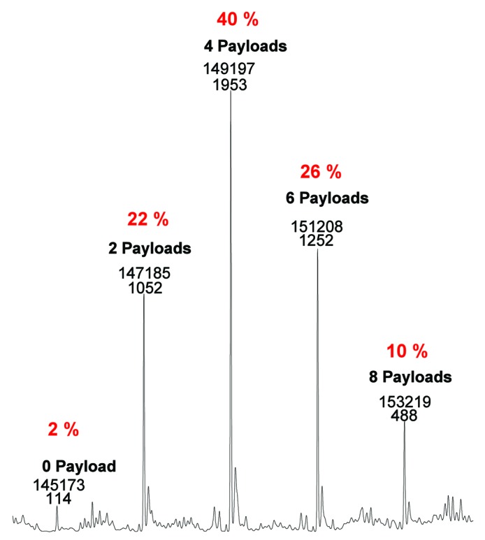
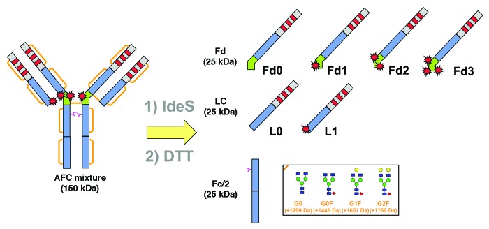
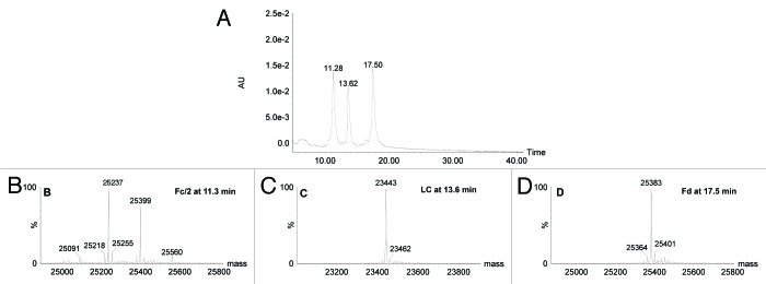
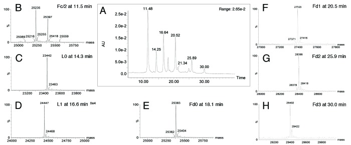
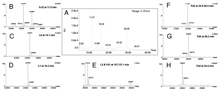
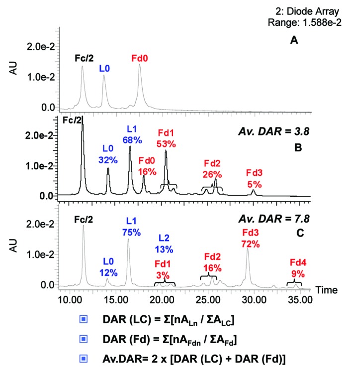
Here we report the design and production of an antibody-fluorophore conjugate (AFC) as a non-toxic model of an antibody-drug conjugate (ADC). This AFC is based on the conjugation of dansyl sulfonamide ethyl amine (DSEA )-linker maleimide on interchain cysteines of trastuzumab used as a reference antibody. The resulting AFC was first characterized by routine analytical methods (SEC, SDS-PAGE, CE-SDS, HIC and native MS), resulting in similar chromatograms,electropherograms and mass spectra to those reported for hinge Cys-linked ADCs. IdeS digestion of the AFC was then performed, followed by reduction and analysis by liquid chromatography coupled to mass spectrometry analysis. Dye loading and distribution on light chain and Fd fragments were calculated, as well as the average dye to antibody ratio (DAR) for both monomeric and multimeric species. In addition, by analyzing the Fc fragment in the same run, full glycoprofiling and demonstration of the absence of additional conjugation was easily achieved. As for naked antibodies and Fc-fusion proteins, IdeS proteolytic digestion may rapidly become a reference analytical method at all stages of ADC discovery, preclinical and clinical development. The method can be routinely used for comparability assays, formulation, process scale-up and transfer, and to define critical quality attributes in a quality-by-design approach.


Figures

None
Figure 1. Structures of (A) linker-cytotoxic (mc_MMAE) and (B) linker-fluorescent (mc_DSEA) payloads.
None
Figure 2. Purification of trastuzumab-mc_DSEA. (A) Preparative SEC. (B) SEC analysis of monomeric AFC.
None
Figure 3. SDS-PAGE of the purified monomeric and multimeric forms of trastuzumab-mc_DSEA in non-reducing (NR) and reducing (R) conditions.
None
Figure 4. CE-SDS-PAGE of the purified (A) monomeric and (B) multimeric forms of trastuzumab-mc_DSEA in non-reducing (NR) and reducing (R) conditions.
None
Figure 5. Drug loading distribution and average DAR calculation by HIC. (A) Monomeric trastuzumab-mc_DSEA. (B) Multimeric trastuzumab-mc_DSEA. Average DAR is calculated by quantifying the various loaded forms based on the peak areas (A) of the UV chromatogram at 210 nm.
None
Figure 6. Drug loading distribution and average DAR calculation by native mass spectrometry.
None
Figure 7. Generation of seven fragments of near 25 kDa after IdeS digestion and DTT reduction of trastuzumab-mc_DSEA.
None
Figure 8. LC-UV-MS of trastuzumab after IdeS digestion and reduction. (A) UV chromatogram at 280 nm. (B‒D) mass spectra of peaks eluting at 11.3, 13.6 and 17.5 min, respectively.
None
Figure 9. LC-UV-MS of monomeric trastuzumab-mc_DSEA after IdeS digestion and reduction. (A) UV chromatogram at 280 nm. (B‒H) Mass spectra of peaks eluting at 11.5, 14.3, 16.6, 18.1, 20.5, 25.9 and 30 min, respectively.
None
Figure 10. LC-UV-MS of multimeric trastuzumab-mc_DSEA after IdeS digestion and reduction. (A) UV chromatogram at 280 nm. (B‒H) Nass spectra of peaks eluting at 11.5, 14.1, 16.3, 19.7–21.1, 24.5–25.4, 29.3 and 34.5 min, respectively.
None
Figure 11. Drug loading distribution and average DAR calculation. (A) Unconjugated trastuzumab. (B) Monomeric. (C) Multimeric trastuzumab-mc_DSEA, respectively. Average DAR is calculated by quantifying the various loaded forms based on the peak areas (A) of the UV chromatogram at 280 nm.

References

    1. Beck A, Wurch T, Bailly C, Corvaia N. Strategies and challenges for the next generation of therapeutic antibodies. Nat Rev Immunol. 2010;10:345–52. doi: 10.1038/nri2747. - DOI - PubMed
    1. Beck A, Senter P, Chari R. World Antibody Drug Conjugate Summit Europe: February 21-23, 2011; Frankfurt, Germany. MAbs. 2011;3:331–7. doi: 10.4161/mabs.3.4.16612. - DOI - PMC - PubMed
    1. Beck A, Lambert J, Sun M, Lin K. Fourth World Antibody-Drug Conjugate Summit: February 29-March 1, 2012, Frankfurt, Germany. MAbs. 2012;4:637–47. doi: 10.4161/mabs.21697. - DOI - PMC - PubMed
    1. Mullard A. Maturing antibody-drug conjugate pipeline hits 30. Nat Rev Drug Discov. 2013;12:329–32. doi: 10.1038/nrd4009. - DOI - PubMed
    1. Beck A, Carter PJ, Gerber HP, Lugovskoy AA, Wurch T, Junutula JR, et al. 8 (th) Annual European Antibody Congress 2012: November 27-28, 2012, Geneva, Switzerland. MAbs. 2013; 5. - PMC - PubMed