

Fibroblast growth factor receptor 1 as a putative therapy target in colorectal cancer
- PMID: 24135816
- PMCID: PMC4186657
- DOI: 10.1159/000355018
Fibroblast growth factor receptor 1 as a putative therapy target in colorectal cancer
Abstract
Background/aims: Resembling a potential therapeutic drug target, fibroblast growth factor receptor 1 (FGFR1) amplification and expression was assessed in 515 human colorectal cancer (CRC) tissue samples, lymph node metastases and CRC cell lines.
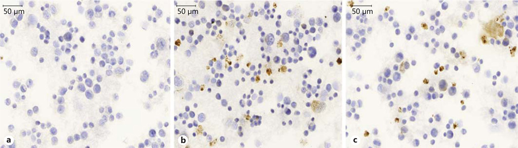
Methods: FGFR1 amplification status was determined using fluorescence in situ hybridization. Additionally, we assessed protein levels employing Western blots and immunohistochemistry. The FGFR1 mRNA localization was analyzed using mRNA in situ hybridization. Functional studies employed the FGFR inhibitor NVP-BGJ398.
Results: Of 454 primary CRCs, 24 displayed FGFR1 amplification. 92/94 lymph node metastases presented the same amplification status as the primary tumor. Of 99 investigated tumors, 18 revealed membranous activated pFGFR1 protein. FGFR1 mRNA levels were independent of the amplification status or pFGFR1 protein occurrence. In vitro, a strong antiproliferative effect of NVP-BGJ398 could be detected in cell lines exhibiting high FGFR1 protein.
Conclusion: FGFR1 is a potential therapeutic target in a subset of CRC. FGFR1 protein is likely to represent a central factor limiting the efficacy of FGFR inhibitors. The lack of correlation between its evaluation at genetic/mRNA level and its protein occurrence indicates that the assessment of the receptor at an immunohistochemical level most likely represents a suitable way to assess FGFR1 as a predictive biomarker for patient selection in future clinical trials.
© 2013 S. Karger AG, Basel.
Conflict of interest statement
The authors have no conflicts of interest to disclose.
Figures


Similar articles
-
FGFR1 Expression Levels Predict BGJ398 Sensitivity of FGFR1-Dependent Head and Neck Squamous Cell Cancers.Clin Cancer Res. 2015 Oct 1;21(19):4356-64. doi: 10.1158/1078-0432.CCR-14-3357. Epub 2015 May 26. Clin Cancer Res. 2015. PMID: 26015511 Free PMC article.
-
Co-active receptor tyrosine kinases mitigate the effect of FGFR inhibitors in FGFR1-amplified lung cancers with low FGFR1 protein expression.Oncogene. 2016 Jul 7;35(27):3587-97. doi: 10.1038/onc.2015.426. Epub 2015 Nov 9. Oncogene. 2016. PMID: 26549034
-
Fibroblast growth factor receptor 1 gene amplification in pancreatic ductal adenocarcinoma.Histopathology. 2013 Aug;63(2):157-66. doi: 10.1111/his.12115. Epub 2013 Jun 28. Histopathology. 2013. PMID: 23808822
-
Fibroblast Growth Factor Receptor 1 (FGFR1) Amplification Detected by Droplet Digital Polymerase Chain Reaction (ddPCR) Is a Prognostic Factor in Colorectal Cancers.Cancer Res Treat. 2020 Jan;52(1):74-84. doi: 10.4143/crt.2019.062. Epub 2019 May 8. Cancer Res Treat. 2020. PMID: 31096734 Free PMC article.
-
FGFR1 amplifications in squamous cell carcinomas of the lung: diagnostic and therapeutic implications.Transl Lung Cancer Res. 2013 Apr;2(2):92-100. doi: 10.3978/j.issn.2218-6751.2013.03.03. Transl Lung Cancer Res. 2013. PMID: 25806220 Free PMC article. Review.
Cited by
-
Honokiol induces apoptosis of lung squamous cell carcinoma by targeting FGF2-FGFR1 autocrine loop.Cancer Med. 2018 Dec;7(12):6205-6218. doi: 10.1002/cam4.1846. Epub 2018 Dec 5. Cancer Med. 2018. PMID: 30515999 Free PMC article.
-
FGFR3 mRNA overexpression defines a subset of oligometastatic colorectal cancers with worse prognosis.Oncotarget. 2018 Aug 14;9(63):32204-32218. doi: 10.18632/oncotarget.25941. eCollection 2018 Aug 14. Oncotarget. 2018. PMID: 30181810 Free PMC article.
-
Identification and validation of an inflammation-related lncRNAs signature for improving outcomes of patients in colorectal cancer.Front Genet. 2022 Sep 30;13:955240. doi: 10.3389/fgene.2022.955240. eCollection 2022. Front Genet. 2022. PMID: 36246600 Free PMC article.
-
Colonic Stent as Bridge to Surgery for Malignant Obstruction Induces Gene Expressional Changes Associated with a More Aggressive Tumor Phenotype.Ann Surg Oncol. 2021 Dec;28(13):8519-8531. doi: 10.1245/s10434-021-10226-4. Epub 2021 Sep 1. Ann Surg Oncol. 2021. PMID: 34467497
-
FGFR1 Expression Levels Predict BGJ398 Sensitivity of FGFR1-Dependent Head and Neck Squamous Cell Cancers.Clin Cancer Res. 2015 Oct 1;21(19):4356-64. doi: 10.1158/1078-0432.CCR-14-3357. Epub 2015 May 26. Clin Cancer Res. 2015. PMID: 26015511 Free PMC article.
References
-
- Liang G, Liu Z, Wu J, Cai Y, Li X. Anticancer molecules targeting fibroblast growth factor receptors. Trends Pharmacol Sci. 2012;33:531–541. - PubMed
-
- Powers CJ, McLeskey SW, Wellstein A. Fibroblast growth factors, their receptors and signaling. Endocr Relat Cancer. 2000;7:165–197. - PubMed
-
- Wesche J, Haglund K, Haugsten EM. Fibroblast growth factors and their receptors in cancer. Biochem J. 2011;437:199–213. - PubMed
Publication types
MeSH terms
Substances
Grants and funding
LinkOut - more resources
Full Text Sources
Other Literature Sources
Medical
Miscellaneous
