. 2013:4:2623.
doi: 10.1038/ncomms3623.

Crossreactivity of a human autoimmune TCR is dominated by a single TCR loop

Affiliations

Crossreactivity of a human autoimmune TCR is dominated by a single TCR loop

Dhruv K Sethi et al. Nat Commun. 2013.

Abstract

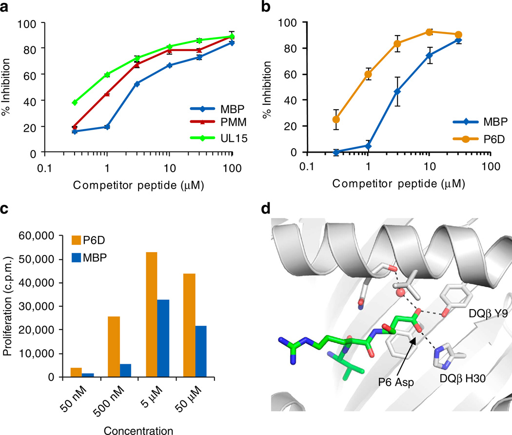
Self-reactive CD4 T cells are thought to have a central role in the pathogenesis of many chronic inflammatory human diseases. Microbial peptides can activate self-reactive T cells, but the structural basis for such crossreactivity is not well understood. The Hy.1B11 T cell receptor (TCR) originates from a patient with multiple sclerosis and recognizes the self-antigen myelin basic protein. Here we report the structural mechanism of TCR crossreactivity with two distinct peptides from human pathogens. The structures show that a single TCR residue (CDR3α F95) makes the majority of contacts with the self-peptide and both microbial peptides (66.7-80.6%) due to a highly tilted TCR-binding topology on the peptide-MHC surface. Further, a neighbouring residue located on the same TCR loop (CDR3α E98) forms an energetically critical interaction with the MHC molecule. These data show how binding by a self-reactive TCR favors crossreactivity between self and microbial antigens.


Figures

Figure 1
Figure 1. Comparison of DQI-bound self and microbial peptides
(a,b) Conformation of microbial peptides in the DQ1 binding site. PMM (yellow, a) and UL15 (green, b) peptides are compared with MBP peptide (magenta; a,b). DQ1 is shown as a surface and the three peptides as stick models; Hy.1B11 TCR has been omitted for clarity. Peptide positions P3 and P8 are indicated (a). (c) Alignment of PMM and UL15 peptides with MBP self peptide. DQ1 anchor residues (P1, P4, P6 and P9) are indicated.
Figure 2
Figure 2. Deep insertion of TCR CDR3α F95 into a pocket created by three peptide residues
(a–c) Interaction of Hy.1B11 TCR with MBP, PMM and UL15 peptides. TCR and peptides are rendered as surfaces, whereas DQ1 has been omitted to improve peptide visualization, MBP (a) PMM (b) and UL15 (c). TCR residues that contact peptides are coloured based on their biochemical properties (hydrophobic—green, polar—orange). Peptide residues are coloured based on charge (acidic—red, basic—blue). Peptide anchor residues (P1, P4, P6 and P9) as well as TCR CDR3α F95 are indicated. (d–f) TCR contact surface for MBP, PMM and UL15 peptides. View is rotated by 90° relative to a–c, and TCR residues contacting MBP (d) PMM (e) and UL15 (f) peptides are indicated.
Figure 3
Figure 3. Stacking interactions by peptide P2 histidine and TCR CDR3α F95
(a,b) Aromatic stacking interactions formed by CDR3α F95. P2 His forms π–π stacking interactions with DQ1 β81 His and TCR CDR3α F95 (residues from MBP structure in orange, UL15 in red and PMM in blue). P2 Leu in PMM peptide disrupts this π–π interaction, thereby affecting the position of both DQ1 β81 His and TCR CDR3α F95. TCR CDR3α F95 forms stacking interactions with P2 His (MBP, UL15) and P3 Phe (all peptides); in addition, it forms hydrophobic interactions with the P5 side chain (Lys/ Arg). View in a from peptide N terminus, in b from peptide C terminus.
Figure 4
Figure 4. Conserved and divergent features of Hy.1B11 TCR interaction with self and microbial peptides
(a–c) Interaction of TCR CDR loops with MBP and microbial peptides. CDR loops that contact MBP (a), PMM (b) and UL15 (c) peptides are shown. TCR residues forming similar contacts with all three peptides are coloured yellow, whereas TCR residues that make different contacts are highlighted in red. Peptide residues are coloured as follows: blue, sequence identity between microbial peptide and MBP; magenta, absence of sequence identity with MBP; green, TCR contact to PMM/UL15 peptides but not MBP peptide.
Figure 5
Figure 5. Similar docking mode of Hy.1B11 TCR on self and microbial peptide/DQ1 complexes
(a,b) Tilted binding mode of Hy.1B11 TCR with DQ1/peptide complexes. Comparison of three crystal structures in which Hy.1B11 TCR recognizes distinct DQ1-bound peptides: MBP self-peptide (orange) as well as microbial peptides PMM (blue) and UL15 (red). Complexes were superimposed using the DQα chain to reveal any differences in TCR positioning. TCR, peptide and MHC are rendered as ribbon diagrams; TCR constant domains have been omitted for clarity. The trimolecular complexes are viewed from the peptide C terminus (top panels) and the DQ β1 helix (90° rotation, bottom panels). TCR Vα and Vβ domains, DQα and DQβ chains as well as TCR CDR1α and CDR2α loops are indicated.
Figure 6
Figure 6. Two residues at the tip of the CDR3α loop are critical for TCR binding to microbial peptides and DQ1
(a–f) Binding of TCR mutants to microbial DQ1/peptide complexes. Binding of Hy.1B11 WT and mutant TCRs (αE98A and αF95A) to DQ1/peptide was examined by surface plasmon resonance, as indicated on each graph. DQ1 proteins were mono-biotinylated on a BirA site located at the DQα C terminus and captured on streptavidin chips (1,500 RU). WT and mutant TCRs were injected over a range of concentrations (2 min at 25°C, 15µl min−1) into flow cells with immobilized DQ1/ CLIP (negative control), DQ1/UL15 and DQ1/PMM. The signal from the DQ1/CLIP control flow cell was subtracted from experimental readings. Equilibrium binding constant Kd was calculated for binding of Hy.1B11 WT TCR to DQ1/PMM (a) and DQ1/UL15 (b) complexes using nonlinear curve fitting. Binding studies were performed at least twice with similar results. (g) Hydrogen bonding network formed by TCR CDR3α E98 with DQα R61 and CDR3 Y30. TCR loops are coloured yellow for CDR3α and red for CDR3β; the DQα chain is coloured blue and the PMM peptide green. (h) Interaction of the CDR3α loop (yellow) with the UL15 peptide (green) and DQ1 (blue, space-filling model). The DQ1α R61 residue forms a salt bridge with CDR3α E98.
Figure 7
Figure 7. Aspartic acid at P6 anchor position enhances DQ1 binding by microbial peptides
(a) UL15 and PMM peptides bind with higher affinity to DQ1 than the MBP peptide. Unlabelled MBP, UL15 or PMM peptides (0.3,1, 3, 10, 30 and 100 µM) were used as competitors for DQ1 binding by a biotinylated MBP peptide (400 nM). Following overnight incubation at RT, DQ1/peptide complexes were captured by an immobilized antibody (9.3.F10), and DQ1-bound biotinylated peptide was detected using europium-labeled streptavidin. Data are shown as ± s.d. Experiment was repeated three times with similar results (b) Competition binding assay performed as in (a) demonstrating enhanced DQ1 binding by MBP peptide with P6 Asn to Asp substitution. Data are shown as ±s.d. The experiment was carried out two times with similar results. (c) Enhanced proliferation of Hy.1B11 T cell clone to MBP P6 Asp analogue (orange) compared with MBP peptide (blue). (d) Interaction of P6 Asp with the P6 pocket of DQ1. Shown is the interaction of the UL15 peptide P6 side Asp side chain with DQβ H30 and DQβ Y9. DQ1 is shown as a cartoon and the peptide (P4–P6 segment) as a stick model.

Similar articles

Cited by

References

    1. Gourraud PA, Harbo HF, Hauser SL, Baranzini SE. The genetics of multiple sclerosis: an up-to-date review. Immunol. Rev. 2012;248:87–103. - PMC - PubMed
    1. Sospedra M, Martin R. Immunology of multiple sclerosis. Annu. Rev. Immunol. 2005;23:683–747. - PubMed
    1. Fujinami RS, Oldstone MB. Amino acid homology between the encephalitogenic site of myelin basic protein and virus: mechanism for autoimmunity. Science. 1985;230:1043–1045. - PubMed
    1. Wucherpfennig KW, Strominger JL. Molecular mimicry in T cell-mediated autoimmunity: viral peptides activate human T cell clones specific for myelin basic protein. Cell. 1995;80:695–705. - PMC - PubMed
    1. Hemmer B, et al. Identification of high potency microbial and self ligands for a human autoreactive class II-restricted T cell clone. J. Exp. Med. 1997;185:1651–1659. - PMC - PubMed

Publication types

MeSH terms