. 2013 Oct 17;4(10):e857.
doi: 10.1038/cddis.2013.340.

Single-cell clones of liver cancer stem cells have the potential of differentiating into different types of tumor cells

Affiliations

Single-cell clones of liver cancer stem cells have the potential of differentiating into different types of tumor cells

H Liu et al. Cell Death Dis. .

Abstract

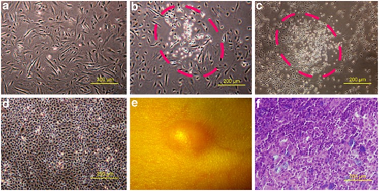
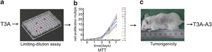
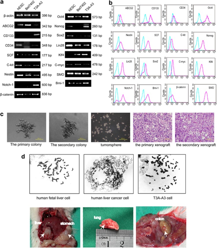
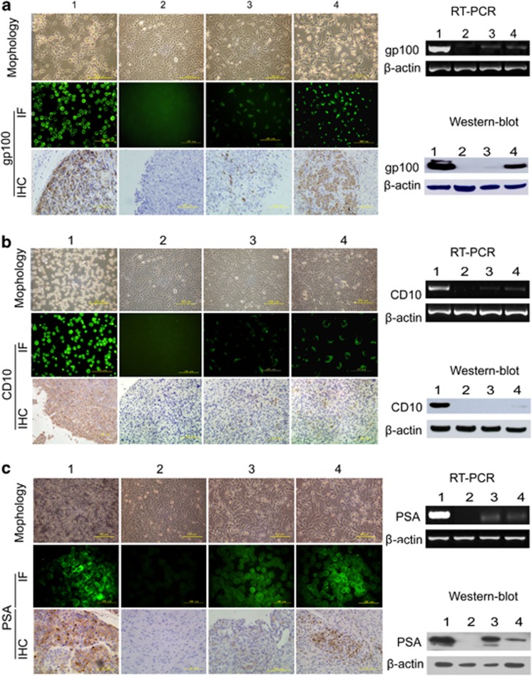
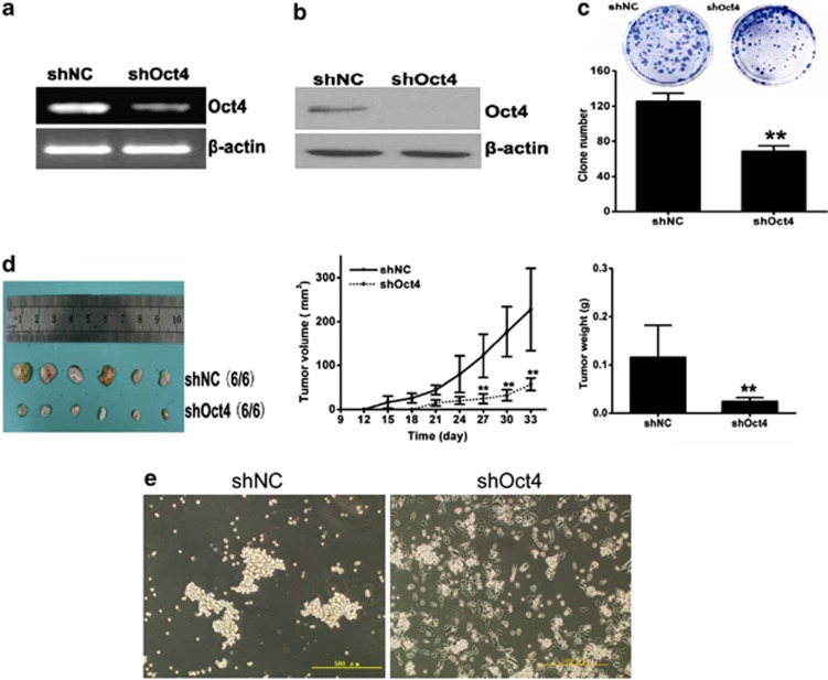
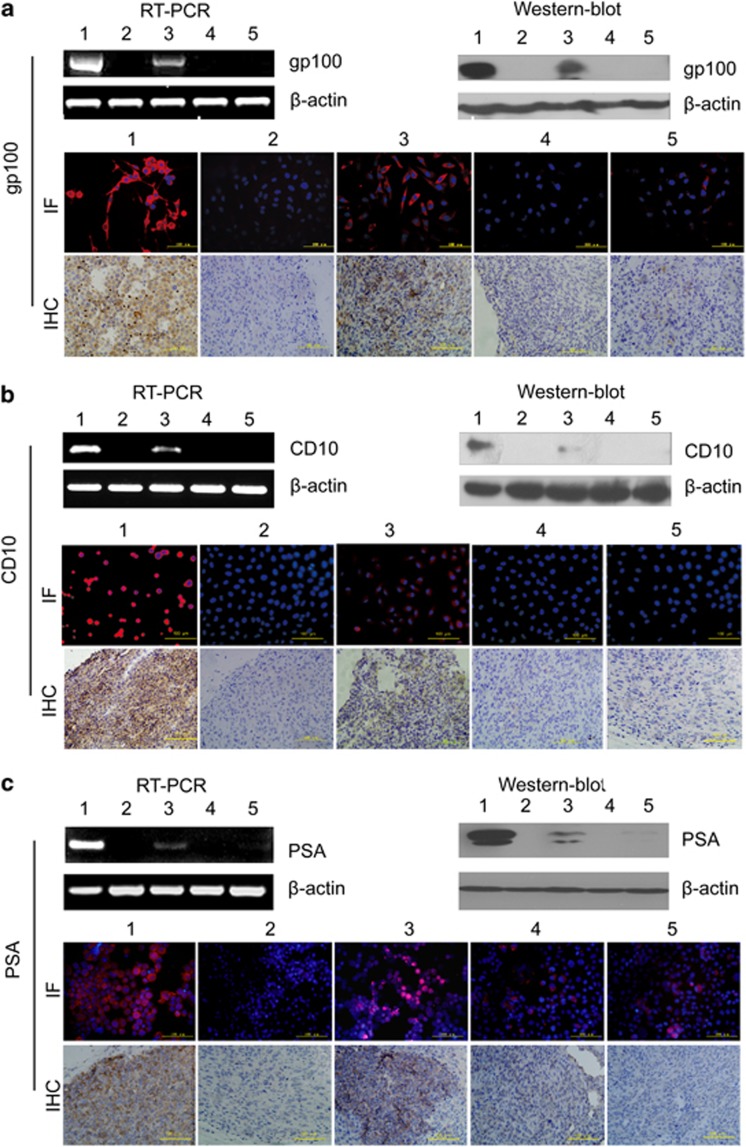
Cancer stem cells (CSCs) are believed to be a promising target for cancer therapy because these cells are responsible for tumor development, maintenance and chemotherapy resistance. Finding out the critical factors regulating CSC fate is the key for target therapy of CSCs. Just as normal stem cells are regulated by their microenvironment (niche), CSCs are also regulated by cells in the tumor microenvironment. However, whether various tumor microenvironments can induce CSCs to differentiate into different cancer cells is not clear. Here, we show that single-cell-cloned CSCs, accidentally obtained from a human liver cancer microvascular endothelial cells, express classic stem cell markers, genes associated with self-renewal and pluripotent factors and possess colony-forming ability in vitro and the ability of serial transplantation in vivo. The single-cell-cloned CSCs treated with the different tumor cell/tissue-derived conditioned culture medium, which is a mimic of carcinoma microenvironment, could differentiate into corresponding tumor cells and express specific markers of the respective type of tumor cells at the gene, protein and cell levels, respectively. Interestingly, this multilineage differentiation potential of single-cell-cloned liver CSCs sharply declined after the specific knockdown of octamer-binding transcription factor 4 (Oct4) alone, even though they were under the same induction conditions (carcinoma microenvironments). These data support the hypothesis that single-cell-cloned liver CSCs have the potential of differentiating into different types of tumor cells, and the tumor microenvironment does play a crucial role in deciding differentiation directions. Simultaneously, Oct4 in CSCs is indispensable in this process. These factors are promising targets for liver CSC-specific therapy.


Figures

Figure 1
Figure 1
Origin and characteristics of T3A. (a) Microvascular endothelial cells derived from human liver cancer. (b) A cell subpopulation (T3A) was observed in cultured human liver cancer microvascular endothelial cells in the eighth generation. (c) The T3A cells rapidly proliferated. (d) T3A cells were purified by subcloning. (e, f) T3A cells formed a solid tumor after a subcutaneous injection into nude mice and the pathological type of tumor tissue was poorly differentiated carcinoma
Figure 2
Figure 2
Generation of the T3A-A3 cells. (a) Single-cell clone screening of T3A cells by limiting-dilution assay. (b) Compared with the proliferation rate of 20 clones in vitro by MTT assay. (c) Compared the tumorigenicity of high and low proliferation rate clones in SCID mice
Figure 3
Figure 3
Identification of T3A-A3 cells. (a) RT-PCR analysis for the expressions of classic stem cell markers and genes associated with the proliferation and self-renewal of stem cells. (b) Flow cytometric analysis for the expressions of classic stem cell markers and genes associated with the proliferation and self-renewal of stem cells. (c) Evaluation of the self-renewing capacity of T3A-A3 cells. Secondary colony formation ability (the first and second panels). Tumor sphere-forming ability (the middle panel). Histopathology of the primary and the secondary grafted tumor (the last two panels). (d) Evaluation of tumor properties of T3A-A3 cells. Comparison of chromosomal karyotype between human fetal liver cells, human liver cancer cells and T3A-A3 cells (upper panels). Evaluation of tumorigenic and metastatic capacities of T3A-A3 cells in SCID mice (bottom panels)
Figure 4
Figure 4
Evaluation of multi-differentiation potential of T3A-A3 cells. The multi-differentiation potential of T3A-A3 cells was analyzed after induction with melanoma, lymphoma and prostate cancer cell- or tissue-derived conditioned culture medium. The morphological changes of T3A-A3 after induction were photographed. The tumor-specific markers were compared between T3A-A3 cells before and after induction by RT-PCR, western blot, immunofluorescence staining in vitro and immunohistochemistry staining on graft tumor tissue in vivo. (a) Morphological changes and expression of gp100 in T3A-A3 after induction by melanoma-derived conditioned culture medium. (b) Morphological changes and expression of CD10 in T3A-A3 after induction by lymphoma-derived conditioned culture medium. (c) Morphological changes and expression of PSA in T3A-A3 after induction by prostate cancer-derived conditioned culture medium. (1: positive control cells (corresponding cancer cell line); 2: T3A-A3 cells; 3: T3A-A3 cells after induction with tumor cell-derived conditioned culture medium; 4: T3A-A3 cells induced with tumor tissue-derived conditioned culture medium)
Figure 5
Figure 5
Effect of Oct4 knockdown on the stem cell properties of T3A-A3. Oct4 in T3A-A3 cells was knocked down by infection with shOct4-carring lentivirus. (a) The expression level of Oct4 was evaluated by RT-PCR. (b) The expression level of Oct4 was evaluated by western blot. (c), The self-renewal ability of shOct4-T3A-A3 was detected by colony formation assay in vitro, **P<0.01 versus shNC, n=3. (d) The tumorigenicity of shOct4-T3A-A3 was examined by grafted tumor growth in vivo **P<0.01 versus shNC, n=6. (e) The sphere assay of shOct4-T3A-A3 was performed on ultra-low adherent plates
Figure 6
Figure 6
Effect of Oct4 knockdown on the multi-differentiation potential of T3A-A3 cells. After Oct4 knockdown in T3A-A3 cells, the committed inductions were repeated using melanoma, lymphoma and prostate cancer tissue-derived conditioned culture medium. The tumor-specific markers were detected by RT-PCR, western blot, immunofluorescence staining in vitro and immunohistochemistry staining on graft tumor tissue in vivo. (a) Detection of the expressions of gp100 in shOct4-T3A-A3 cells induced by melanoma tissue-derived conditioned culture medium. (b) Detection of CD10 in shOct4-T3A-A3 cells induced by lymphoma tissue-derived conditioned culture medium. (c) Detection of PSA in shOct4-T3A-A3 induced by prostate cancer tissue-derived conditioned culture medium. 1: Positive control (corresponding cancer cell line); 2: shNC-T3A-A3 cells; 3: induced shNC-T3A-A3 cells; 4: shOct4-T3A-A3 cells; 5: induced shOct4-T3A-A3 cells)

References

    1. Rountree CB, Mishra L, Willenbring H. Stem cells in liver diseases and cancer: recent advances on the path to new therapies. Hepatology. 2012;55:298–306. - PMC - PubMed
    1. O'Brien CA, Pollett A, Gallinger S, Dick JE. A human colon cancer cell capable of initiating tumour growth in immunodeficient mice. Nature. 2007;445:106–110. - PubMed
    1. Reya T, Morrison SJ, Clarke MF, Weissman IL. Stem cells, cancer and cancer stem cells. Nature. 2001;414:105–111. - PubMed
    1. Kryczek I, Liu S, Roh M, Vatan L, Szeliga W, Wei S, et al. Expression of aldehyde dehydrogenase and CD133 defines ovarian cancer stem cells. International journal of cancer Journal international du cancer. 2012;130:29–39. - PMC - PubMed
    1. Schneider M, Huber J, Hadaschik B, Siegers GM, Fiebig HH, Schuler J. Characterization of colon cancer cells: a functional approach characterizing CD133 as a potential stem cell marker. BMC cancer. 2012;12:96. - PMC - PubMed

Publication types

Substances