. 2013 Oct 17;4(10):e865.
doi: 10.1038/cddis.2013.395.

Chemotherapy-mediated p53-dependent DNA damage response in clear cell renal cell carcinoma: role of the mTORC1/2 and hypoxia-inducible factor pathways

Affiliations

Chemotherapy-mediated p53-dependent DNA damage response in clear cell renal cell carcinoma: role of the mTORC1/2 and hypoxia-inducible factor pathways

J Selvarajah et al. Cell Death Dis. .

Abstract

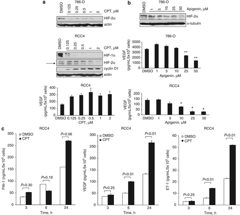
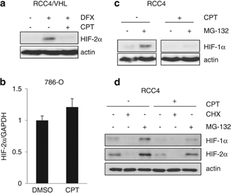
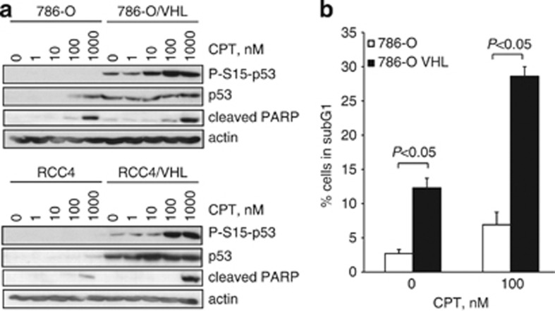
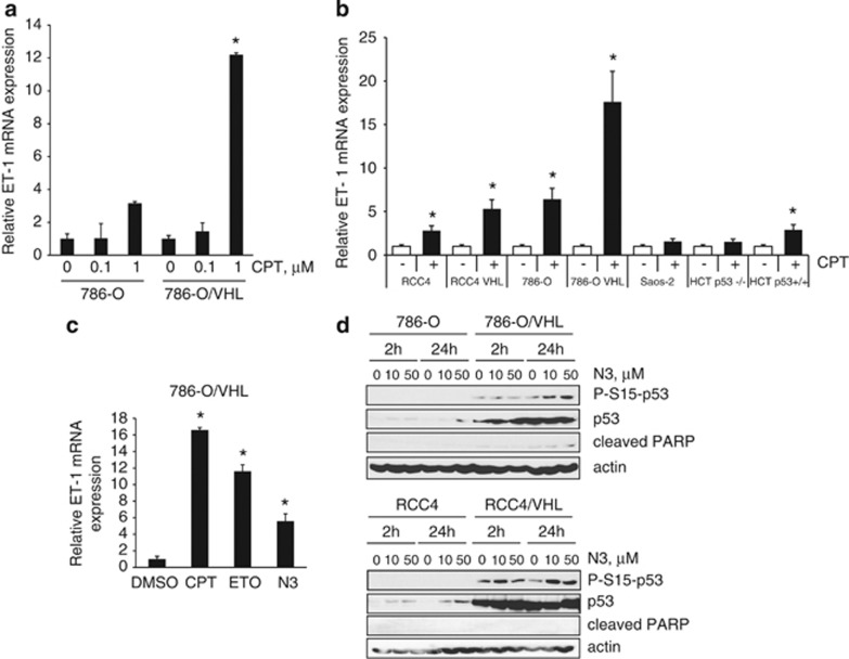
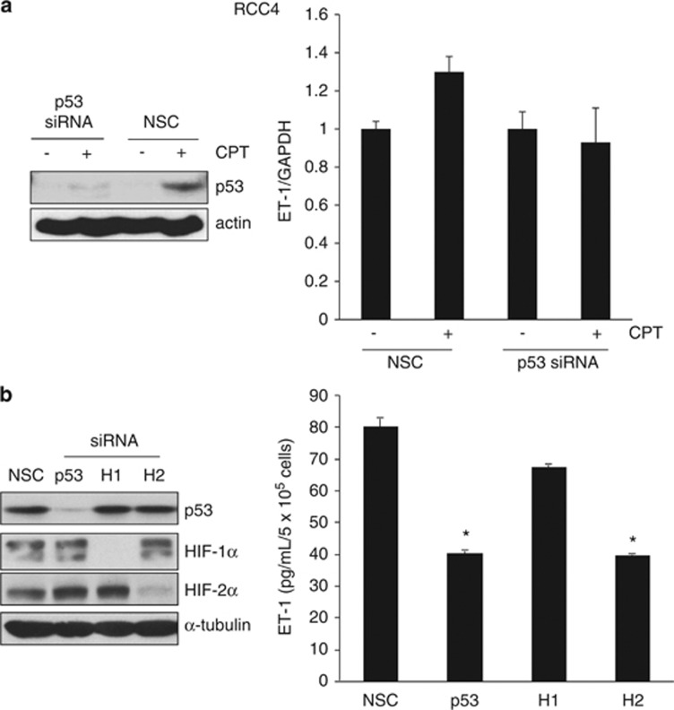
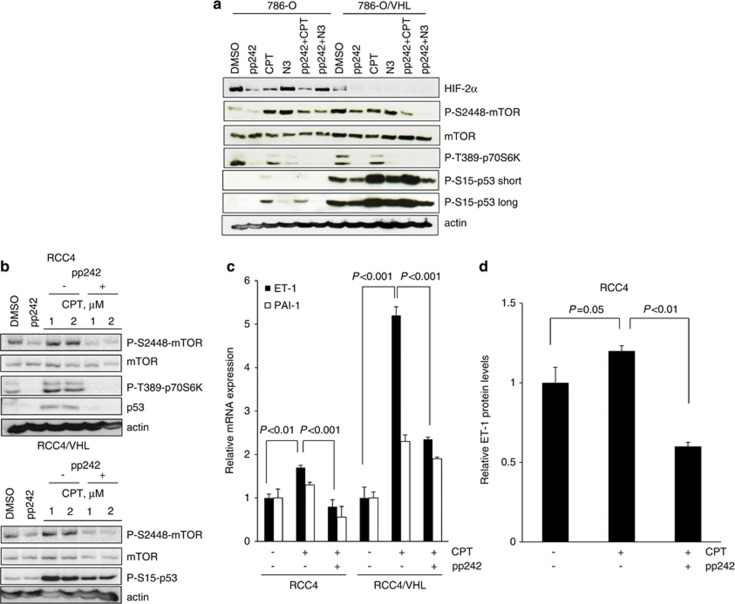
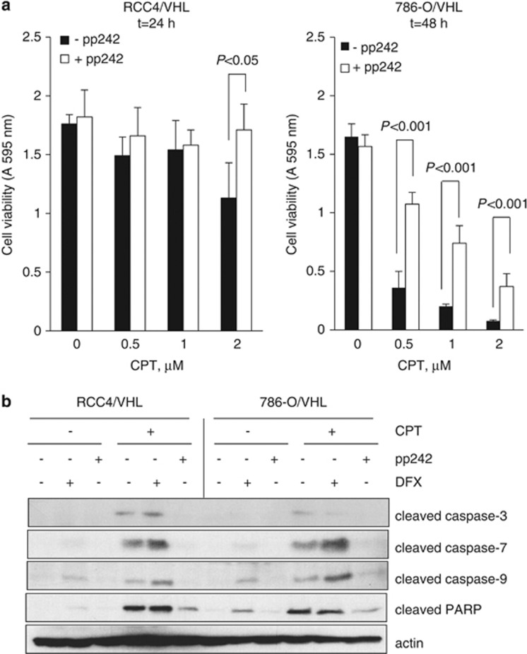
The DNA-damaging agent camptothecin (CPT) and its analogs demonstrate clinical utility for the treatment of advanced solid tumors, and CPT-based nanopharmaceuticals are currently in clinical trials for advanced kidney cancer; however, little is known regarding the effects of CPT on hypoxia-inducible factor-2α (HIF-2α) accumulation and activity in clear cell renal cell carcinoma (ccRCC). Here we assessed the effects of CPT on the HIF/p53 pathway. CPT demonstrated striking inhibition of both HIF-1α and HIF-2α accumulation in von Hippel-Lindau (VHL)-defective ccRCC cells, but surprisingly failed to inhibit protein levels of HIF-2α-dependent target genes (VEGF, PAI-1, ET-1, cyclin D1). Instead, CPT induced DNA damage-dependent apoptosis that was augmented in the presence of pVHL. Further analysis revealed CPT regulated endothelin-1 (ET-1) in a p53-dependent manner: CPT increased ET-1 mRNA abundance in VHL-defective ccRCC cell lines that was significantly augmented in their VHL-expressing counterparts that displayed increased phosphorylation and accumulation of p53; p53 siRNA suppressed CPT-induced increase in ET-1 mRNA, as did an inhibitor of ataxia telangiectasia mutated (ATM) signaling, suggesting a role for ATM-dependent phosphorylation of p53 in the induction of ET-1. Finally, we demonstrate that p53 phosphorylation and accumulation is partially dependent on mTOR activity in ccRCC. Consistent with this result, pharmacological inhibition of mTORC1/2 kinase inhibited CPT-mediated ET-1 upregulation, and p53-dependent responses in ccRCC. Collectively, these data provide mechanistic insight into the action of CPT in ccRCC, identify ET-1 as a p53-regulated gene and demonstrate a requirement of mTOR for p53-mediated responses in this tumor type.


Figures

Figure 1
Figure 1
Effect of CPT and apigenin on HIF-1α, HIF-2α and HIF-α target genes in RCC4 and 786-O cells. (a and b) 786-O or RCC4 cells were treated with CPT or apigenin at the concentrations indicated or vehicle control (DMSO). Panels, whole-cell lysates were assayed by western blot for HIF-1α, HIF-2α and cyclin D1 proteins. Actin and/or tubulin were used as loading controls. Graphs, conditioned media were harvested after 24 h and secreted protein levels of VEGF were determined by ELISA and normalized to cell number. (c) RCC4 cells were treated with 2 μM CPT or DMSO vehicle control for the times indicated. Conditioned media were harvested and secreted protein levels of VEGF, PAI-1 and ET-1 were determined by ELISA and normalized to cell number. Mean±S.E. of duplicate values of one representative experiment is shown. *P<0.05, **P<0.01, t-test compared with control
Figure 2
Figure 2
CPT inhibits HIF-1α and HIF-2α protein synthesis. (a) RCC4/VHL cells were incubated with 500 μM desferrioxamine (DFX) in the absence or presence of 2 μM CPT for 24 h as indicated. Whole-cell lysates were assayed by western blot for HIF-2α. Actin was used as a loading control. (b) The 786-O cells were incubated with 2 μM CPT or vehicle control (DMSO) for 24 h and analyzed for mRNA expression of HIF-2α by real-time quantitative PCR relative to GAPDH. (c and d) RCC4 cells were incubated with or without 2 μM CPT for 6 h in the absence or presence of 10 μM MG-132 or 10 μM cycloheximide (CHX) for the final 3 h as indicated. Whole-cell lysates were assayed by western blot for HIF-1α and HIF-2α. Actin was used as a loading control
Figure 3
Figure 3
CPT-mediated increase in p53 levels and apoptosis is augmented in VHL-competent cells. (a) The 786-O and RCC4 cells and their VHL-expressing counterparts were treated with increasing concentrations of CPT for 24 h. Whole-cell lysates were assayed by western blot for p53 and phosphorylated-S15-p53 proteins and cleaved PARP. Actin was used as a loading control. (b) The 786-O and 786-O/VHL cells were treated with 100 nM CPT or vehicle for 24 h, fixed, stained with propidium iodide and the percentage (%) of cells in subG1 was assessed by flow cytometry. Mean±S.E. of three replicates is shown
Figure 4
Figure 4
DNA-damaging agents increase ET-1 mRNA abundance in a p53-dependent manner. (a) The 786-O and 786-O/VHL cells were treated with increasing concentrations of CPT for 24 h and analyzed for mRNA expression of ET-1 by real-time quantitative PCR relative to GAPDH. (b) Cells as indicated were treated with 2 μM CPT (+) or vehicle DMSO (−) for 24 h and analyzed for mRNA expression of ET-1 by real-time quantitative PCR relative to GAPDH. (c) The 786-O/VHL cells were treated with vehicle DMSO, 2 μM CPT, 100 μM etoposide (ETO) or 10 μM nutlin-3a (N3) and analyzed for mRNA expression of ET-1 by real-time quantitative PCR relative to GAPDH. (d) The 786-O and RCC4 cells and their VHL-expressing counterparts were treated with vehicle control, 10 μM or 50 μM nutlin-3a (N3) for 2 or 24 h. Whole-cell lysates were assayed by western blot for phosphorylated-S15-p53, p53 and cleaved PARP proteins. Actin was used as a loading control. In the graphs, mean±S.E. of duplicate values of one representative experiment is shown. * P<0.05 as compared with control
Figure 5
Figure 5
The p53-dependent regulation of ET-1 in RCC4 cells. (a) RCC4 cells were transfected with 10 nM siRNA to p53 or nonsilencing control (NSC) duplex for 24 h before addition of 2 μM CPT (+) or DMSO vehicle control (−) for a further 24 h. Panels, whole-cell lysates were assayed by western blot for p53 protein. Actin was used as a loading control. Graph, mRNA expression of ET-1 by real-time quantitative PCR relative to GAPDH. Mean±S.E. of duplicate values of one representative experiment is shown. (b) RCC4 cells were transfected with 10 nM siRNA to p53, HIF-1α (H1), or HIF-2α (H2) or NSC duplex for 24 h. Panels, whole-cell lysates were assayed by western blot for p53, HIF-1α and HIF-2α proteins. Tubulin was used as a loading control. Graph, conditioned media were harvested and secreted protein levels of ET-1 were determined by ELISA and normalized to cell number. Mean±S.E. of duplicate values of one representative experiment is shown. *P<0.05, t test compared with control
Figure 6
Figure 6
DNA damage response is suppressed in HIF-α-expressing cells. (a) RCC4 and RCC4/VHL cells were treated with 2 μM CPT, 100 μM etoposide (ETO) or 400 nM of the mTORC1/2 kinase inhibitor pp242 for 24 h. Whole-cell lysates were assayed by western blot for phosphorylated p53 (S15), CHK1 (S317, S345), γH2AX (S139), total p53 and CHK1 proteins. Actin was used as a loading control. (b) The 786-O and 786-O/VHL cells were transfected with siRNA to ATR or nonsilencing control (NSC) for 24 h before addition of 2 μM CPT for a further 24 h. mRNA expression of ET-1 was assessed by real-time quantitative PCR relative to GAPDH. Mean±S.E. of duplicate values of one representative experiment is shown. (c) RCC4/VHL cells were treated with 10 μM ATM inhibitor (ATMi), 400 nM pp242 or vehicle control (DMSO) in the absence (−) or presence (+) of 2 μM CPT for 24 h. Whole-cell lysates were assayed by western blot for phosphorylated p53 (S15), and p53 proteins. Short and long exposure times are shown for P-S15-p53. Actin was used as a loading control. (d) RCC4/VHL and 786-O/VHL cells were preincubated with 10 μM ATMi for 1 h before addition of 2 μM CPT for a further 24 h and mRNA expression of ET-1 was assessed by real-time quantitative PCR relative to GAPDH. Mean±S.E. of duplicate values of one representative experiment is shown
Figure 7
Figure 7
Attenuation of mTORC1/2 kinase ablates CPT-induced p53- and p53-dependent responses in ccRCC. (a) The 786-O and 786-O/VHL cells were treated with 400 nM pp242, 2 μM CPT, 10 μM nutlin-3a (N3) or vehicle control (DMSO) alone for 24 h, or preincubated with 400 nM pp242 for 1 h before addition of 2 μM CPT or 10 μM N3 for a further 24 h. Whole-cell lysates were assayed by western blot for HIF-2α, mTOR, phosphorylated p53 (S15), mTOR (S2448) and p70S6K (T389) proteins. Actin was used as a loading control. Short and long exposure times are shown for P-S15-p53. (b) RCC4 and RCC4/VHL cells were treated with 400 nM pp242 or vehicle control (DMSO) alone or preincubated with 400 nM pp242 for 1 h before addition of either 1 μM or 2 μM CPT for 24 h. Whole-cell lysates were assayed by western blot for mTOR, p53, phosphorylated p53 (S15), mTOR (S2448) and p70S6K (T389) proteins. Actin was used as a loading control. (c) RCC4 and RCC4/VHL cells were preincubated with 400 nM pp242 for 1 h before addition of 2 μM CPT for 24 h and mRNA expression of ET-1 and PAI-1 were assessed by real-time quantitative PCR relative to GAPDH. Mean±S.E. of duplicate values of one representative experiment is shown. (d) RCC4 cells were pretreated with 400 nM pp242 for 1 h before addition of 2 μM CPT for 24 h. Conditioned media were harvested and assessed for secreted ET-1 protein levels by ELISA and normalized to total protein levels. Mean±S.E. of duplicate values of one representative experiment is shown
Figure 8
Figure 8
mTORC1/2 kinase inhibition reverses CPT-induced cell death in ccRCC. (a) RCC4/VHL and 786-O/VHL cells were incubated with (open bars) or without (solid bars) 400 nM pp242 for 1 h before addition of increasing concentrations of CPT as indicated for 24 or 48 h. Cell viability was determined by reduction of MTT at A 595 nm. Mean±S.E. of triplicate values is shown. (b) RCC4/VHL and 786-O/VHL cells were incubated with 400 nM pp242 or 500 μM DFX for 1 h before addition of 2 μM CPT as indicated for 48 h. Whole-cell lysates were assayed for cleaved caspases 3, 7 and 9 and cleaved PARP. Actin was used as a loading control

Similar articles

Cited by

References

    1. Young AC, Craven RA, Cohen D, Taylor C, Booth C, Harnden P, et al. Analysis of VHL gene alterations and their relationship to clinical parameters in sporadic conventional renal cell carcinoma. Clin Cancer Res. 2009;15:7582–7592. - PMC - PubMed
    1. Gore ME, Larkin JM. Challenges and opportunities for converting renal cell carcinoma into a chronic disease with targeted therapies. Br J Cancer. 2011;104:399–406. - PMC - PubMed
    1. Rini BI. New strategies in kidney cancer: therapeutic advances through understanding the molecular basis of response and resistance. Clin Cancer Res. 2010;16:1348–1354. - PubMed
    1. Rini BI, Atkins MB. Resistance to targeted therapy in renal-cell carcinoma. Lancet Oncol. 2009;10:992–1000. - PubMed
    1. Wander SA, Hennessy BT, Slingerland JM. Next-generation mTOR inhibitors in clinical oncology: how pathway complexity informs therapeutic strategy. J Clin Invest. 2011;121:1231–1241. - PMC - PubMed

Publication types

MeSH terms

Substances