. 2014 Mar;23(3):552-9.
doi: 10.1007/s00586-013-2953-8. Epub 2013 Oct 18.

Reciprocal changes in cervical spine alignment after corrective thoracolumbar deformity surgery

Affiliations

Reciprocal changes in cervical spine alignment after corrective thoracolumbar deformity surgery

Yoon Ha et al. Eur Spine J. 2014 Mar.

Abstract

Purpose: To identify changes in cervical alignment parameters following surgical correction of thoracolumbar deformity and then assess the preoperative parameters which induce changes in cervical alignment following corrective thoracolumbar deformity surgery.

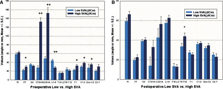
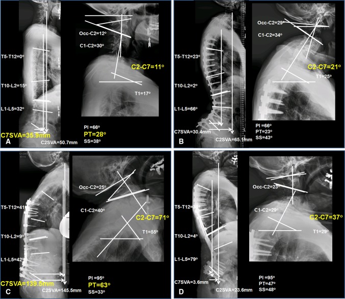
Methods: A retrospective study of 49 patients treated for thoracolumbar deformity with preoperative planning of an acceptably aligned coronal and sagittal plane in each case. We compared cervical spine parameters in two distinct low [preoperative C7 sagittal vertical axis (SVA) ≤ 6 cm] and high (preoperative C7 SVA ≥ 9 cm) C7 SVA groups. Multilinear regression analysis was performed and revealed the relationship between postoperative cervical lordosis and preoperative spinopelvic parameters and surgical plans.

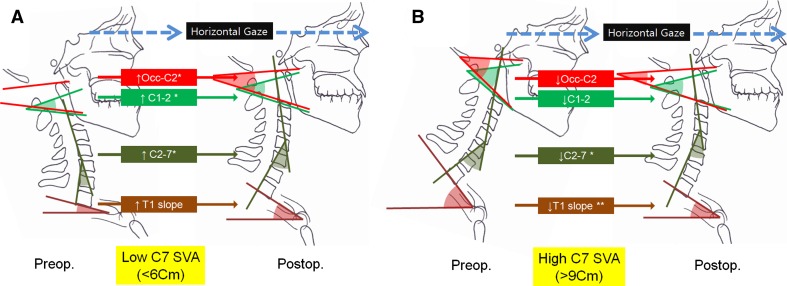
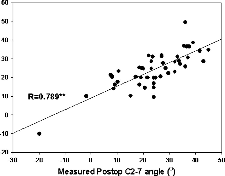
Results: In the lower C7 SVA group, cervical lordosis was significantly increased after thoracic/lumbar deformity correction (p < 0.01). In contrast, the high C7 SVA group showed decreased cervical lordosis postoperatively (p < 0.01). Multilinear regression analysis demonstrated the preoperative parameters (preoperative C2-7 angle, T1 slope, surgical plan for PT and C7 SVA), which determine the postoperative cervical lordosis.

Conclusion: In spinal deformity procedures, preoperative spinal alignment parameters, and surgical plans could affect postoperative cervical spine alignment.


Figures

Fig. 1
Fig. 1
Comparison of preoperative and postoperative sagittal radiographic measurements. a The high C7 SVA group demonstrates higher PT, L1–5, T1, C1–2, and Occ–C2. b The high C7 SVA group, compared to the low C7 SVA group, has higher PT and T5–T12. (*p < 0.05, **p < 0.01). PI pelvic incidence, PT pelvic tilt, SS sacral slope, SVA sagittal vertical axis
Fig. 2
Fig. 2
a Reciprocal changes in cervical spine alignment of flattened spinal curve with low C7 SVA (<6 cm). Cervical lordosis parameters increased after thoracolumbar correction deformity surgery. Occ–C2 angle*(preop 31.8 ± 1.5° vs. postop 35.0 ± 2.5°), C1–C2 anlge*(preop 36.0 ± 1.8° vs. 38.6 ± 1.8°), C2–C7 angle*(preop 25.1 ± 2.2° vs. postop 30.5 ± 4.5°) and T1 slope (preop 27.3 ± 2.3° vs. postop 32.2 ± 3.0°) increase postoperatively. b High C7 SVA patient group (mean C7 SVA = 132.4 mm) patients depicting higher cervical lordosis parameters preoperatively (left). These cervical lordosis parameters decrease after thoracolumbar correction deformity surgery (right). Occ–C2 angle (Preop 35.0 ± 2.5° vs. postop 34.6 ± 2.0°), C1–C2 angle (preop 38.6 ± 1.8° vs. 36.9 ± 1.8°), C2–C7 angle*(preop 30.5 ± 4.5° vs. postop 23.0 ± 3.1°), and T1 slope**(preop 32.2 ± 3.0° vs. postop 25.5 ± 1.8°) decrease postoperatively (*p < 0.05, **p < 0.01)
Fig. 3
Fig. 3
Case illustrations. Low C7 SVA. a Sagittal preoperative radiographs (left full-length spine lateral radiograph, right cervical spine lateral radiograph) of a patient presenting a low C7 SVA (C7 SVA = 35.9 mm). b Sagittal postoperative radiograph of the same patient after thoracolumbar deformity surgery. Angles reflecting cervical lordosis (Occ–C2, C1–2, C2–7, and T1) are increased postoperatively. High C7 SVA. c Sagittal preoperative radiographs (left whole spine lateral, right cervical spine lateral) of a patient presenting a high C7 SVA (C7 SVA = 139.5 mm). d Sagittal postoperative radiograph of the same patient after pedicle subtraction osteotomy. Angles reflecting cervical lordosis (Occ–C2, C1–2, C2–7 and T1) are decreased postoperatively. PI pelvic incidence, PT pelvic tilt, SS sacral slope, SVA sagittal vertical axis
Fig. 4
Fig. 4
Graphical representation of the relationship between predicted and true postoperative values of cervical lordosis (n = 49)

References

    1. Boulay C, Tardieu C, Hecquet J, Benaim C, Mouilleseaux B, Marty C, Prat-Pradal D, Legaye J, Duval-Beaupere G, Pelissier J. Sagittal alignment of spine and pelvis regulated by pelvic incidence: standard values and prediction of lordosis. Eur Spine J. 2006;15:415–422. doi: 10.1007/s00586-005-0984-5. - DOI - PMC - PubMed
    1. Hilibrand AS, Tannenbaum DA, Graziano GP, Loder RT, Hensinger RN. The sagittal alignment of the cervical spine in adolescent idiopathic scoliosis. J Pediatr Orthop. 1995;15:627–632. doi: 10.1097/01241398-199509000-00015. - DOI - PubMed
    1. Lafage V, Schwab F, Vira S, Hart R, Burton D, Smith JS, Boachie-Adjei O, Shelokov A, Hostin R, Shaffrey CI, Gupta M, Akbarnia BA, Bess S, Farcy JP. Does vertebral level of pedicle subtraction osteotomy correlate with degree of spinopelvic parameter correction? J Neurosurg Spine. 2011;14:184–191. doi: 10.3171/2010.9.SPINE10129. - DOI - PubMed
    1. Lafage V, Schwab F, Vira S, Patel A, Ungar B, Farcy JP. Spino-pelvic parameters after surgery can be predicted: a preliminary formula and validation of standing alignment. Spine (Phila Pa 1976) 2011;36:1037–1045. doi: 10.1097/BRS.0b013e3181eb9469. - DOI - PubMed
    1. Glassman SD, Bridwell K, Dimar JR, Horton W, Berven S, Schwab F. The impact of positive sagittal balance in adult spinal deformity. Spine (Phila Pa 1976) 2005;30:2024–2029. doi: 10.1097/01.brs.0000179086.30449.96. - DOI - PubMed

Publication types