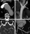
Multilevel assessment of atherosclerotic extent using a 40-section multidetector scanner after transient ischemic attack or ischemic stroke
- PMID: 24136645
- PMCID: PMC7964709
- DOI: 10.3174/ajnr.A3760
Multilevel assessment of atherosclerotic extent using a 40-section multidetector scanner after transient ischemic attack or ischemic stroke
Abstract
Background and purpose: The first part of this study assessed the potential of MDCT with a CTA examination of the aorta and the coronary, cervical, and intracranial vessels in the etiologic work-up of TIA or ischemic stroke compared with established imaging methods. The objective of the second part of this study was to assess the atherosclerotic extent by use of MDCT in these patients.
Materials and methods: From August 2007 to August 2011, a total of 96 patients with ischemic stroke or TIA without an evident cardioembolic source were enrolled. All patients underwent MDCT. Atherosclerotic extent was classified in 0, 1, 2, 3, and 4 atherosclerotic levels according to the number of arterial territories (aortic arch, coronary, cervical, intracranial) affected by atherosclerosis defined as ≥ 50% cervical, intracranial, or coronary stenosis or ≥ 4-mm aortic arch plaque.
Results: There were 91 patients who had an interpretable MDCT. Mean age was 67.4 years (± 11 years), and 75 patients (83.3%) were men. The prevalence of 0, 1, 2, 3, and 4 atherosclerotic levels was 48.3%, 35.2%, 12.1%, 4.4%, and 0%, respectively. Aortic arch atheroma was found in 47.6% of patients with 1 atherosclerotic level. The combination of aortic arch atheroma and cervical stenosis was found in 63.6% of patients with ≥ 2 atherosclerotic levels. Patients with ≥ 2 atherosclerotic levels were older than patients with < 2 atherosclerotic levels (P = .04) in univariate analysis.
Conclusions: MDCT might be useful to assess the extent of atherosclerosis. It could help to screen for high-risk patients who could benefit from a more aggressive preventive strategy.
Figures
Similar articles
-
Significance of aortic atherosclerotic disease in possibly embolic stroke: 64-multidetector row computed tomography study.J Neurol. 2010 May;257(5):699-705. doi: 10.1007/s00415-009-5391-0. Epub 2009 Nov 24. J Neurol. 2010. PMID: 19937048
-
Influence of Age Ranges on Relationship of Complex Aortic Plaque With Cervicocephalic Atherosclerosis in Ischemic Stroke.J Stroke Cerebrovasc Dis. 2019 Jun;28(6):1586-1596. doi: 10.1016/j.jstrokecerebrovasdis.2019.03.001. Epub 2019 Mar 28. J Stroke Cerebrovasc Dis. 2019. PMID: 30928215
-
Characterization of the infarct pattern caused by vulnerable aortic arch atheroma: DWI and multidetector row CT study.Cerebrovasc Dis. 2012;33(6):549-57. doi: 10.1159/000338018. Epub 2012 Jun 8. Cerebrovasc Dis. 2012. PMID: 22688060
-
Population-based study of the relationship between atherosclerotic aortic debris and cerebrovascular ischemic events.Mayo Clin Proc. 2006 May;81(5):609-14. doi: 10.4065/81.5.609. Mayo Clin Proc. 2006. PMID: 16706257
-
Strength of evidence relating periodontal disease and atherosclerotic disease.Compend Contin Educ Dent. 2009 Sep;30(7):430-9. Compend Contin Educ Dent. 2009. PMID: 19757736 Free PMC article. Review.
Cited by
-
One-stop combined coronary-craniocervical computed tomography angiography with low-dose body coverage using artificial intelligence iterative reconstruction: a clinically feasible solution to multi-territorial atherosclerosis diagnosis.Quant Imaging Med Surg. 2025 Feb 1;15(2):1516-1527. doi: 10.21037/qims-24-1545. Epub 2025 Jan 10. Quant Imaging Med Surg. 2025. PMID: 39995738 Free PMC article.
-
Asymptomatic coronary artery disease in ischaemic stroke survivors: A systematic review and meta-analysis.Eur Stroke J. 2024 Sep;9(3):540-554. doi: 10.1177/23969873241231702. Epub 2024 Feb 15. Eur Stroke J. 2024. PMID: 38357886 Free PMC article.
References
-
- Nonent M, Serfaty JM, Nighoghossian N, et al. . Concordance rate differences of 3 noninvasive imaging techniques to measure carotid stenosis in clinical routine practice: results of the CARMEDAS multicenter study. Stroke 2004;35:682–86 - PubMed
-
- Hamon M, Morello R, Riddell JW, et al. . Coronary arteries: diagnostic performance of 16- versus 64-section spiral CT compared with invasive coronary angiography–meta-analysis. Radiology 2007;245:720–31 - PubMed
-
- Nguyen-Huynh MN, Wintermark M, English J, et al. . How accurate is CT angiography in evaluating intracranial atherosclerotic disease? Stroke 2008;39:1184–88 - PubMed
-
- Ko Y, Park JH, Yang MH, et al. . Significance of aortic atherosclerotic disease in possibly embolic stroke: 64-multidetector row computed tomography study. J Neurol 2010;257:699–705 - PubMed
-
- Wardlaw JM, Stevenson MD, Chappell F, et al. . Carotid artery imaging for secondary stroke prevention: both imaging modality and rapid access to imaging are important. Stroke 2009;40:3511–17 - PubMed
Publication types
MeSH terms
LinkOut - more resources
Full Text Sources
Other Literature Sources
Medical


