. 2013 Oct 14:7:127.
doi: 10.3389/fnbeh.2013.00127. eCollection 2013.

Stress affects theta activity in limbic networks and impairs novelty-induced exploration and familiarization

Affiliations

Stress affects theta activity in limbic networks and impairs novelty-induced exploration and familiarization

Luis R Jacinto et al. Front Behav Neurosci. .

Abstract

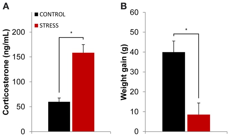
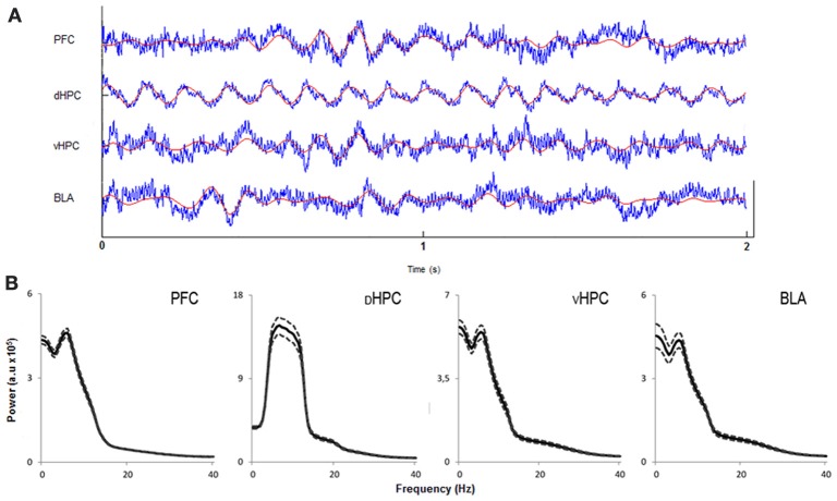
Exposure to a novel environment triggers the response of several brain areas that regulate emotional behaviors. Here, we studied theta oscillations within the hippocampus (HPC)-amygdala (AMY)-medial prefrontal cortex (mPFC) network in exploration of a novel environment and subsequent familiarization through repeated exposures to that same environment; in addition, we assessed how concomitant stress exposure could disrupt this activity and impair both behavioral processes. Local field potentials (LFP) were simultaneously recorded from dorsal and ventral hippocampus (dHPC and vHPC, respectively), basolateral amygdala (BLA) and mPFC in freely behaving rats while they were exposed to a novel environment, then repeatedly re-exposed over the course of 3 weeks to that same environment and, finally, on re-exposure to a novel unfamiliar environment. A longitudinal analysis of theta activity within this circuit revealed a reduction of vHPC and BLA theta power and vHPC-BLA theta coherence through familiarization which was correlated with a return to normal exploratory behavior in control rats. In contrast, a persistent over-activation of the same brain regions was observed in stressed rats that displayed impairments in novel exploration and familiarization processes. Importantly, we show that stress also affected intra-hippocampal synchrony and heightened the coherence between vHPC and BLA. In summary, we demonstrate that modulatory theta activity in the aforementioned circuit, namely in the vHPC and BLA, is correlated with the expression of anxiety in novelty-induced exploration and familiarization in both normal and pathological conditions.

Keywords: amygdala; anxiety; local field potentials; pre-frontal cortex; stress; ventral hippocampus.


Figures

Figure 1
Figure 1
Efficacy of the stress protocol. (A) Plasmatic corticosterone levels measured on the day following the final exposure to the OF arena (which also corresponded to the day after the stress protocol ended). Stressed animals showed higher levels of plasmatic corticosterone than controls at the end of the stress protocol. (B) Body weight gain measured as the difference between the animal's weight at the beginning and ending of the stress protocol for control and stressed animals. Stressed animals showed higher levels of plasmatic corticosterone than controls at the end of the stress protocol as well as reduced body weight gain. *p < 0.05 for unpaired Wilcoxon rank sum test comparison of average plasmatic corticosterone and body weight gain between control and stress groups. Error bars: ± s.e.m.
Figure 2
Figure 2
Evolution of behavioral parameters through familiarization from first and last exposures to the OF arena. (A) Representative trajectories from one rat from each group (control left; stress right) between the first (top) and last (bottom) exposures. (B) Comparison of exploratory activity, as measured by the total number of crossings between the squares on which the arena was divided for behavioral analysis, between first (left) and last (right) exposure to the OF arena on control (black bar) and stress (red bar) groups. (C) Comparison of time spent in the center of the OF arena between first (left) and last (right) exposure on control (black bar) and stress (red bar) groups. Rats from both groups significantly decreased exploratory activity between the first and last exposures to the arena, although the observed decrease was five times greater in stress group animals. Control rats significantly increased the time spent in the center of the arena from the first to the last exposure while stressed rats did not. #p < 0.05 for paired Wilcoxon sign rank test comparison of behavioral parameters within the same group between exposures; *p < 0.05 for unpaired Wilcoxon rank sum test comparison of behavioral parameters between groups on each exposure. Error bars: ± s.e.m.
Figure 3
Figure 3
Characterization of local field potentials recorded in the OF arena. (A) Representative traces from local field potentials simultaneously recorded from the medial prefrontal cortex (PFC), dorsal hippocampus (dHPC), ventral hippocampus (vHPC), and basolateral amygdala (BLA) in one rat exploring the OF arena during the first exposure. Raw traces are plotted in blue and filtered theta traces (5–12 Hz) are overlayed in red. Presented segment duration is 2 s. Voltage scale (bottom right) is −0.2 to 0.2 mV for PFC, vHPC, and BLA; and −0.4 to 0.4 mV for dHPC. (B) Power spectra for PFC, dHPC, vHPC, and BLA. Spectra are average of multitaper spectrum estimates for all animals (n = 14) during the first exposure to the OF arena within the 10–25 cm/s speed range. Dotted lines are ± s.e.m. a.u, arbitrary units.
Figure 4
Figure 4
Temporal evolution of theta power variation from initial exposure with familiarization. Theta power variation during all exposures to the OF arena (day 5, day 9, day 13, day 17, and day 21) relative to theta power in the initial exposure (day 1, origin) from activity recorded from ventral hippocampus (vHPC) (A), basolateral amygdala (BLA) (B), dorsal hippocampus (dHPC) (C), and medial prefrontal cortex (PFC) (D). Theta power decreased in the vHPC and BLA in control rats with familiarization; the same decrease was not observed in stressed rats. No differences were observed between the groups for dHPC and PFC theta power variation with familiarization despite a trend for PFC theta power decrease on both groups. Also note that the second exposure to the OF arena (which corresponded to 4 days of exposure to stressors on stress group rats) showed an incremental effect of stress on theta power as vHPC activity increased from the first exposure on stressed rats only. Error bars: ± s.e.m.
Figure 5
Figure 5
Theta power variation between first and last exposures to the OF arena. (A) Theta power variation between the initial exposure (unfamiliar environment, day 1, origin) and the final exposure (familiar environment, day 21) to the OF arena from activity recorded from ventral hippocampus (vHPC), basolateral amygdala (BLA), dorsal hippocampus (dHPC), and medial prefrontal cortex (PFC). Control rats significantly reduced theta power in vHPC and BLA through familiarization between the first and last exposure to the OF arena while stressed rats failed to display a similar reduction. (B) Correlation of vHPC theta power variation between first and last exposures to the OF arena and exploratory activity, as measured by the total number of crossings between the squares on which the arena was divided for behavioral analysis, on the last exposure to the OF arena. (C) Correlation of vHPC theta power variation between first and last exposures to the OF arena and time spent in the center on the last exposure to the OF arena. Rats that presented the biggest reduction of vHPC theta power between the first and last exposure to the OF arena were the same that presented higher exploratory activity and spent more time in the center on the last exposure to the arena. (D) Correlation of vHPC theta power variation between first and last exposures to the OF arena and serum corticosterone measured on the day following the last exposure to the arena. Rats that presented the lowest levels of serum corticosterone on the day following the last exposure to the OF arena were also the same that presented the highest reduction of vHPC theta power between the first and last exposure to the OF arena. #p < 0.05 for paired Wilcoxon sign rank test comparison of within group theta power estimates between exposures; *p < 0.05 for unpaired Wilcoxon rank sum test comparison of average theta power variation between groups on each exposure. Error bars: ± s.e.m. Plotted r and p values for correlations are for Pearson correlation.
Figure 6
Figure 6
Theta coherence variation between first and last exposures to the OF arena. Theta coherence variation between the between the first exposure to the OF arena (unfamiliar environment, origin) and the final exposure to the same environment (familiar environment, day 21) for the following brain area pairs: vHPC-BLA, vHPC-PFC, dHPC-vHPC and BLA-PFC. Note that theta coherence variation presented for the novel environment is relative to theta coherence on the first exposure (day 1) to the OF arena. Controls presented a significant decrease of vHPC-BLA theta coherence and a tendency for decrease of vHPC-PFC and BLA-PFC theta coherence between first and last exposures to the OF arena; while stressed rats presented an absence of variation of theta coherence between the same brain area pairs. Control rats also presented increased dHPC-vHPC theta coherence through familiarization between first and last exposures to the OF arena while stress rats failed to display a similar variation. #p < 0.05 for paired Wilcoxon sign rank test comparison of within group theta coherence estimates between exposures; *p < 0.05 for unpaired Wilcoxon rank sum test comparison of average theta coherence variation between groups on each exposure. Error bars: ± sem.
Figure 7
Figure 7
Theta power and coherence variation between final exposure to the OF and novel unfamiliar environment. (A) Theta power variation between last exposure to the OF arena (familiar environment) and the second novel unfamiliar arena from activity recorded from ventral hippocampus (vHPC), basolateral amygdala (BLA), dorsal hippocampus (dHPC), and medial prefrontal cortex (PFC). Note that theta power variation presented for the novel environment is relative to theta power in the last exposure (day 21) to the OF arena. When rats from both groups were exposed to the new unfamiliar arena a significant theta power increase in vHPC and BLA was observed in controls but not on stressed rats. No significant variations were observed in dHPC and PFC theta power between plotted exposures or between groups on each exposure. (B) Theta coherence variation between the last exposure to the OF arena (familiar environment, day 21) and the second novel unfamiliar arena (day 24) for the following brain area pairs vHPC-BLA, vHPC-PFC, dHPC-vHPC, and BLA-PFC. When all rats were moved to the new unfamiliar environment control rats presented a significant increase of BLA-PFC theta coherence only; while stressed rats presented a significant increase of vHPC-BLA theta coherence. On dHPC-vHPC theta coherence, control rats presented a decrease whereas stressed rats showed no variation. #p < 0.05 for paired Wilcoxon sign rank test comparison of within group theta power and theta coherence estimates between exposures; *p < 0.05 for unpaired Wilcoxon rank sum test comparison of average theta power and theta coherence variation between groups on each exposure. Error bars: ± s.e.m.

References

Supplemental reference

    1. Paxinos G., Watson C. (2006). The Rat Brain in Stereotaxic Coordinates, 6th Edn. London: Elsevier

References

    1. Adhikari A., Topiwala M. A., Gordon J. A. (2010). Synchronized activity between the ventral hippocampus and the medial prefrontal cortex during anxiety. Neuron 65, 257–269 10.1016/j.neuron.2009.12.002 - DOI - PMC - PubMed
    1. Behrendt R. F. (2011). Neuroanatomy of Social Behaviour: an Evolutionary and Psychoanalytic Perspective. London: Karnac
    1. Bessa J. M., Mesquita A. R., Oliveira M., Pêgo J. M., Cerqueira J. J., Palha J. A., et al. (2009). A trans-dimensional approach to the behavioral aspects of depression. Front. Behav. Neurosci. 3:1. 10.3389/neuro.08.001.2009 - DOI - PMC - PubMed
    1. Blanchard D. C., Griebel G., Pobbe R., Blanchard R. J. (2011). Risk assessment as an evolved threat detection and analysis process. Neurosci. Biobehav. Rev. 35, 991–998 10.1016/j.neubiorev.2010.10.016 - DOI - PubMed
    1. Bueno C. H., Zangrossi H., Jr. Viana M. B. (2005). The inactivation of the basolateral nucleus of the rat amygdala has an anxiolytic effect in the elevated T-maze and light/dark transition tests. Braz. J. Med. Biol. Res. 38, 1697–1701 10.1590/S0100-879X2005001100019 - DOI - PubMed

LinkOut - more resources