. 2013 Sep 30:4:307.
doi: 10.3389/fimmu.2013.00307. eCollection 2013.

CD90(+) Stromal Cells are Non-Professional Innate Immune Effectors of the Human Colonic Mucosa

Affiliations

CD90(+) Stromal Cells are Non-Professional Innate Immune Effectors of the Human Colonic Mucosa

Benjamin M J Owens et al. Front Immunol. .

Erratum in

Abstract

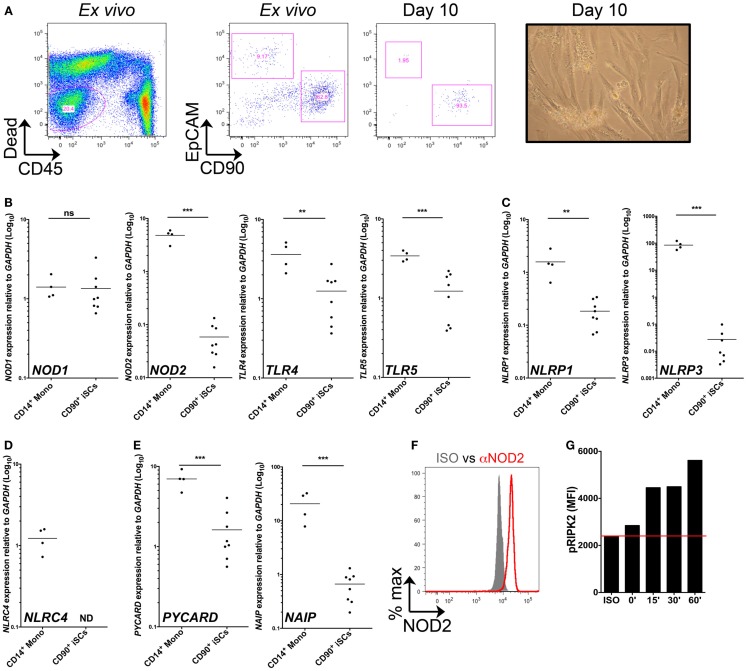
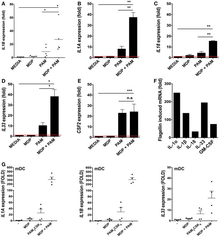
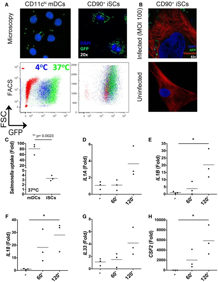
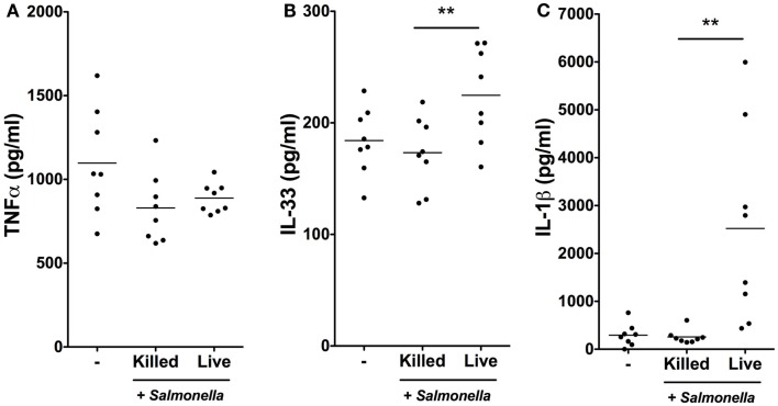
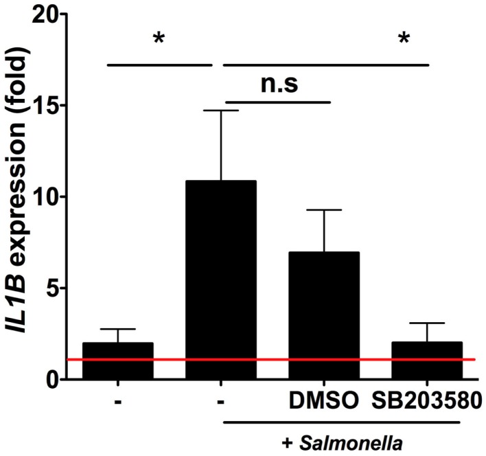
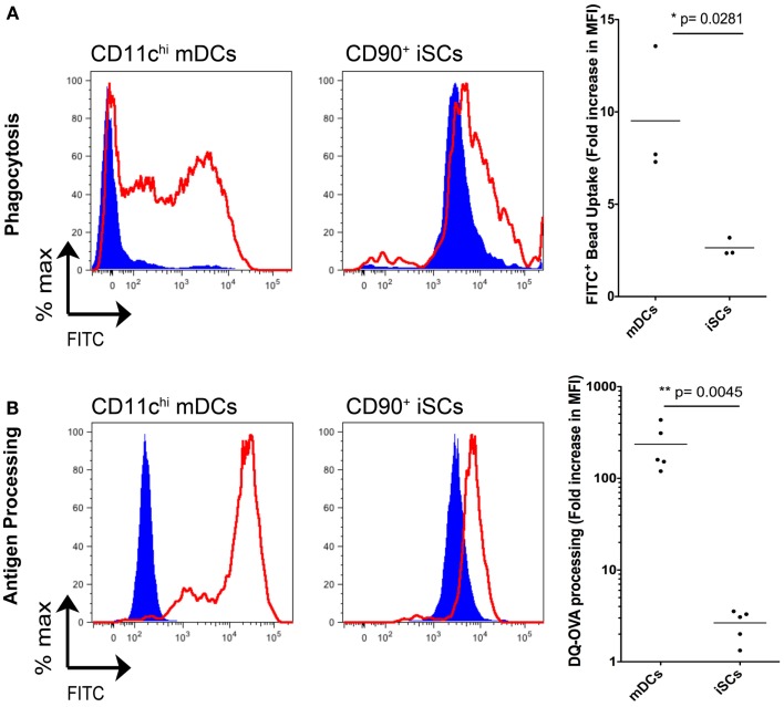
Immune responses at the intestinal mucosa must allow for host protection whilst simultaneously avoiding inappropriate inflammation. Although much work has focused on the innate immune functionality of hematopoietic immune cells, non-hematopoietic cell populations - including epithelial and stromal cells - are now recognized as playing a key role in innate defense at this site. In this study we examined the innate immune capacity of primary human intestinal stromal cells (iSCs). CD90(+) iSCs isolated from human colonic mucosa expressed a wide array of innate immune receptors and functionally responded to stimulation with bacterial ligands. iSCs also sensed infection with live Salmonella typhimurium, rapidly expressing IL-1 family cytokines via a RIPK2/p38MAPK-dependent signaling process. In addition to responding to innate immune triggers, primary iSCs exhibited a capacity for bacterial uptake, phagocytosis, and antigen processing, although to a lesser extent than professional APCs. Thus CD90(+) iSCs represent an abundant population of "non-professional" innate immune effector cells of the human colonic mucosa and likely play an important adjunctive role in host defense and immune regulation at this site.

Keywords: innate immunity; intestinal homeostasis; mucosal immunology; stromal cells.


Figures

Figure 1
Figure 1
CD90+ colonic stromal cells express diverse PRRs and functional NOD2. (A) Shows representative flow cytometric staining of collagenase-digested colonic tissue ex vivo and cultured stromal cells after 10 days in culture, revealing iSCs as viable, CD45EpCAMCD90+ cells; and a representative image of day 10 cultured stromal cells obtained by light microscopy. (B–E) Show quantification of the expression of the indicted genes in monocytes and cultured iSCs by qRT-PCR, expressed as arbitrary units (AU) relative to GAPDH. (F) Shows quantification of NOD2 expression by intracellular flow cytometry; gray filled histogram indicates isotype control, red open histogram indicting specific staining. (G) Shows accumulation of phosphorylated RIPK2 in cultured iSCs determined by flow cytometry after stimulation for the indicated time periods with MDP. Data in (A) are representative of tissue, (B–E) are from monocytes isolated from n = 4 healthy peripheral blood donors and iSCs cultured from colonic tissue of n = 8 uninflamed surgical patients. Data in (F,G) are from iSCs of one patient, representative of four patients with similar results. **p < 0.01, ***p < 0.001.
Figure 2
Figure 2
Bacterial ligands regulate IL-1 family member cytokine expression in iSCs. (A–F) Show changes in expression of the indicated cytokines by cultured iSCs, (G) shows the same by monocyte-derived dendritic cells (mDCs), determined by qRT-PCR after 24 h of in vitro stimulation with the PRR ligands indicated. Data in (A–E) show mean fold changes ±SEM in iSCs from colonic tissue of n = 5 surgical patients, (F) shows the mean induction of expression of the indicated genes by iSCs from one patient after stimulation with flagellin, representative of three experiments with similar results. (G) Shows mean fold changes ±SEM in expression of the indicated mRNA in stimulated mDCs generated from n = 4 peripheral blood donors. *p < 0.05, **p < 0.01, ***p < 0.001.
Figure 3
Figure 3
Rapid internalization and sensing of live Salmonella by iSCs. (A) Shows presence of GFP+ Salmonella in CD11c+ mDCs and CD90+stromal cells by representative fluorescence microscopy images (upper panels) and flow cytometry plots (lower panels) after 2 h of bacterial exposure. (B) (upper panel) shows the presence of GFP-expressing Salmonella in Vimentin+ iSCs infected for 4 h at an MOI of 100; uninfected iSCs shown for comparison (lower panel). (C) Shows quantification of Salmonella uptake/internalization by mDCs and iSCs exposed to bacteria as in (A), expressed as the mean fold change in GFP MFI determined by flow cytometry. (D–H) Show the mean fold increases in expression of the indicated genes after exposure of iSCs to Salmonella at an MOI of 10 for the indicated time periods, relative to unstimulated cells. (A,B) shows representative images and FACS plots, data in (C) quantified from mDCs generated from peripheral blood of n = 3 healthy donors, compared to iSCs from colonic tissue of n = 3 surgical patients. Data in (D–H) are from iSCs isolated from colonic tissue of n = 3 patients. *p < 0.05, other p values as indicated.
Figure 4
Figure 4
Salmonella infection stimulates IL-1β and IL-33 production by iSCs. (A–C) show production levels of the indicated cytokines over a 24 h period by iSCs, determined by ELISA. iSCs were left unstimulated (−) or exposed to live or heat-killed Salmonella at an MOI of 10 for 4 h, followed by addition of Gentamycin for the subsequent 20 h. Data are mean levels of cytokine produced by iSCs isolated from colonic tissue of n = 8 surgical patients. **p < 0.01.
Figure 5
Figure 5
Intestinal stromal cell responses to Salmonella require RIPK2/p38MAPK signaling. Figure 5 shows relative fold changes in IL1B expression determined by qRT-PCR in iSCs left uninfected or after 6 h of infection with live Salmonella at an MOI of 10, in the presence of bacteria alone (−), DMSO or SB203580. Data show mean ± SEM fold increases in IL1B expression by iSCs under the indicated conditions isolated from n = 6 surgical patients. *p < 0.05.
Figure 6
Figure 6
Intestinal stromal cells have a limited capacity for phagocytosis and antigen processing. (A) shows the quantification of FITC-labeled bead uptake by CD11c+ mDCs or CD90+ cultured iSCs, determined by flow cytometry at either 4°C (blue filled histograms) or 37°C (red open histograms), expressed as the fold increase in FITC MFI expression by cells at 37°C vs. 4oC. (B) Shows quantification of antigen processing by CD11c+ mDCs or CD90+ iSCs, determined by flow cytometry after pulsing with DQ-OVA at either 4°C (blue filled histograms) or 37°C (red open histograms), expressed as the fold increase in FITC MFI expression by cells at 37°C vs. 4°C. Data in (A,B) show mean fold increases in FITC MFI by mDCs and iSCs from n = 3–5 patients. P values as indicated.

Similar articles

Cited by

References

    1. Owens BM, Simmons A. Intestinal stromal cells in mucosal immunity and homeostasis. Mucosal Immunol (2013) 6(2):224–3410.1038/mi.2012.125 - DOI - PubMed
    1. Powell DW, Pinchuk IV, Saada JI, Chen X, Mifflin RC. Mesenchymal cells of the intestinal lamina propria. Annu Rev Physiol (2011) 73:213–3710.1146/annurev.physiol.70.113006.100646 - DOI - PMC - PubMed
    1. Otte JM, Rosenberg IM, Podolsky DK. Intestinal myofibroblasts in innate immune responses of the intestine. Gastroenterology (2003) 124(7): 1866–7810.1016/S0016-5085(03)00403-7 - DOI - PubMed
    1. Kim YG, Kamada N, Shaw MH, Warner N, Chen GY, Franchi L, et al. The Nod2 sensor promotes intestinal pathogen eradication via the chemokine CCL2-dependent recruitment of inflammatory monocytes. Immunity (2011) 34(5):769–8010.1016/j.immuni.2011.04.013 - DOI - PMC - PubMed
    1. Tada H, Aiba S, Shibata K, Ohteki T, Takada H. Synergistic effect of Nod1 and Nod2 agonists with toll-like receptor agonists on human dendritic cells to generate interleukin-12 and T helper type 1 cells. Infect Immun (2005) 73(12): 7967–7610.1128/IAI.73.12.7967-7976.2005 - DOI - PMC - PubMed