

Biocompatibility study of a silk fibroin-chitosan scaffold with adipose tissue-derived stem cells in vitro
- PMID: 24137218
- PMCID: PMC3786727
- DOI: 10.3892/etm.2013.1185
Biocompatibility study of a silk fibroin-chitosan scaffold with adipose tissue-derived stem cells in vitro
Abstract
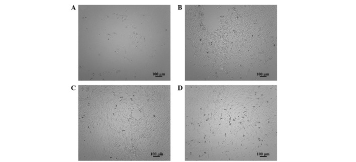
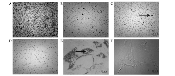
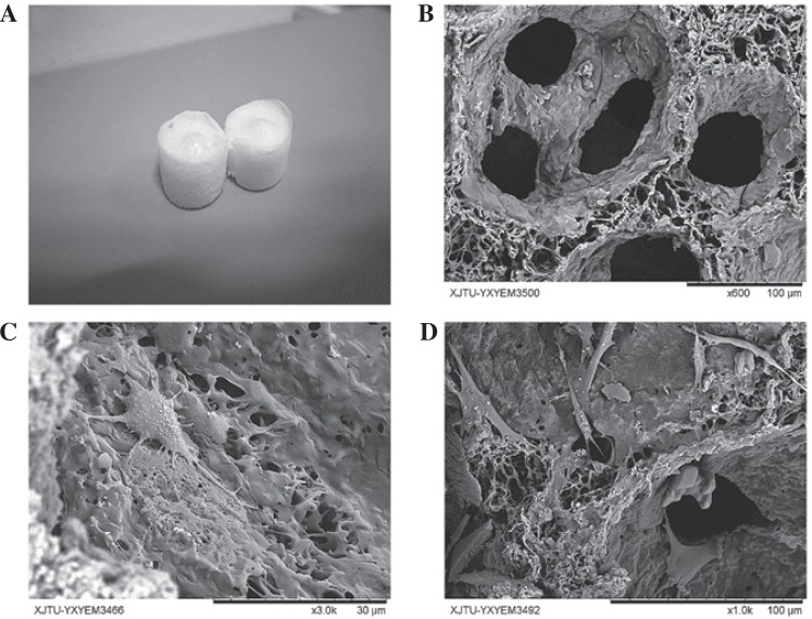
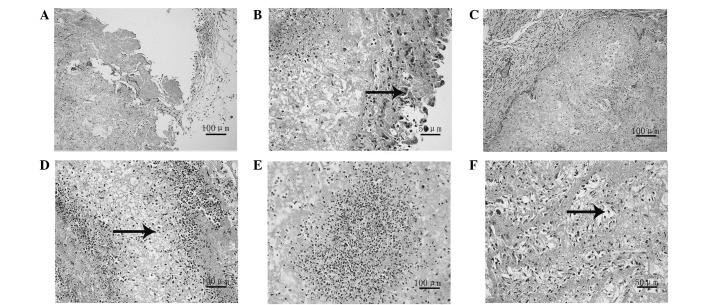
The use of tissue engineering technology in the repair of spinal cord injury (SCI) is a topic of current interest. The success of the repair of the SCI is directly affected by the selection of suitable seed cells and scaffold materials with an acceptable biocompatibility. In this study, adipose tissue-derived stem cells (ADSCs) were incorporated into a silk fibroin-chitosan scaffold (SFCS), which was constructed using a freeze-drying method, in order to assess the biocompatibility of the ADSCs and the SFCS and to provide a foundation for the use of tissue engineering technology in the repair of SCI. Following the seeding of the cells onto the scaffold, the adhesion characteristics of the ADSCs and the SFCS were assessed. A significant difference was observed between the experimental group (a composite of the ADSCs with the SFCS) and the control group (ADSCs without the scaffold) following a culture period of 6 h (P<0.05). The differences in the results at the following time-points were statistically insignificant (P>0.05). The use of an MTT assay to assess the proliferation of the cells on the scaffold revealed that there were significant differences between the experimental and control groups following culture periods of 2 and 4 days (P<0.05). However, the results at the subsequent time-points were not statistically significantly different (P>0.05). Scanning electron microscopy (SEM), using hematoxylin and eosin (H&E) staining, was used to observe the cellular morphology following seeding, and this revealed that the cells displayed the desired morphology. The results indicate that ADSCs have a good biocompatibility with a SFCS and thus provide a foundation for further studies using tissue engineering methods for the repair of SCI.
Keywords: adipose stem cells; biocompatibility; silk fibroin-chitosan scaffold.
Figures
Similar articles
-
Selected suitable seed cell, scaffold and growth factor could maximize the repair effect using tissue engineering method in spinal cord injury.World J Exp Med. 2016 Aug 20;6(3):58-62. doi: 10.5493/wjem.v6.i3.58. eCollection 2016 Aug 20. World J Exp Med. 2016. PMID: 27622154 Free PMC article. Review.
-
Protective effect of brain-derived neurotrophic factor and neurotrophin-3 overexpression by adipose-derived stem cells combined with silk fibroin/chitosan scaffold in spinal cord injury.Neurol Res. 2020 May;42(5):361-371. doi: 10.1080/01616412.2020.1735819. Epub 2020 Mar 9. Neurol Res. 2020. PMID: 32149594
-
Self-assembly model, hepatocytes attachment and inflammatory response for silk fibroin/chitosan scaffolds.Biomed Mater. 2009 Aug;4(4):045014. doi: 10.1088/1748-6041/4/4/045014. Epub 2009 Aug 11. Biomed Mater. 2009. PMID: 19671956
-
[Biocompatibility of silk fibroin nanofibers scaffold with olfactory ensheathing cells].Zhongguo Xiu Fu Chong Jian Wai Ke Za Zhi. 2009 Nov;23(11):1365-70. Zhongguo Xiu Fu Chong Jian Wai Ke Za Zhi. 2009. PMID: 19968182 Chinese.
-
[Application of silk fibroin scaffold in bone tissue engineering].Zhongguo Xiu Fu Chong Jian Wai Ke Za Zhi. 2014 Oct;28(10):1307-10. Zhongguo Xiu Fu Chong Jian Wai Ke Za Zhi. 2014. PMID: 25591313 Review. Chinese.
Cited by
-
Effects of brain‑derived neurotrophic factor and neurotrophin‑3 on the neuronal differentiation of rat adipose‑derived stem cells.Mol Med Rep. 2015 Oct;12(4):4981-8. doi: 10.3892/mmr.2015.4099. Epub 2015 Jul 20. Mol Med Rep. 2015. PMID: 26239042 Free PMC article.
-
Compatibility of Porous Chitosan Scaffold with the Attachment and Proliferation of human Adipose-Derived Stem Cells In Vitro.J Stem Cells Regen Med. 2016 Nov 29;12(2):79-86. doi: 10.46582/jsrm.1202012. eCollection 2016. J Stem Cells Regen Med. 2016. PMID: 28096632 Free PMC article.
-
The combined strategy of mesenchymal stem cells and tissue-engineered scaffolds for spinal cord injury regeneration.Exp Ther Med. 2017 Oct;14(4):3355-3368. doi: 10.3892/etm.2017.4939. Epub 2017 Aug 16. Exp Ther Med. 2017. PMID: 29042919 Free PMC article.
-
Characterization of Silk Fibroin/Chitosan 3D Porous Scaffold and In Vitro Cytology.PLoS One. 2015 Jun 17;10(6):e0128658. doi: 10.1371/journal.pone.0128658. eCollection 2015. PLoS One. 2015. PMID: 26083846 Free PMC article.
-
Selected suitable seed cell, scaffold and growth factor could maximize the repair effect using tissue engineering method in spinal cord injury.World J Exp Med. 2016 Aug 20;6(3):58-62. doi: 10.5493/wjem.v6.i3.58. eCollection 2016 Aug 20. World J Exp Med. 2016. PMID: 27622154 Free PMC article. Review.
References
-
- Silva NA, Salgado AJ, Sousa RA, et al. Development and characterization of a novel hybrid tissue engineering-based scaffold for spinal cord injury repair. Tissue Eng Part A. 2010;16:45–54. - PubMed
-
- Anghileri E, Marconi S, Pignatelli A, et al. Neuronal differentiation potential of human adipose-derived mesenchymal stem cells. Stem Cells Dev. 2008;17:909–916. - PubMed
LinkOut - more resources
Full Text Sources
Other Literature Sources