

Microsurgical treatment for parasagittal meningioma in the central gyrus region
- PMID: 24137410
- PMCID: PMC3789089
- DOI: 10.3892/ol.2013.1429
Microsurgical treatment for parasagittal meningioma in the central gyrus region
Abstract
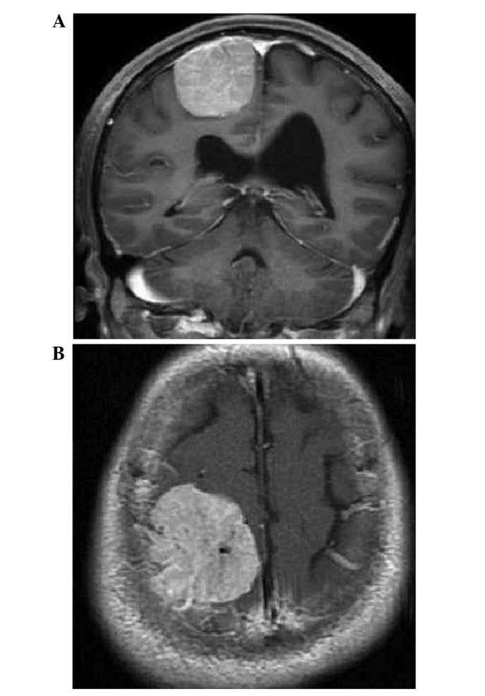
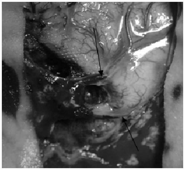
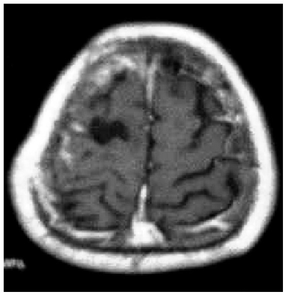
The aim of the present study was to determine the efficacy of microsurgery treatment for parasagittal meningioma in the central gyrus region. A microsurgical technique was used to treat 26 patients with large parasagittal meningioma in the central gyrus region. The Rolandic and draining veins and the peritumoral normal brain tissue were retained, and the associated sagittal sinus was appropriately protected. A Simpson grade I, II or III resection was performed in 8 (30.8%), 12 (46.2%) and 6 (23.1%) patients, respectively, with no post-operative mortalities. Following treatment, 9 patients exhibited hemiparalysis. No tumor recurrence was found in 21 patients during the follow-up examination. The treatment protocol described in the current study included sufficient pre-operative imaging evaluations, a skilled microsurgical technique, improved protection of the Rolandic vein and treatment of the sagittal sinus, and was found to significantly increase the total tumor removal rate and decrease post-operative recurrence.
Keywords: Rolandic vein; central gyrus region; microsurgery; parasagittal meningioma.
Figures
Similar articles
-
Microsurgical treatment for central gyrus region meningioma with epilepsy as primary symptom.J Craniofac Surg. 2014 Sep;25(5):1773-5. doi: 10.1097/SCS.0000000000000889. J Craniofac Surg. 2014. PMID: 24999673 Free PMC article.
-
Multimodal treatment of parasagittal meningiomas: a single-center experience.J Neurosurg. 2017 Dec;127(6):1249-1256. doi: 10.3171/2016.9.JNS161859. Epub 2017 Feb 3. J Neurosurg. 2017. PMID: 28156245
-
Tumor recurrence in parasagittal and falcine atypical meningiomas invading the superior sagittal sinus.Rom J Morphol Embryol. 2020 Apr-Jun;61(2):385-395. doi: 10.47162/RJME.61.2.08. Rom J Morphol Embryol. 2020. PMID: 33544790 Free PMC article.
-
Meningiomas engaging major venous sinuses.World Neurosurg. 2014 Jan;81(1):116-24. doi: 10.1016/j.wneu.2013.01.095. Epub 2013 Jan 30. World Neurosurg. 2014. PMID: 23376533 Review.
-
Parasagittal meningiomas.Handb Clin Neurol. 2020;170:93-100. doi: 10.1016/B978-0-12-822198-3.00031-8. Handb Clin Neurol. 2020. PMID: 32586512 Review.
Cited by
-
Microsurgical treatment for central gyrus region meningioma with epilepsy as primary symptom.J Craniofac Surg. 2014 Sep;25(5):1773-5. doi: 10.1097/SCS.0000000000000889. J Craniofac Surg. 2014. PMID: 24999673 Free PMC article.
-
Meningiomas of the rolandic region: risk factors for motor deficit and role of intra-operative monitoring.Acta Neurochir (Wien). 2023 Jul;165(7):1707-1716. doi: 10.1007/s00701-023-05630-6. Epub 2023 Jun 5. Acta Neurochir (Wien). 2023. PMID: 37277557 Free PMC article. Review.
-
Optimal surgical strategy for meningiomas involving the superior sagittal sinus: a systematic review.Neurosurg Rev. 2020 Apr;43(2):525-535. doi: 10.1007/s10143-018-1026-1. Epub 2018 Aug 31. Neurosurg Rev. 2020. PMID: 30171502
-
Long-term follow-up of motor function deterioration following microsurgical resection of middle third parasagittal and falx meningioma.Egypt J Neurol Psychiatr Neurosurg. 2018;54(1):9. doi: 10.1186/s41983-018-0013-3. Epub 2018 Apr 25. Egypt J Neurol Psychiatr Neurosurg. 2018. PMID: 29780229 Free PMC article.
References
-
- Wang ZC, editor. Neurosurgery. Hubei Science and Technology Press; 2005. Diagnosis of neurological diseases; pp. 595–597. (In Chinese)
-
- Caroli E, Orlando ER, Mastronardi L, Ferrante L. Meningiomas infiltrating the superior sagittal sinus: surgical considerations of 328 cases. Neurosurg Rev. 2006;29:236–241. - PubMed
-
- Chen GZ, Chen GM, Song ZH. Microneurosurgery treatment of parasagittal meningiomas. Zhong Hua Xian Wei Wai Ke Za Zhi Bian Ji Bu. 2006;29:72–74. (In Chinese)
-
- Nowak A, Marchel A. Surgical treatment of parasagittal and falx meningiornas. Neurol Neurochir Pol. 2007;41:306–314. - PubMed
LinkOut - more resources
Full Text Sources
Other Literature Sources