. 2014 Mar 1;23(5):515-29.
doi: 10.1089/scd.2013.0263. Epub 2014 Jan 20.

Generation of highly purified neural stem cells from human adipose-derived mesenchymal stem cells by Sox1 activation

Affiliations

Generation of highly purified neural stem cells from human adipose-derived mesenchymal stem cells by Sox1 activation

Nianhua Feng et al. Stem Cells Dev. .

Abstract

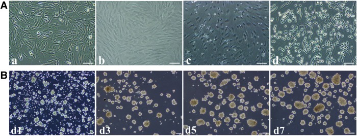
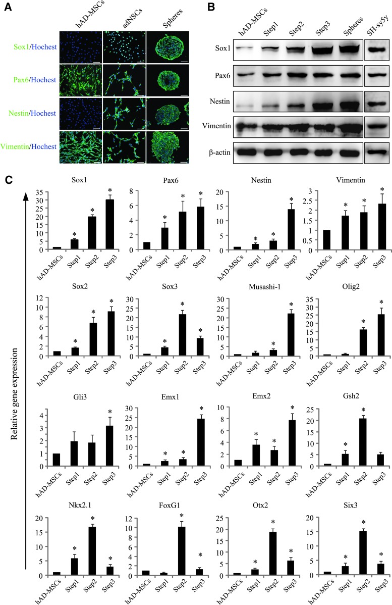
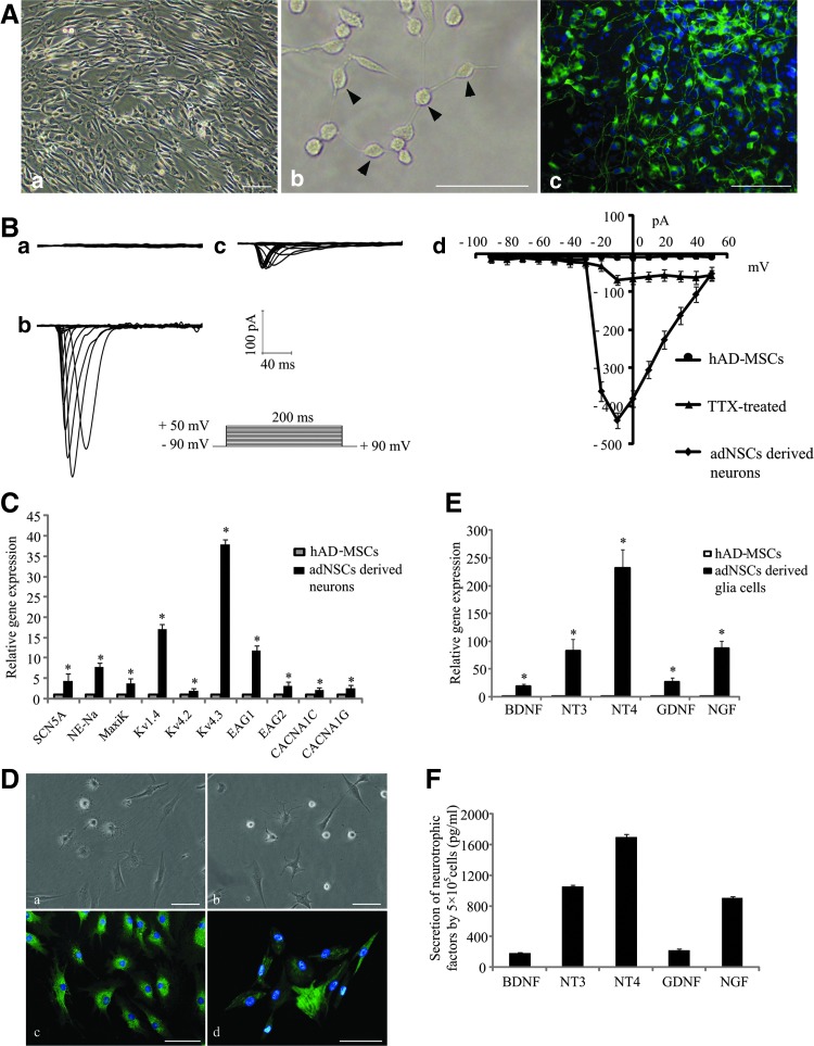
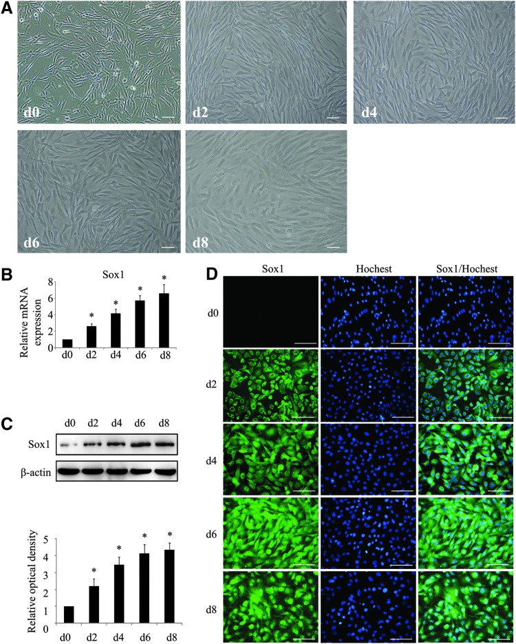
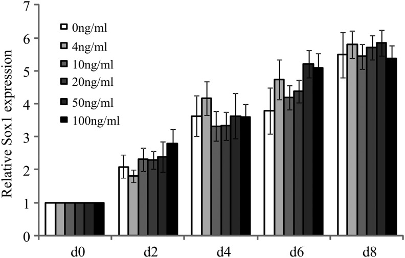
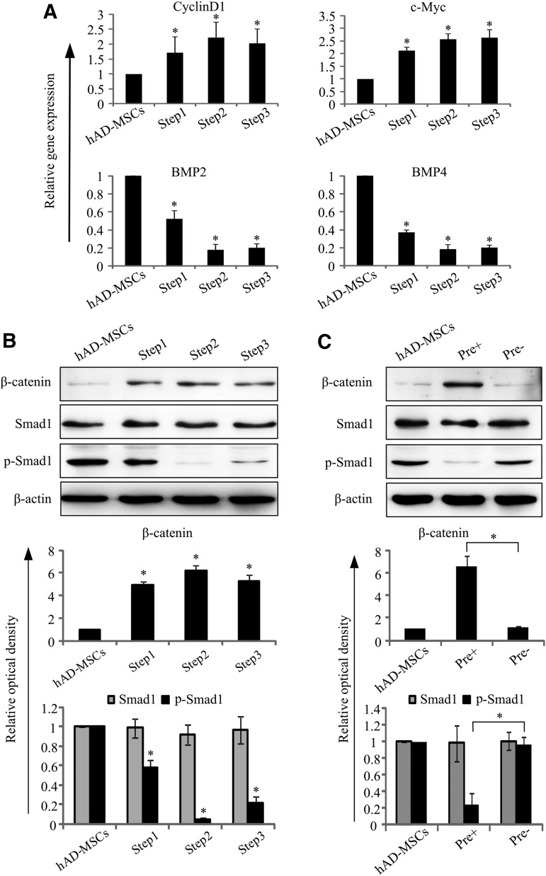
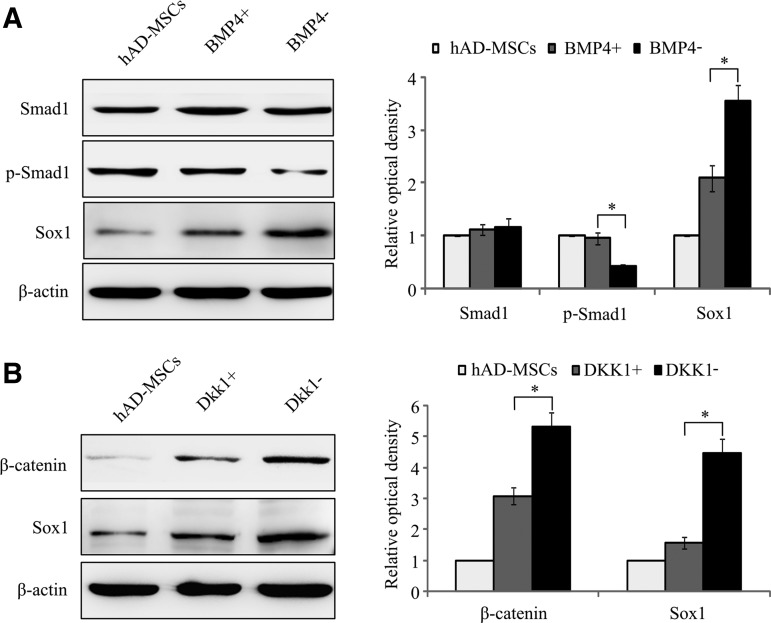
Neural stem cells (NSCs) are ideal candidates in stem cell-based therapy for neurodegenerative diseases. However, it is unfeasible to get enough quantity of NSCs for clinical application. Generation of NSCs from human adipose-derived mesenchymal stem cells (hAD-MSCs) will provide a solution to this problem. Currently, the differentiation of hAD-MSCs into highly purified NSCs with biological functions is rarely reported. In our study, we established a three-step NSC-inducing protocol, in which hAD-MSCs were induced to generate NSCs with high purity after sequentially cultured in the pre-inducing medium (Step1), the N2B27 medium (Step2), and the N2B27 medium supplement with basic fibroblast growth factor and epidermal growth factor (Step3). These hAD-MSC-derived NSCs (adNSCs) can form neurospheres and highly express Sox1, Pax6, Nestin, and Vimentin; the proportion was 96.1% ± 1.3%, 96.8% ± 1.7%, 96.2% ± 1.3%, and 97.2% ± 2.5%, respectively, as detected by flow cytometry. These adNSCs can further differentiate into astrocytes, oligodendrocytes, and functional neurons, which were able to generate tetrodotoxin-sensitive sodium current. Additionally, we found that the neural differentiation of hAD-MSCs were significantly suppressed by Sox1 interference, and what's more, Step1 was a key step for the following induction, probably because it was associated with the initiation and nuclear translocation of Sox1, an important transcriptional factor for neural development. Finally, we observed that bone morphogenetic protein signal was inhibited, and Wnt/β-catenin signal was activated during inducing process, and both signals were related with Sox1 expression. In conclusion, we successfully established a three-step inducing protocol to derive NSCs from hAD-MSCs with high purity by Sox1 activation. These findings might enable to acquire enough autologous transplantable NSCs for the therapy of neurodegenerative diseases in clinic.


Figures

<b>FIG. 1.</b>
FIG. 1.
Differentiation procedure from human adipose-derived mesenchymal stem cells (hAD-MSCs) to neural stem cells (NSCs) (hAD-MSC-derived NSCs, adNSCs). hAD-MSCs (Sox1low/Nestinlow) were cultured in the pre-inducing medium for 8 days to activate Sox1 expression. Then, Sox1moderate/Nestinlow cells were cultured in the N2B27 medium for 7 days to promote Sox1 expression. Finally, the medium was changed to the N2B27 medium containing basic fibroblast growth factor (bFGF) and epidermal growth factor (EGF), and cells were cultured for another 7 days to generate Sox1high/Nestinhigh adNSCs.
<b>FIG. 2.</b>
FIG. 2.
Differentiation of hAD-MSCs into adNSCs. (A) Cell morphology of hAD-MSCs (a), after pre-induction (Step1) (b), cultured in the N2B27 medium (Step2) for 7 days (c), and cultured in the N2B27 medium containing bFGF and EGF (Step3) (adNSCs) for another 7 days (d). (B) Expansion of neurospheres. Spheres cultured in suspension at d1, d3, d5, d7 after passage. Bar=100 μm.
<b>FIG. 3.</b>
FIG. 3.
Identification of adNSCs. (A) Immunostaining of NSCs markers Sox1, Pax6, Nestin, and Vimentin in undifferentiated hAD-MSCs, adNSCs culture monolayer and neurospheres. adNSCs we obtained expressed NSCs markers both in monolayer and in neurospheres. Bar=100 μm. (B) Western blot analysis. (C) Expression of genes associated with embryonic neural development during induction (*P<0.05 compared with hAD-MSCs).
<b>FIG. 4.</b>
FIG. 4.
Terminal differentiation of adNSCs. (A) adNSC-derived neurons (a, b) and characterization of neurons by immunostaining for mature neurons marker MAP2 (green) (c). Bar=100 μm. (B) Electrophysiological analysis for inward sodium current. No inward sodium current was detected in hAD-MSCs (a). Voltage-dependent sodium current was detected in adNSC-derived neurons (b), this current can be blocked by 500 nM tetrodotoxin (TTX) (c). The peak current–voltage relationship was plotted against the voltages (d). (C) Gene expression of ion channel markers. Gene expression of ion channel markers increased significantly compared with hAD-MSCs (*P<0.05). (D) Glia differentiation of adNSCs. adNSCs can differentiate into astrocytes (a) and oligodendrocytes (b). GFAP expression (green) in astrocytes (c) and O4 expression (green) in oligodendrocytes (d) by immnostaining. Bar=100 μm. (E) Examination of gene expression of neurotrophic factors in adNSC-derived glia cells. Gene expression of neurotrophic factors significantly increased compared with hAD-MSCs (*P<0.05). (F) Secretion of neurotrophic factors in 24 h in the culture surpernanant of adNSC-derived glia cells by enzyme-linked immunosorbent assay analysis. All data represent mean±standard deviation, n=3.
<b>FIG. 5.</b>
FIG. 5.
Sox1 inhibition suppresses the differentiation of hAD-MSCs into NSCs. (A) Real-time PCR analysis of the Sox1 mRNA level after transfection with small-interfering RNAs (siSox1 or siNC). (B) Protein level of Sox1 after transfection with siSox1 or siNC by western blot analysis, relative optical density was measured. (C) mRNA expression of NSC markers of adNSCs after Sox1 or NC transfection. (D) Protein level of NSCs markers after transfection with siSox1 or siNC by western blot analysis, relative optical density was measured. (All data displayed as mean±standard deviation, n=3.*P<0.05 compared with the siNC group).
<b>FIG. 6.</b>
FIG. 6.
(A) Cell morphology in the pre group for which pre-inducing step was omitted. hAD-MSCs (a), cells cultured in N2B27 for 7 days (b), then cultured in the N2B27 medium containing bFGF and EGF for 7 days (c). Bar=100 μm. (B) Immunostaining for NSCs markers in cells of pre group. Western blot (C) and qPCR analysis (D) of NSCs markers in cells finally obtained in the pre+ group and pre group. These results showed that when pre-inducing step was omitted (pre group), the expression of NSCs markers greatly decreased in both mRNA and protein levels compared to the pre+ group (*P<0.05).
<b>FIG. 7.</b>
FIG. 7.
Nuclear translocation of Sox1 during pre-inducing process. (A) Cell morphorlogy at d0, d2, d4, d6, and d8 after cultured in the pre-inducing medium. Real-time PCR (B), western blot analysis (C), and immunostaining (D) of Sox1 expression. Bar=100 μm. All data represent mean±standard deviation, n=3 (*P<0.05 compared with d0).
<b>FIG. 8.</b>
FIG. 8.
mRNA expression of Sox1 in cells cultured in the pre-inducing medium supplemented with different concentrations of bFGF at different time points. There were no differences at same time point between groups with different bFGF concentration.
<b>FIG. 9.</b>
FIG. 9.
Activation state of Wnt/β-catenin and bone morphogenetic protein (BMP) signal pathways in differentiating process. (A) Expression of genes related with Wnt/β-catenin (Cyclin D1 and c-Myc) and BMP (BMP2 and BMP4) signal pathways during differentiation (*P<0.05 compared with hAD-MSCs, n=3). (B) Western blot analysis of β-catenin, Smad1, and p-Smad1, optical density of each band was analyzed. Results showed that protein level of β-catenin increased while p-Smad1 decreased after differentiation (*P<0.05 compared with hAD-MSCs, n=3). (C) Comparison of Wnt/β-catenin and BMP signal pathways in cells finally obtained in the pre+ group and pre group by western blot. Protein level of β-catenin and p-Smad1 significantly decreased compared with the pre group (*P<0.05, n=3).
<b>FIG. 10.</b>
FIG. 10.
Expression of Sox1 was influenced by BMP activation (BMP4+) (A) and Wnt/β-catenin inhibition (DKK1+) (B) in pre-inducing process. Expression of Sox1 was suppressed after treated with BMP4 compared to the BMP group or after treated with DKK1 compared to the DKK1 group (*P<0.05, n=3).

References

    1. Feng Z. and Gao F. (2012). Stem cell challenges in the treatment of neurodegenerative disease. CNS Neurosci Ther 18:142–148 - PMC - PubMed
    1. Brown RC, Lockwood AH. and Sonawane BR. (2005). Neurodegenerative diseases: an overview of environmental risk factors. Environ Health Perspect 113:1250–1256 - PMC - PubMed
    1. Honig LS. and Rosenberg RN. (2000). Apoptosis and neurologic disease. Am J Med 108:317–330 - PubMed
    1. Mujtaba T, Piper DR, Kalyani A, Groves AK, Lucero MT. and Rao MS. (1999). Lineage-restricted neural precursors can be isolated from both the mouse neural tube and cultured ES cells. Dev Biol 214:113–127 - PubMed
    1. Riess P, Zhang C, Saatman KE, Laurer HL, Longhi LG, Raghupathi R, Lenzlinger PM, Lifshitz J, Boockvar J, et al. (2002). Transplanted neural stem cells survive, differentiate, and improve neurological motor function after experimental traumatic brain injury. Neurosurgery 51:1043–1052; discussion 1052–1054. - PubMed

Publication types

MeSH terms

Substances

LinkOut - more resources