Review
. 2014 Jan;171(1):38-54.
doi: 10.1111/bph.12472.

SERCA2a gene therapy in heart failure: an anti-arrhythmic positive inotrope

Affiliations
Review

SERCA2a gene therapy in heart failure: an anti-arrhythmic positive inotrope

Markus B Sikkel et al. Br J Pharmacol. 2014 Jan.

Abstract

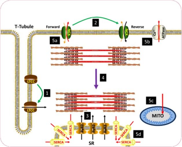
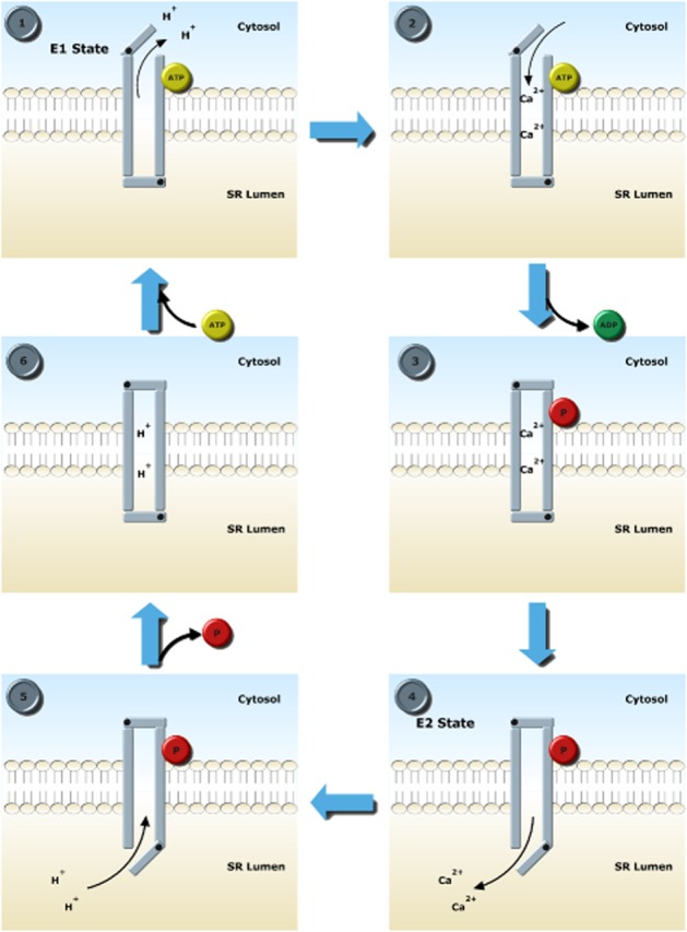
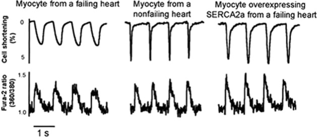
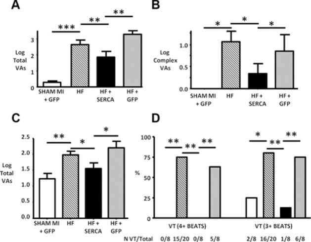
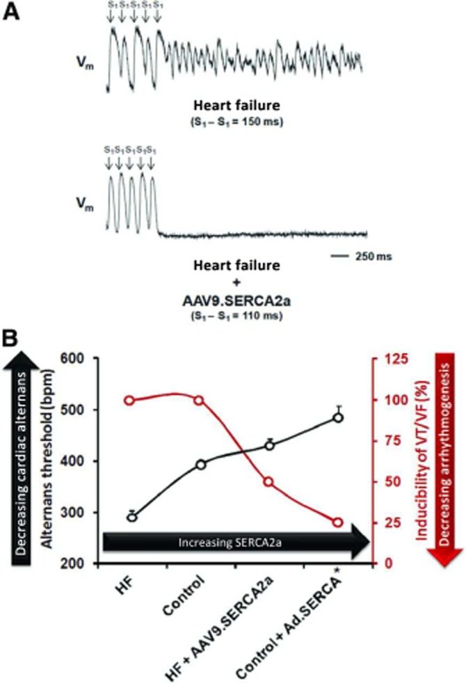
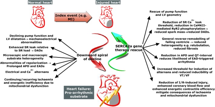
Therapeutic options that directly enhance cardiomyocyte contractility in chronic heart failure (HF) therapy are currently limited and do not improve prognosis. In fact, most positive inotropic agents, such as β-adrenoreceptor agonists and PDE inhibitors, which have been assessed in HF patients, cause increased mortality as a result of arrhythmia and sudden cardiac death. Cardiac sarcoplasmic reticulum Ca(2)(+) -ATPase2a (SERCA2a) is a key protein involved in sequestration of Ca(2)(+) into the sarcoplasmic reticulum (SR) during diastole. There is a reduction of SERCA2a protein level and function in HF, which has been successfully targeted via viral transfection of the SERCA2a gene into cardiac tissue in vivo. This has enhanced cardiac contractility and reduced mortality in several preclinical models of HF. Theoretical concerns have been raised regarding the possibility of arrhythmogenic adverse effects of SERCA2a gene therapy due to enhanced SR Ca(2)(+) load and induction of SR Ca(2)(+) leak as a result. Contrary to these concerns, SERCA2a gene therapy in a wide variety of preclinical models, including acute ischaemia/reperfusion, chronic pressure overload and chronic myocardial infarction, has resulted in a reduction in ventricular arrhythmias. The potential mechanisms for this unexpected beneficial effect, as well as mechanisms of enhancement of cardiac contractile function, are reviewed in this article.

Keywords: anti-arrhythmic; arrhythmia; gene therapy; heart failure.


Figures

Figure 1
Figure 1
Schematic diagram of excitation-contraction coupling in the ventricular cardiomyocyte. (1) Sarcolemmal depolarization causes L-type Ca2+ channels (LTCC) to open, resulting in influx of Ca2+ during systole. Ca2+ enters initially through the LTCC. (2) During the early stages of the action potential, the Na+–Ca2+ exchanger (NCX) also transitions from functioning in forward mode (Na+ in Ca2+ out) to reverse mode (Ca2+ in Na+ out) to further enhance the rise in [Ca2+]i. (3) The initial rise in [Ca2+]i induces a larger release of Ca2+ from the sarcoplasmic reticulum (SR) via ryanodine receptors (RyR2), resulting in a large rise in [Ca2+]i. (4) The large rise in Ca2+ causes contraction of the myofilaments. (5) [Ca2+] is subsequently reduced to bring about diastole through movement out of the cytosol (a) via NCX in forward mode and (b) the sarcolemmal Ca2+-ATPase. (c) Ca2+ is also buffered by mitochondria. (d) The majority of Ca2+ movement in diastole is into the SR via SERCA2a. Green arrows denote transitions brought about by sarcolemmal depolarization. Black arrows denote routes of influx of Ca2+ to the cytosol during systole. Purple arrow denotes contraction of myofilaments. Red arrows denote routes of efflux of Ca2+ during diastole.
Figure 2
Figure 2
E1-E2 Ca transport scheme via which SERCA2a transports Ca into the sarcoplasmic reticulum (SR) lumen. Stage 1: In the E1 conformation, the high-affinity Ca2+ binding sites are accessible by the cytosol. Two protons leave the pump as ATP binds to allow access to the cytosol. Stage 2: 2 Ca2+ ions bind with high affinity. Stage 3: Bound ATP is used to phosphorylate Asp351, causing a conformational change which occludes Ca2+. At this point, the pump is in the high-energy unstable phosphointermediate condition. Stage 4: A further conformational change brings about the E2 form of SERCA2a where the SR lumen is accessible to Ca2+. Ca2+ ions are released because of a lower affinity in the E2 state. Stage 5: Protons replace Ca2+. Stage 6: Asp351 is dephosphorylated, bringing the pump into a low-energy intermediate form, which is unstable and returns to the E1 state.
Figure 3
Figure 3
Rescue of human failing ventricular cardiomyocyte contraction and relaxation by in vitro Ad.SERCA2a gene transfer. Analysis of % shortening (above) and Fura-2-reported Ca2+ transients (below) reveals lower amplitude, slower contractions and Ca2+ transients in heart failure cells. This was rescued by SERCA2a gene transfer via an adenoviral vector. Reproduced with permission from Wolters Kluwer Health: Circulation [del Monte et al. (1999)]. © 1999.
Figure 4
Figure 4
SERCA2a gene therapy rescues pro-arrhythmic phenotype of myocardial infarction (MI)-induced heart failure (HF) in the rat. (A) Simple spontaneous ventricular arrhythmias (VAs) [i.e. ventricular ectopic (VE) beat incidence] during a 24 h study period were significantly increased in HF versus sham animals and rescued in SERCA2a-treated animals. Control virus (HF + GFP) group showed no improvement over HF group. (B) Complex spontaneous VAs [i.e. multiple sequential VEs or ventricular tachycardia (VT)] showed a similar pattern. (C) Total isoprenaline-induced VAs during the 60 min recording period after injection were reduced by SERCA2a gene transfer. (D) The effect on isoprenaline-induced VT was even more marked with 15/20 HF animals suffering isoprenaline-induced VT (4 + beats), but none of the SERCA2a-treated HF animals. *P < 0.05, **P < 0.01, and ***P < 0.001. N VT/Total – number of rats that suffered VT versus total rats assessed with isoprenaline challenge. Reproduced with permission from Wolters Kluwer Health: Circulation Arrhythmia & Electrophysiology [Lyon et al. (2011)]. © 2011.
Figure 5
Figure 5
Modulation of alternans-mediated arrhythmia by SERCA2a gene transfer. (A) Optical mapping of a Langendorff-perfused heart failure (HF) heart reveals marked action potential duration (APD) alternans with subsequent induction of ventricular fibrillation (VF). In a HF heart treated with SERCA2a gene therapy (HF + AAV9.SERCA2a), such alternans does not occur despite an even shorter basic cycle length. (B) Graph of increasing threshold for alternans and decreasing inducibility of VT/VF in the presence of higher SERCA2a expression from lowest (HF) to highest (SERCA2a-treated controls – control + Ad.SERCA). Reproduced with permission from Wolters Kluwer Health: Circulation [Cutler et al. (2012b)]. © 2012.
Figure 6
Figure 6
Multiple mechanisms lead to pro-arrhythmic heart failure phenotype and are rescued by SERCA2a gene therapy. An index event causes a degree of cardiac damage and leads to a downward spiral of events which result in impaired pump function and a pro-arrhythmic substrate. Pathological changes leading to arrhythmia, and how they are rescued by SERCA2a gene therapy, are highlighted. APD, action potential duration; CaMKII, Ca2+/calmodulin-dependent protein kinase II; DAD, delayed afterdepolarization; EAD, early afterdepolarization; I/R, ischaemia/reperfusion; LV, left ventricle; MI, myocardial infarction; RyR2, cardiac ryanodine receptor; SR, sarcoplasmic reticulum; VF, ventricular fibrillation; VT, ventricular tachycardia.

References

    1. Ai X, Curran JW, Shannon TR, Bers DM, Pogwizd SM. Ca2+/calmodulin protein kinase modulates cardiac ryanodine receptor phosphorylation and sarcoplasmic reticulum Ca2+ leak in heart failure. Circ Res. 2005;97:1314–1322. - PubMed
    1. Anderson ME, Braun AP, Wu Y, Lu T, Wu Y, Schulman H, et al. KN-93, an inhibitor of multifunctional Ca++/calmodulin-dependent protein kinase, decreases early afterdepolarizations in rabbit heart. J Pharmacol Exp Ther. 1998;287:996–1006. - PubMed
    1. Andersson KB, Birkeland JAK, Finsen AV, Louch WE, Sjaastad I, Wang Y, et al. Moderate heart dysfunction in mice with inducible cardiomyocyte-specific excision of the Serca2 gene. J Mol Cell Cardiol. 2009;47:180–187. - PubMed
    1. Antipenko AY, Spielman AI, Kirchberger MA. Interactions of 6-gingerol and ellagic acid with the cardiac sarcoplasmic reticulum Ca2+-ATPase. J Pharmacol Exp Ther. 1999;290:227–234. - PubMed
    1. Aracena P, Tang W, Hamilton SL, Hidalgo C. Effects of S-glutathionylation and S-nitrosylation on calmodulin binding to triads and FKBP12 binding to type 1 calcium release channels. Antioxid Redox Signal. 2005;7:870–881. - PubMed

Publication types

MeSH terms

Substances