

The relationship between lymphatic vascular density and vascular endothelial growth factor A (VEGF-A) expression with clinical-pathological features and survival in pancreatic adenocarcinomas
- PMID: 24138811
- PMCID: PMC3816792
- DOI: 10.1186/1746-1596-8-170
The relationship between lymphatic vascular density and vascular endothelial growth factor A (VEGF-A) expression with clinical-pathological features and survival in pancreatic adenocarcinomas
Abstract
Background: Pancreatic cancer is a rare tumor with an extremely low survival rate. Its known risk factors include the chronic use of tobacco and excessive alcohol consumption and the presence of chronic inflammatory diseases, such as pancreatitis and type 2 diabetes. Angiogenesis and lymphangiogenesis, which have been the focus of recent research, are considered prognostic factors for cancer development. Knowing the angiogenic and lymphangiogenic profiles of a tumor may provide new insights for designing treatments according to the different properties of the tumor. The aim of this study was to evaluate the density of blood and lymphatic vessels, and the expression of VEGF-A, in pancreatic adenocarcinomas, as well as the relationship between blood and lymphatic vascular density and the prognostically important clinical-pathological features of pancreatic tumors.
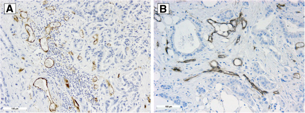
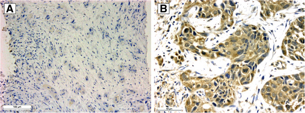
Methods: Paraffin blocks containing tumor samples from 100 patients who were diagnosed with pancreatic cancer between 1990 and 2010 were used to construct a tissue microarray. VEGF expression was assessed in these samples by immunohistochemistry. To assess the lymphatic and vascular properties of the tumors, 63 cases that contained sufficient material were sectioned routinely. The sections were then stained with the D2-40 antibody to identify the lymphatic vessels and with a CD34 antibody to identify the blood vessels. The vessels were counted individually with the Leica Application Suite v4 program. All statistical analyses were performed using SPSS 18.0 (Chicago, IL, USA) software, and p values ≤ 0.05 were considered significant.
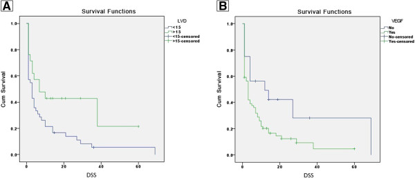
Results: In the Cox regression analysis, advanced age (p=0.03) and a history of type 2 diabetes (p=0.014) or chronic pancreatitis (p=0.02) were shown to be prognostic factors for pancreatic cancer. Blood vessel density (BVD) had no relationship with clinical-pathological features or death. Lymphatic vessel density (LVD) was inversely correlated with death (p=0.002), and by Kaplan-Meyer survival analysis, we found a significant association between low LVD (p=0.021), VEGF expression (p=0.023) and low patient survival.
Conclusions: Pancreatic carcinogenesis is related to a history of chronic inflammatory processes, such as type 2 diabetes and chronic pancreatitis. In pancreatic cancer development, lymphangiogenesis can be considered an early event that enables the dissemination of metastases. VEGF expression and low LVD can be considered as poor prognostic factors as tumors with this profile are fast growing and highly aggressive.
Virtual slides: The virtual slide(s) for this article can be found here: http://www.diagnosticpathology.diagnomx.eu/vs/5113892881028514.
Figures
Similar articles
-
Analysis of tumor-induced lymphangiogenesis and lymphatic vessel invasion of pancreatic carcinoma in the peripheral nerve plexus.Cancer Sci. 2012 Oct;103(10):1756-63. doi: 10.1111/j.1349-7006.2012.02364.x. Epub 2012 Aug 27. Cancer Sci. 2012. PMID: 22716017 Free PMC article.
-
Different significance between intratumoral and peritumoral lymphatic vessel density in gastric cancer: a retrospective study of 123 cases.BMC Cancer. 2010 Jun 17;10:299. doi: 10.1186/1471-2407-10-299. BMC Cancer. 2010. PMID: 20565772 Free PMC article.
-
Expressions of COX-2 and VEGF-C in gastric cancer: correlations with lymphangiogenesis and prognostic implications.J Exp Clin Cancer Res. 2011 Jan 28;30(1):14. doi: 10.1186/1756-9966-30-14. J Exp Clin Cancer Res. 2011. PMID: 21272377 Free PMC article.
-
Molecular control of lymphatic metastasis.Ann N Y Acad Sci. 2008;1131:225-34. doi: 10.1196/annals.1413.020. Ann N Y Acad Sci. 2008. PMID: 18519975 Review.
-
Mechanisms of lymphatic metastasis in human colorectal adenocarcinoma.J Pathol. 2009 Apr;217(5):608-19. doi: 10.1002/path.2517. J Pathol. 2009. PMID: 19253334 Review.
Cited by
-
Tumor-associated lymphatic vessel density is a postoperative prognostic biomarker of hepatobiliary cancers: a systematic review and meta-analysis.Front Immunol. 2025 Jan 7;15:1519999. doi: 10.3389/fimmu.2024.1519999. eCollection 2024. Front Immunol. 2025. PMID: 39840048 Free PMC article.
-
Overexpression of both platelet-derived growth factor-BB and vascular endothelial growth factor-C and its association with lymphangiogenesis in primary human non-small cell lung cancer.Diagn Pathol. 2014 Jun 27;9:128. doi: 10.1186/1746-1596-9-128. Diagn Pathol. 2014. PMID: 24972450 Free PMC article.
-
SPHK1 Promotes Pancreatic Cancer Lymphangiogenesis Through the Activation of ERK in LECs.Mol Biotechnol. 2025 Jun;67(6):2246-2253. doi: 10.1007/s12033-024-01192-9. Epub 2024 Jun 11. Mol Biotechnol. 2025. PMID: 38861202
-
Selecting Tumor-Specific Molecular Targets in Pancreatic Adenocarcinoma: Paving the Way for Image-Guided Pancreatic Surgery.Mol Imaging Biol. 2016 Dec;18(6):807-819. doi: 10.1007/s11307-016-0959-4. Mol Imaging Biol. 2016. PMID: 27130234 Free PMC article.
-
Exploration of the System-Level Mechanisms of the Herbal Drug FDY003 for Pancreatic Cancer Treatment: A Network Pharmacological Investigation.Evid Based Complement Alternat Med. 2022 May 10;2022:7160209. doi: 10.1155/2022/7160209. eCollection 2022. Evid Based Complement Alternat Med. 2022. PMID: 35591866 Free PMC article.
References
-
- Vainio H. Targeting angiogenesis – a novel mode in cancer chemoprevention. Asian Pacific J Cancer Prev. 2003;4:83–86. - PubMed
-
- Whitehurst B, Flister MJ, Bagaitkar J, Volk L, Bivens CM, Pickett B, Castro-Rivera E, Brekken RA, Gerard RD, Ran S. Anti-VEFG-A therapy reduces lymphatic vessel density and expression of VEGFR-3 in an orthotopic breast tumor model. Int J Cancer. 2007;121:2182–2191. - PubMed
Publication types
MeSH terms
Substances
LinkOut - more resources
Full Text Sources
Other Literature Sources
Medical
