. 2013 Dec;54(6):387-94.
doi: 10.1016/j.ceca.2013.09.001. Epub 2013 Sep 24.

α1-Adrenergic receptors mediate coordinated Ca2+ signaling of cortical astrocytes in awake, behaving mice

Affiliations

α1-Adrenergic receptors mediate coordinated Ca2+ signaling of cortical astrocytes in awake, behaving mice

Fengfei Ding et al. Cell Calcium. 2013 Dec.

Abstract

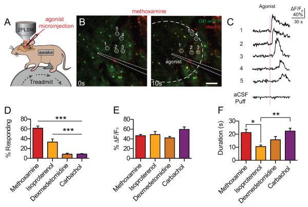
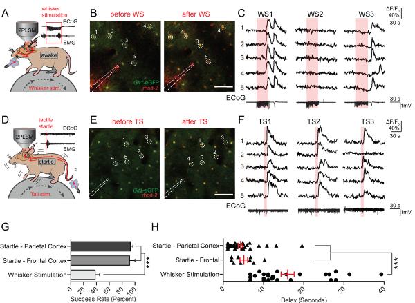
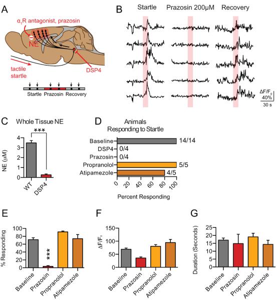
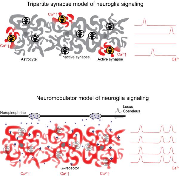
Astrocyte Ca2+ signals in awake behaving mice are widespread, coordinated and differ fundamentally from the locally restricted Ca2+ transients observed ex vivo and in anesthetized animals. Here we show that the synchronized release of norepinephrine (NE) from locus coeruleus (LC) projections throughout the cerebral cortex mediate long-ranging Ca2+ signals by activation of astrocytic α1-adrenergic receptors. When LC output was triggered by either physiological sensory (whisker) stimulation or an air-puff startle response, astrocytes responded with fast Ca2+ transients that encompassed the entire imaged field (positioned over either frontal or parietal cortex). The application of adrenergic inhibitors, including α1-adrenergic antagonist prazosin, potently suppressed both evoked, as well as the frequently observed spontaneous astroglial Ca2+ signals. The LC-specific neurotoxin N-(2-chloroethyl)-N-ethyl-2-bromobenzylamine (DSP-4), which reduced cortical NE content by >90%, prevented nearly all astrocytic Ca2+ signals in awake mice. The observations indicate that in adult, unanesthetized mice, astrocytes do not respond directly to glutamatergic signaling evoked by sensory stimulation. Instead astrocytes appear to be the primary target for NE, with astrocytic Ca2+ signaling being triggered by the α1-adrenergic receptor. In turn, astrocytes may coordinate the broad effects of neuromodulators on neuronal activity.

Keywords: Astrocyte; Awake; Calcium; Norepinephrine; Startle.


Figures

Fig. 1
Fig. 1
Astrocytes respond to α1- and β-AR agonists with increases in intracellular Ca2+. (A) Cortical astrocytes loaded with rhod-2 (AM) were imaged in layers I/II to detect changes in [Ca2+]i. Agonists were injected using a microelectrode loaded with ACSF and the tracer Alexa 488. (B) Representative images of astrocytic [Ca2+]i responses to the α1-AR agonist, methoxamine, in awake Glt-1-eGFP transgenic mice. Astrocyte-specific loading of rhod-2 (AM) was confirmed by colocalization with Glt-1-eGFP. Scale bar = 100μM. (C) rhod-2 (AM) ΔF/F0 traces from (B), normalized to Glt-1-eGFP fluorescence. Representative aCSF-microinjection trace at the bottom. (D) Astrocytic [Ca2+]i responses to adrenergic and acetylcholinergic receptor agonists, measured by rhod-2 (AM) ΔF/F0. Bar graph showing the percent of astrocytes within the puff radius responding with a [Ca2+]i increase. *** p<0.001, one-way ANOVA, Bonferroni correction; [n= 25 trials in 6 animals (methoxamine); 14 trials in 4 animals (isoproterenol); 24 trials in 5 animals (dexmedetomidine); 23 trials in 5 animals (carbachol)] (E and F) Bar graphs showing average rhod-2 (AM) ΔF/F0 and response duration from trials with responding cells. ** p < 0.01; * p < 0.05, one-way ANOVA, Bonferroni correction; [n = 25 trials (methoxamine), 13 trials (isoproterenol), 17 trials (dexmedetomidine), 20 trials (carbachol).] Data are shown as mean ± SEM.
Fig. 2
Fig. 2
Astrocytes respond reliably to startle stimulation with widespread Ca2+ waves. (A) Astrocytic Ca2+ transients measured by rhod-2 (AM) were detected in response to 30s of 3Hz whisker stimulation. Representative LFP and EMG recordings are shown. (B) Representative images of rhod-2 (AM) fluorescence increases during whisker stimulation in a Glt-1-eGFP animal. Scale bar = 100μM. (C) Selected cells from (B). rhod-2 ΔF/F0 was normalized to Glt-1-eGFP fluorescence. ECoG traces corresponding to rhod-2 ΔF/F0 are shown below. (D) Air pulses were directed at the face or tail of the animal to elicit a startle response. Representive ECoG and EMG traces are shown with no apparent evoked ECoG response, and strong EMG activity – indicative of a startle response. (E and F) Representative images and corresponding rhod-2 dF/F0 traces show stable and repeatable astrocytic [Ca2+]i transients after startle stimulations. Bottom, representative ECoG traces from startle stimulation are shown. (G) Bar graph showing the response rate of cortical astrocytes to whisker and startle stimulation, averaged across animals. *** p<0.001, one-way ANOVA, Bonferroni correction; [n = 11 animals (parietal cortex startle and whisker stimulation), 4 animals (frontal cortex startle)] (H) Scatter diagram with superimposed mean and SEM for the delay from the onset of whisker/startle stimulation to the beginning of astrocytic rhod-2 ΔF/F0 responses. Average delay from each successful trial is shown. *** p<0.001, one-way ANOVA, Bonferroni correction; [n= 28 trials in 14 animals (whisker stimulation), 38 trials in 13 animals (parietal cortex startle), and 9 trials in 4 animals (frontal cortex startle)]. Data are shown as mean ± SEM.
Fig. 3
Fig. 3
Astrocytic Ca2+ responses are dependent upon activation of the α1-AR. (A) Schematic illustrating diffuse NE release from the LC across the cortex in response to startle stimulation. LC-specific neurotoxin DSP4 and prazosin are shown blocking astrocytic Ca2+ responses, as measured by rhod-2 ΔF/F0. (B) Representative traces of astrocytic rhod-2 ΔF/F0 before, during, and after application of the α1-AR antagonist prazosin. (C) Bar graphs of cortical NE content in whole brain homogenate determined by HPLC showing depletion following DSP-4 injection. *** p<0.001, unpaired, two-tailed t-test [n=5 animals per group]. (D) Histogram presenting the percent of animals exhibiting at least one response to startle stimulation (>1/3 of cells exhibiting a rhod-2 ΔF/F0 response) for DSP4 and adrenergic antagonist treated animals. (E) Bar graph showing the average percent of cells responding with an increase in rhod-2 ΔF/F0 in the imaging field *** p<0.001, paired t-test; [prazosin: 20 trials baseline, 16 trials with drug in 4 animals; atipamezole: n = 13 trials baseline, 13 trials with drug in 5 animals; propranolol: 14 trials baseline, 14 trials with drug in 5 animals.] (F and G) Bar graphs showing the average peak amplitude increase in astrocytic rhod-2 ΔF/F0 and the duration of the response, presented as the average responses from each trial with at least 1 cell responding. paired t-test; [n = 13 trials baseline, 4 trials with drug (prazosin); 13 trials baseline, 11 trials with drug (atipamezole); 14 trials baseline, 14 trials with drug (propranolol)] Baseline data are presented as a pooled average. Data are shown as mean ± SEM.
Fig. 4
Fig. 4
Spontaneous astrocytic Ca2+ activity in awake mice is dependent upon activation of the α1-AR. (A) Representative astrocytic rhod-2 fluorescence traces show frequent widespread and coordinated astrocytic (Ca2+)i waves in awake animals, which were almost completely abrogated by the α1-AR antagonist, prazosin, and LC toxin, DSP4. (B) Bar graph showing that prazosin and DSP4 treatment causes a significant decrease in the frequency of coordinated astrocytic [Ca2+]i waves (quantified by counting each time >1/3 of cells responded within the imaging field) ** p<0.01, * p<0.05, paired t-test (Control versus prazosin), unpaired t-test (control versus DSP4); [n=4 animals (prazosin, control), 5 animals (DSP4)] Data are shown as mean ± SEM.
Fig. 5
Fig. 5
Diagram illustrating two possible mechanisms driving astrocytic Ca2+ responses in awake, head-restrained mice. (A) The tripartite synapse model: Here, astrocytes respond to local neuronal activity and neurotransmitter release with spotty, asynchronous increases in [Ca2+]i. (B) The neuromodulator model: Following novel or noxious behavioral stimuli, neuromodulators are released throughout the cortex to rapidly alter network connectivity. Our data suggests that in the startle response, the release of NE from the LC drives a coordinated increase in astrocytic [Ca2+]i in multiple brain regions. This suggests that coordinated astrocytic calcium transients in the awake brain are integrally related to widespread neuromodulation in response to behaviorally salient stimuli – placing them at the center of the network response to these stimuli.

References

    1. Nedergaard M, Verkhratsky A. Artifact versus reality--how astrocytes contribute to synaptic events. Glia. 2012;60:1013–1023. - PMC - PubMed
    1. Verkhratsky A, Orkand RK, Kettenmann H. Glial calcium: homeostasis and signaling function. Physiol Rev. 1998;78:99–141. - PubMed
    1. Benedetti B, Matyash V, Kettenmann H. Astrocytes control GABAergic inhibition of neurons in the mouse barrel cortex. J Physiol. 2011;589:1159–1172. - PMC - PubMed
    1. Di Castro MA, Chuquet J, Liaudet N, et al. Local Ca2+ detection and modulation of synaptic release by astrocytes. Nat Neurosci. 2011;14:1276–1284. - PubMed
    1. Panatier A, Vallee J, Haber M, Murai KK, Lacaille JC, Robitaille R. Astrocytes are endogenous regulators of basal transmission at central synapses. Cell. 2011;146:785–798. - PubMed

Publication types

MeSH terms