. 2013 Oct 16;80(2):415-28.
doi: 10.1016/j.neuron.2013.10.015.

RNA toxicity from the ALS/FTD C9ORF72 expansion is mitigated by antisense intervention

Affiliations

RNA toxicity from the ALS/FTD C9ORF72 expansion is mitigated by antisense intervention

Christopher J Donnelly et al. Neuron. .

Erratum in

  • Neuron. 2013 Nov 20;80(4):1102. Heusler, Aaron R [corrected to Haeusler, Aaron R]

Abstract

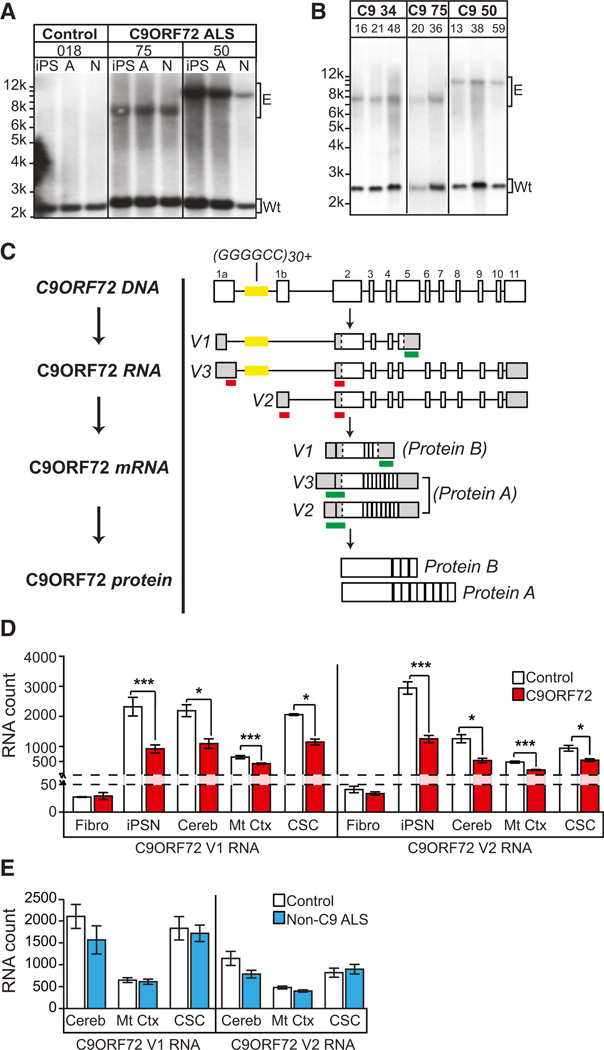
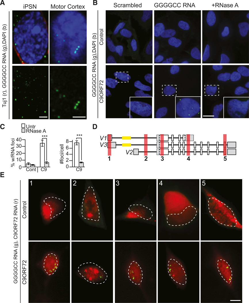
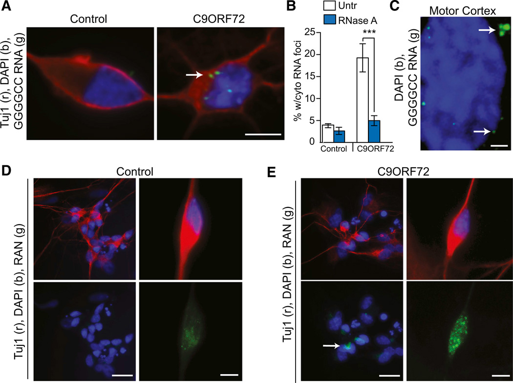
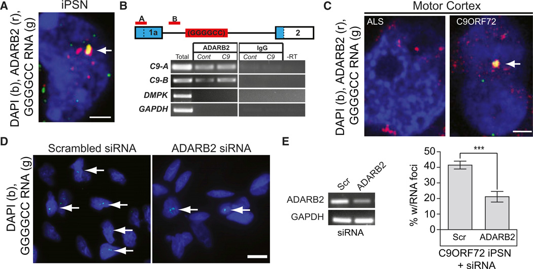
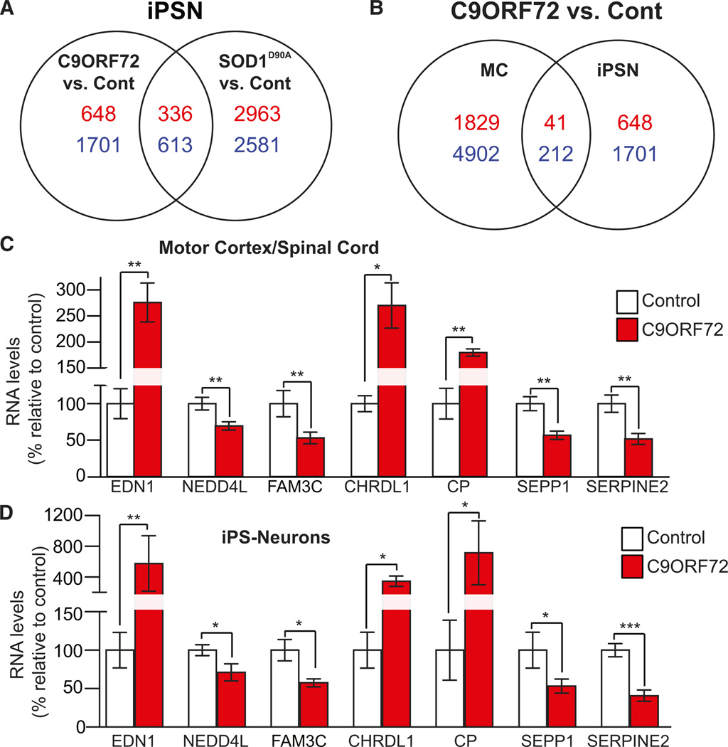
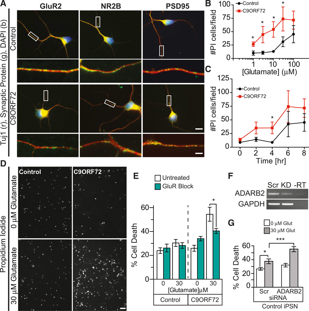
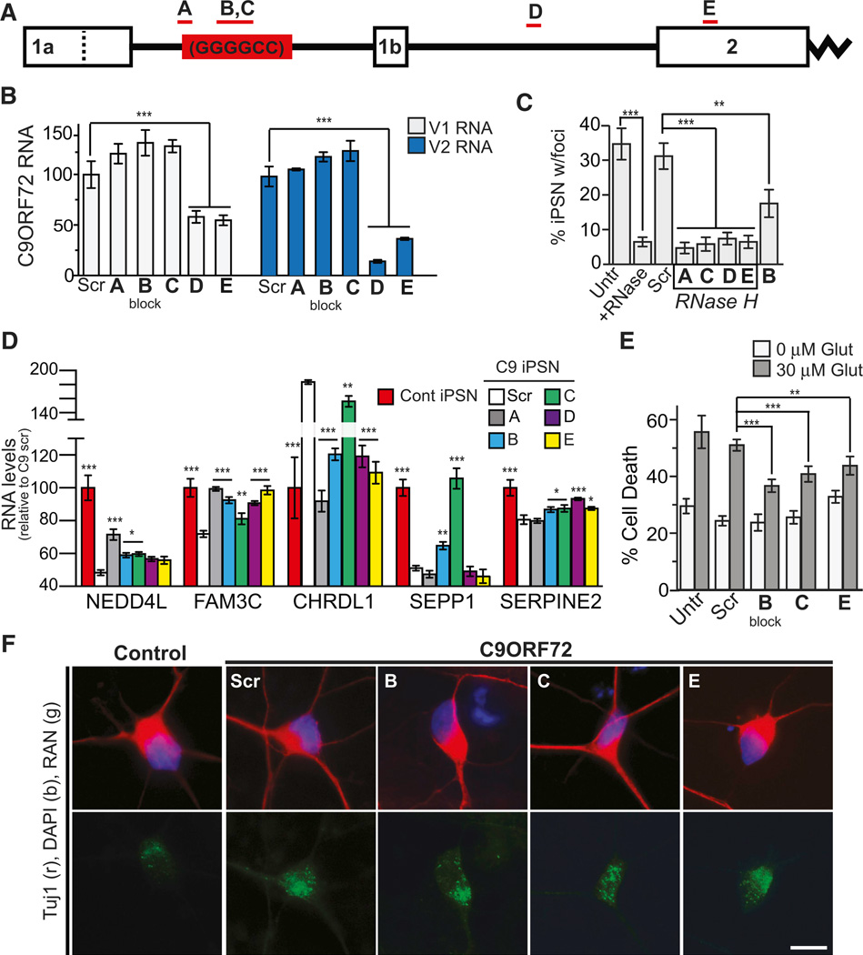
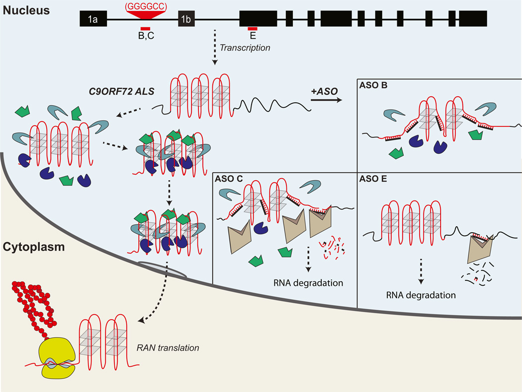
A hexanucleotide GGGGCC repeat expansion in the noncoding region of the C9ORF72 gene is the most common genetic abnormality in familial and sporadic amyotrophic lateral sclerosis (ALS) and frontotemporal dementia (FTD). The function of the C9ORF72 protein is unknown, as is the mechanism by which the repeat expansion could cause disease. Induced pluripotent stem cell (iPSC)-differentiated neurons from C9ORF72 ALS patients revealed disease-specific (1) intranuclear GGGGCCexp RNA foci, (2) dysregulated gene expression, (3) sequestration of GGGGCCexp RNA binding protein ADARB2, and (4) susceptibility to excitotoxicity. These pathological and pathogenic characteristics were confirmed in ALS brain and were mitigated with antisense oligonucleotide (ASO) therapeutics to the C9ORF72 transcript or repeat expansion despite the presence of repeat-associated non-ATG translation (RAN) products. These data indicate a toxic RNA gain-of-function mechanism as a cause of C9ORF72 ALS and provide candidate antisense therapeutics and candidate human pharmacodynamic markers for therapy.


Figures

Figure 1
Figure 1. C9ORF72 ALS iPSNs Exhibit GGGGCCexp Repeat Allele and Reduced C9ORF72
(A) Southern blot of C9ORF72 ALS fibroblasts and subsequent iPSCs for lines used in this study. All C9ORF72 lines contained normal length allele (Wt) and an expanded ‘‘GGGGCC’’ repeat allele (E) that is maintained during differentiation to iPSN (N) or astrocytes (A). (B) Repeat length of iPSCs was maintained over several iPSC cell passage numbers (ranging from p13 to p59). (C) Schematic of the C9ORF72 gene, location of the repeat sequence (yellow), validated RNA variants, and location of the nanostring probesets. For detection of nanostring probe, each individual probe (red) must bind in tandem for detection (green). Probes to C9ORF72 V1 will detect pre-mRNA and mRNA; probes to V2 and V3 will only detect C9ORF72 mRNA. (D) C9ORF72 ALS iPSNs and CNS tissue exhibit reduced C9ORF72 RNA levels (n for C9ORF72 versus Control: n = (fibroblast, Fibro) 5 versus 5; (iPSN) 3 versus 3; (cerebellum, Cereb) 4 versus 6; (motor cortex, Mt Ctx) 10 versus 6; (cervical spinal cord, CSC) 4 versus 6). (E) Non-C9ORF72 ALS patients do not show attenuated levels of C9ORF72 RNA. (*p < 0.05; ***p < 0.001). See also Figures S1 and S2; Tables S1, S2, and S3.
Figure 2
Figure 2. C9ORF72 ALS iPSN Exhibit Pathological Nuclear GGGGCCexp RNA Foci
(A) Single optical plane (∼0.3 µm) of C9ORF72 iPS-differentiated neurons processed for GGGGCC RNA FISH exhibit clear intranuclear foci (left) similar to those found in C9ORF72 ALS motor cortex (right). (B and C) GGGGCC probe is specific to C9ORF72 RNA foci and foci are RNase A sensitive (n = 30 fields of view). (D) Schematic of RNA FISH probe location relative to C9ORF72 RNA variants. (E) Probes to exonic and intronic regions upstream and downstream of the repeat (red) do not form nuclear foci, do not consistently overlap with GGGGCC RNA foci (green), and exhibit similar localization in control and C9ORF72 ALS iPSN lines. Dotted line outlines cell nucleus. Data in (C) indicate mean ± SEM (p < 0.001). Scale bar = 2 mm for (A), 20 mm for (B), and 5 mm for (E). See also Figure S3A.
Figure 3
Figure 3. C9RF72 ALS Cells Show Cytoplasmic RAN Translation Peptides
(A) C9ORF72 iPSN cells contain cytoplasmic GGGGCC RNA foci, while control iPSNs do not. (B) Quantification of cytoplasmic RNA foci reveals that cytoplasmic foci are RNase A sensitive. (C) Single-optical plane (∼0.3 µm) image of C9ORF72 ALS motor cortex also shows cytoplasmic GGGGCC RNA foci similar to iPSNs. (D) RAN protein shown by IF using C9RANT antibody that preferentially detects the poly-(Gly-Pro) RAN product in C9ORF72 iPSNs but not control iPSNs. ***p < 0.001. Scale bar = 5µm for (A), (D), and (E), right panels; 1 mm for (C), and 20 µm for (D) and (E), left panels. See also Figure S3B.
Figure 4
Figure 4. ADARB2 Protein Binds to the GGGGCCexp of C9ORF72 RNA
(A) Single optical plane (0.34 µm) colocalization of GGGGCC RNA foci with ADARB2 protein signal in C9ORF72 iPSN. (B) ADARB2 co-IP with bound RNA was performed; RNA was isolated from ADARB2 protein co-IP in control and C9ORF72 iPSNs. RT-PCR of the co-IP RNA using two primer sets (red) upstream of the C9ORF72 GGGGCC repeat (schematic, blue = UTR) indicates that ADARB2 binds to the C9ORF72 RNA in both control and C9ORF72 iPSNs. Amplification of DMPK and GAPDH were used to test for immunoprecipitation specificity (lower panel). (C) Single optical plane (∼0.3µm) image demonstrating colocalization of GGGGCC RNA foci and ADARB2 in C9ORF72 patient postmortem motor cortex tissue, layers 3–5. (D and E) siRNA knockdown of ADARB2 results in a significant reduction in the percent of iPSNs with nuclear RNA foci (arrows). Data in (E) indicate mean ±SEM (***p < 0.001). Scale = 2 mm for (A) and (C), and 15 mm for (D). See also Figure S4 and S5; Tables S4 and S5.
Figure 5
Figure 5. C9ORF72 ALS-Specific Gene Expression
(A) Venn diagrams comparing the differentially expressed, statistically significant genes in C9ORF72 iPSNs versus control iPSNs and SOD1D90A iPSN versus control iPSNs indicate vast differences in global gene expression. Red indicates upregulated genes; blue indicates down-regulated genes. (B) Gene expression comparison of common, statistically significant gene expression in C9ORF72 iPSNs versus control (negating any genes significant in SOD1D90A versus control iPSNs) and C9ORF72 human motor cortex tissue (versus healthy control motor cortex). p < 0.05. (C and D) Targeted gene expression analysis of aberrantly expressed secreted genes, chosen by microarray results, in human motor cortex and spinal cord revealed dysregulation of seven genes that were also found to be similarly dysregulated in C9ORF72 iPSNs (versus control). Data in (C) and (D) indicate mean ± SEM. *p < 0.05; **p < 0.01; ***p < 0.001. See also Figure S6 and S7; Tables S6–S16.
Figure 6
Figure 6. C9ORF72 ALS iPSC Neurons Are Highly Susceptible to Glutamate Toxicity
(A) Immunofluorescent staining of control and C9ORF72 iPSN cultures show expression of glutamate receptors GluR2, NR2B, and post-synaptic density protein PSD95 at comparable levels as determined by qualitative analysis. Box indicates region of high magnification seen below each image (scale = 10 mm [top] and 2.5 mm [bottom]). (B) Dose response curve of control and C9ORF72 iPSC neurons revealed that C9ORF72 iPSNs are highly susceptible to glutamate excitotoxicity at 1, 3, 10, and 30 µM concentrations after 8 hr of treatment by popidium iodide staining. (C) Glutamate-induced excitotoxicity of C9ORF72 iPSNs shows statistically significant cell death after 4 hr of 30 µM glutamate treatment when compared to control iPSNs. (D) Representative image of propidium iodide staining of control and C9ORF72 ALS iPSN after 4 hr of 0 and 30 µM glutamate treatment; note the increased prodium iodide signal in the C9ORF72 iPSNs as compared to the control iPSNs. (E) Blocking glutamate receptors prevents glutamate-induced C9ORF72 iPSN cell death (4 hr, 30 µM glutamate). (F and G) Knockdown of ADARB2 via siRNA treatment resulted in a statistically significant increased susceptibility to glutamate-induced excitotoxicity in control non-C9ORF72 iPSNs at 4 hr, 30 µM glutamate treatment. Data in (B), (C), (E), and (G) indicate mean ± SEM (*p < 0.05; ***p < 0.001). See also Figure S8.
Figure 7
Figure 7. Antisense Oligonucleotides Rescue RNA Toxicity Observed in C9ORF72 iPSC Neurons
(A) Map of the C9ORF72 gene indicating the ASOs utilized in this study. ASO B is endonuclease resistant and designed to block the repeat while the remaining are gapmers, which mediate RNA degradation through an RNase H-based mechanism. Dotted line indicates end of V1 Exon 1A sequence. (B) ASOs that target the repeat sequence do not statistically alter C9ORF72RNA levels in C9ORF72 iPSNs, while ASOs downstream of the repeat significantly reduce the levels of C9ORF72 variants 1 and 2. (C) All ASOs significantly reduced the number of nuclear GGGGCC RNA foci observed in C9ORF72 iPSNs to levels comparable to the RNase A-treated cells while treatment with a nontargeting scrambled probe did not affect the percent of cells with nuclear foci. (D) ASO treatment was able to rescue the aberrant expression of NEDD4IL, FAM3C, CHRDL1, SEPP1, and SERPINE2 in a statistically significant manner. (E) ASO B, C, and E rescued the increased susceptibility of C9ORF72 iPSNs to glutamate exposure (30 µM glutamate, 5 hr). (F) ASO treatment for 3 days does not dramatically reduce the levels of cytoplasmic RAN proteins. Data in (B), (C), and (E) indicate mean ± SEM; data in (D) indicates mean ± SD (*p < 0.05; **p < 0.01; ***p < 0.001). See also Figure S9.
Figure 8
Figure 8. Pathogenesis Resulting from the C9ORF72 Noncoding GGGGCC Expansion in ALS/FTD
Top. Representation of the human C9ORF72 gene, showing hexanucleotide expansion (red) and location of various antisense oligonucleotide (ASO) probes (B, C, E). Hexanucleotide expansion is located between the noncoding exons 1a and 1b. The expanded RNA is predicted to form a series of G-quartet tertiary structures. At least three different pathophysiological pathways may results from the mutation: (1) RNA toxicity resulting from excessive binding of various different RNA binding proteins (green, blue, gray polygons) leading to sequestration of these RBPs, and loss of their activity resulting in anomalous downstream events; (2) export of the expansion to cytoplasm with repeat-associated translation (RAN) (in the sense or antisense directions) and the formation of excessive high-molecular-weight cytoplasmic peptides (red), which may be toxic or protective; and (3) loss of C9ORF72 mRNA leading to loss of protein function (not shown). ASOs may disrupt C9ORF72 RNA toxicity by several pathways. ASO B: an endonuclease-resistant oligonucleotide that is predicted to disrupt the expansion, interfering with the G-quartet structure and preventing RBP interaction/sequestration. ASO C; a gapmer ASO that binds to the expansion targeting it RNase H-mediated cleavage and subsequent RNA degradation by nuclear endogenous nucleases, since this ASO targets the repeat it will also disrupt the repeat sequence structure prior to cleavage and prevent RBP sequestration. ASO E: a gapmer that targets the C9ORF72 RNA coding sequence marking the RNA for RNase H cleavage, thus resulting in the degradation of wild-type and the expanded C9ORF72 RNA; this will further reduce the already attenuated levels of cellular C9ORF72 RNA. Notably, ASOs B and C do not reduce cellular C9ORF72 RNA.

Comment in

Similar articles

Cited by

References

    1. Almeida S, Gascon E, Tran H, Chou HJ, Gendron TF, Degroot S, Tapper AR, Sellier C, Charlet-Berguerand N, Karydas A, et al. Modeling key pathological features of frontotemporal dementia with C9ORF72 repeat expansion in iPSC-derived human neurons. Acta Neuropathol. 2013;126:385–399. - PMC - PubMed
    1. Ash PE, Bieniek KF, Gendron TF, Caulfield T, Lin WL, Dejesus-Hernandez M, van Blitterswijk MM, Jansen-West K, Paul JW, 3rd, Rademakers R, et al. Unconventional translation of C9ORF72 GGGGCC expansion generates insoluble polypeptides specific to c9FTD/ALS. Neuron. 2013;77:639–646. - PMC - PubMed
    1. Brouwer JR, Willemsen R, Oostra BA. Microsatellite repeat instability and neurological disease. Bioessays. 2009;31:71–83. - PMC - PubMed
    1. Chen CX, Cho DS, Wang Q, Lai F, Carter KC, Nishikura K. A third member of the RNA-specific adenosine deaminase gene family, ADAR3, contains both single- and double-stranded RNA binding domains. RNA. 2000;6:755–767. - PMC - PubMed
    1. Ciura S, Lattante S, Le Ber I, Latouche M, Tostivint H, Brice A, Kabashi E. Loss of function of C9orf72 causes motor deficits in a zebrafish model of Amyotrophic Lateral Sclerosis. Ann. Neurol. 2013 May 30; - PubMed

Publication types

MeSH terms