. 2013 Oct 16;80(2):458-69.
doi: 10.1016/j.neuron.2013.09.005.

Establishment of neurovascular congruency in the mouse whisker system by an independent patterning mechanism

Affiliations

Establishment of neurovascular congruency in the mouse whisker system by an independent patterning mechanism

Won-Jong Oh et al. Neuron. .

Abstract

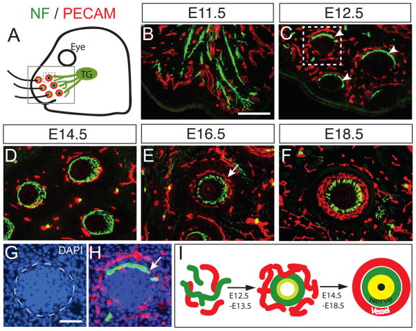
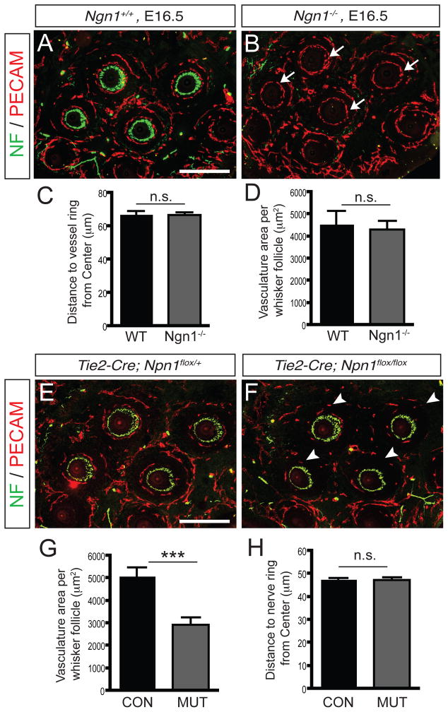
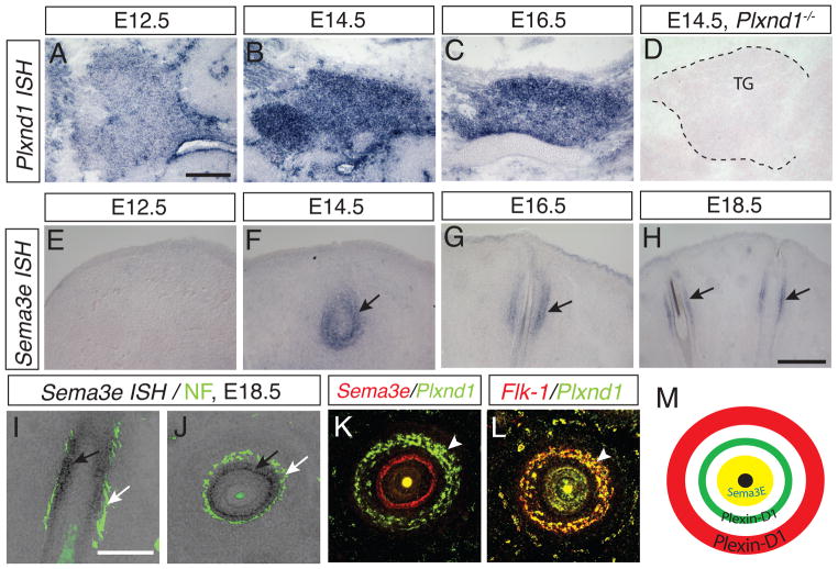
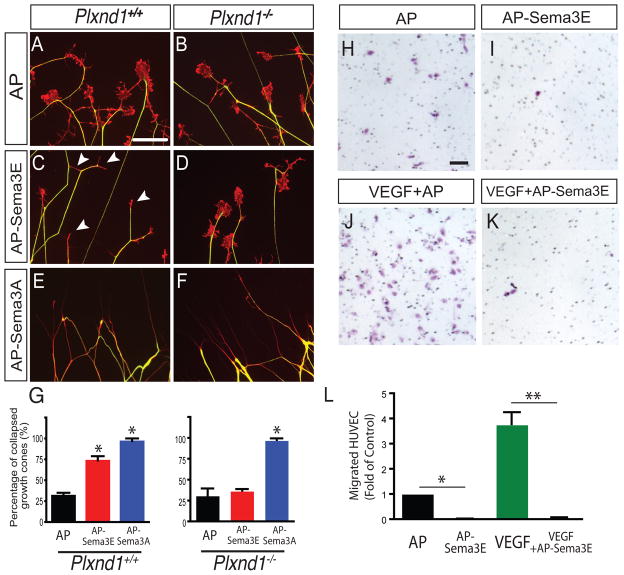
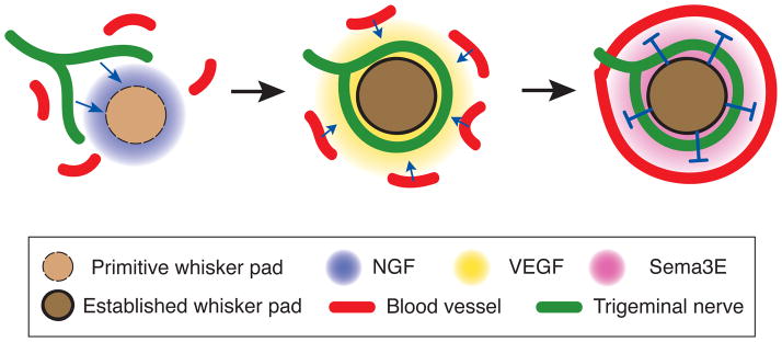
Nerves and vessels often run parallel to one another, a phenomenon that reflects their functional interdependency. Previous studies have suggested that neurovascular congruency in planar tissues such as skin is established through a "one-patterns-the-other" model, in which either the nervous system or the vascular system precedes developmentally and then instructs the other system to form using its established architecture as a template. Here, we find that, in tissues with complex three-dimensional structures such as the mouse whisker system, neurovascular congruency does not follow the previous model but rather is established via a mechanism in which nerves and vessels are patterned independently. Given the diversity of neurovascular structures in different tissues, guidance signals emanating from a central organizer in the specific target tissue may act as an important mechanism to establish neurovascular congruency patterns that facilitate unique target tissue function.


Figures

Figure 1
Figure 1. Nerve and blood vessels are organized into a “double ring” structure in the whisker follicle during development
(A–F) Developmental profile of trigeminal axon innervation and blood vessel patterning in the whisker follicles. Trigeminal axons and blood vessels are visualized by neurofilament (green) and PECAM (red), respectively, in the snout area (boxed region in Fig. 1A). Co-immunostained tangential sections of whisker follicles at E11.5 (B), E12.5 (C), E14.5 (D), E16.5 (E), E18.5 (F) show the dynamic processes that lead to the stereotypic organization of a double ring structure. The trigeminal axons form a ring-like structure as early as E12.5 (arrowheads in C), and are then surrounded by a blood vessel ring that results in the formation of a double ring structure around each follicle at E16.5, with the nerve ring located inside and the vessel ring outside (arrow in E). (G–H) One whisker follicle in C (dotted box) is overlaid by DAPI staining to show follicle primordium (dotted circle in G). Nerve terminals surround the outside of the primordium (arrow in H). (I) Schematic illustration of nerve and vessel ring organization in the whisker follicle. Scale bar: 100 μm (B–F), 50μm (G–H). See also Figure S1 and Movie S1–S3.
Figure 2
Figure 2. Nerve ring and vessel ring are patterned independent of each other in the whisker follicle
(A–B) Nerve/vessel immunostaining of wild-type littermate control exhibits normal double ring patterning (A). Nerve/vessel immunostaining of the Ngn1 knockout shows no trigeminal innervation in the whisker follicles, but blood vessel rings are normally organized at each whisker follicle (arrows in B, C–D). (C–D) For the quantification, 10 pairs of whisker follicles were analyzed and the mean distance from the center (C) and vasculature area surrounding the follicle (D) ± SEM is shown. (E–F) Endothelial specific-Npn1 deletion shows less vasculature formation around whisker follicle (white arrowheads in F and G) compared to the vasculature of control (E), but nerve rings are organized normally (H). (G–H) Vasculature area surrounding follicle (G) and mean distance from center to nerve ring (H) ± SEM is shown (n=14). Paired student t-test; ***, P<0.001; n.s, not significant. Scale bar: 200 μm (A–B and E–F)
Figure 3
Figure 3. Complementary expression pattern of Plexin-D1 in the TG and blood vessels and Sema3E in the whisker follicle
(A–D) Plxnd1 in situ hybridization (ISH) on sagittal sections of wild-type embryos at E12.5 (A), E14.5 (B), and E16.5 (C) shows that Plxnd1 mRNA is detected at very low level as early as E12.5 and is significantly increased at E14.5 and continues to E16.5 as the double ring structure develops. Plxnd1 mRNA is not detectable in sections of the trigeminal ganglion from plexin-D1 knockout animals at E14.5 (TG outlined by dashed line in D). (E–H) Sema3e mRNA ISH on coronal sections parallel to the whisker follicles at E12.5 (E), E14.5 (F), E16.5 (G) and E18.5 (H) shows that Sema3e mRNA is expressed in the mesenchymal sheath surrounding the hair follicles starting at E14.5 and continues at E16.5 and E18.5 (black arrows). Scale bar: 100 μm in A applies to G, and 200 μm (H) (I–J) Neurofilament immunostaining (white arrows) after Sema3e ISH on the same sections shows that Sema3e (black arrows) is expressed inside of the nerve rings (coronal image in I and tangential image in J). (K–L) Double fluorescence ISH with Plxnd1 and Sema3e (K) or Flk-1 (L) shows that Plxnd1 mRNA is expressed in the endothelial cells that form the vessel ring (white arrowhead in K and L) and Sema3e is expressed inside of both nerve and vessel rings. (M) Summary of Plxnd1 and Sema3e mRNA expression pattern in the whisker follicle. Sema3e is expressed in mesenchymal tissue (yellow) and Plxnd1 is expressed in blood vessel (red) and in TG therefore Plexin-D1 protein probably in nerve (green). Scale bar: 100 μm in A applies to G, 200 μm (H), and 100 μm in I applies to L.
Figure 4
Figure 4. Sema3E-Plexin-D1 signaling provides a repulsive guidance cue to both TG axons and blood vessels in vitro
(A–G) Sema3E-Plexin-D1 signaling serves as repulsive cue for cultured TG neurons. Trigeminal ganglia were isolated from wild type (A, C, E) or Plexin-D1 null mice (B, D, F) at E14.5 and a growth cone collapse assay was performed. Axons (green) and growth cones (red) were visualized by anti-neurofilament and phalloidin staining, respectively. After incubation with 2 nM of AP (A, B), AP-Sema3E (C, D), or AP-Sema3A (E, F) for 30 min, >200 growth cones were scored for each experimental condition. Sema3E treatment induced significant growth cone collapse (white arrowheads in C), but growth cone collapse was absent in Plexin-D1 mutant TG neurons (D). (G) Quantification of growth cone collapse assay (n=3, data shown as mean ± SD, ANOVA; *, P<0.001). (H–L) Sema3E inhibits endothelial cell migration. VEGF-induced HUVEC transwell migration assay was performed in the presence (K) or absence (J) of Sema3E (AP-Sema3E, 0.5 nM). After a 5 hr incubation, migrated cells were fixed and visualized by Nissl staining. Sema3E prevents HUVEC migration induced by VEGF as well as the basal level of migration (I). (L) Quantification of migrated HUVECs (data shown as mean ± SEM, n=3, ANOVA; *, P<0.05; **, P<0.001). Scale bar: 50 μm (A–F, H–K). See also Figure S2.
Figure 5
Figure 5. Plexin-D1 protein expression is selectively down-regulated in the target terminal of TG axons
(A–C) Plexin-D1 protein expression is visualized by AP-Sema3E binding. Tangential sections of tissue were cut at E14.5 and then incubated with 1 nM AP-Sema3E. Binding was then detected by reaction with an AP substrate. Plexin-D1 (black arrows in A) is highly expressed within the TG (dotted red outline) and the trigeminal nerves that project both centrally toward the brainstem and peripherally towards the snout (right side from dotted green outline). But Plexin-D1 protein is extremely low in nerves when they reach the whisker follicle (white arrowhead in B and C). In contrast, Plexin-D1 protein is strongly detected in the blood vessel ring (white arrows in B and C). Yellow arrowhead (in B and C) indicates non-specific signals in membrane structure of the whisker follicle. (C) To identify the relative location of nerve and blood vessel rings, adjacent sections were immunostained with NF (green) and PECAM (red) and overlaid onto the AP-stained sections. (D–E) Neuropilin-1 protein expression is detected by AP-Sema3A (2 nM) binding in the terminal nerve ring (arrowheads in D-E), which completely overlaps with NF-positive nerve ring on the same sections after AP-staining (E). (F–I) Plexin-D1 protein expression is visualized by immunohistochemistry with anti-Plexin-D1. In the TG nerve terminal surrounding the whisker follicles, Plexin-D1 protein expression is extremely low (arrowheads in F and G). In contrast, Plexin-D1 protein is strongly detected in the blood vessel ring (arrows in H and I). Anti-Plexin-D1 displays non-specific signals in the hair sheath and glassy membrane of the whisker follicle (asterisks in F and H). Scale bar: 1 mm (A), 50 μm (BE, F-I). See also Figure S3.
Fig 6
Fig 6. Plexin-D1 and Sema3E is required for the stereotypic double ring nerve/vessel patterning in the whisker pad in vivo
(A–C) Plexin-D1 null mice exhibit severe nerve/vessel organization defects. The whisker follicle region of Plexin-D1 mutants (B) and their littermate controls (A) at E16.5 were sectioned and immunostained with NF and PECAM. In the Plexin-D1 mutant, nerve and vessel rings were abnormally close, with some areas showing complete vessel/nerve intermingling (arrow in B). Radii of the vessel ring (R) and nerve ring (r) depicted in I were measured and their ratio was calculated for each follicle (C). (D–F) Sema3E null mice also show the same nerve/vessel patterning phenotype (white arrow in E) compared to their littermate controls (D) and r/R ratio measurement is shown in F. For the quantification, 6–8 whisker follicles per animal from three different pairs of embryos in each experimental condition were measured and the mean ratios ± SEM are shown (C, F). Paired student t-test; *, P<0.05 (G–I) The detail patterning phenotype was analyzed in coronal view of whisker follicles using confocal imaging process after neurofilament and VE-cadherin whole mount staining. The blood vessel layer (white arrows in G) is clearly separated from the TG nerve layer in wild-type (G), but in the Plxnd1 mutant, vessels (yellow arrows in H) are intermingled with nerve layers (H). White arrowhead (in G and H) indicates non-specific signals from the whisker sheath marked in white asterisks. Scale bar: 100 μm (A, B, D, and E) and 50 μm (G and H). See also Figure S4.
Figure 7
Figure 7. Model of how stereotypic double ring neurovascular structure is established during development
(Left) During early development (E11–E12), NGF is expressed in tissue surrounding the primitive whisker follicle and attracts trigeminal axons to the whisker target area. At this stage, the blood vessel plexus is dispersed in the whisker follicle and is not organized. (Middle) As development progress, around E13–E14 when trigeminal nerve forms a nerve ring structure, VEGF expression in the whisker follicle region begins to recruit blood vessel close to the nerve ring. (Right) Around E14–E16, Sema3E starts to be expressed in the mesenchymal sheath of the whisker follicle and repels incoming vessels through its interaction with Plexin-D1, which then settle outside of nerve ring. Although Plexin-D1 is also expressed in the trigeminal nerve at this stage, its expression in the axon terminals is selectively down-regulated to a level that is not sufficient to respond to Sema3E, thereby maintaining the location of the nerve ring close to the whisker follicle. See also Figures S5 and S6.

Comment in

Similar articles

Cited by

References

    1. Adams RH, Eichmann A. Axon guidance molecules in vascular patterning. Cold Spring Harb Perspect Biol. 2010;2:a001875. - PMC - PubMed
    1. Armulik A, Genove G, Mae M, Nisancioglu MH, Wallgard E, Niaudet C, He L, Norlin J, Lindblom P, Strittmatter K, et al. Pericytes regulate the blood-brain barrier. Nature. 2010;468:557–561. - PubMed
    1. Bai G, Pfaff SL. Protease regulation: the Yin and Yang of neural development and disease. Neuron. 2011;72:9–21. - PMC - PubMed
    1. Bandtlow CE, Heumann R, Schwab ME, Thoenen H. Cellular localization of nerve growth factor synthesis by in situ hybridization. The EMBO journal. 1987;6:891–899. - PMC - PubMed
    1. Bates D, Taylor GI, Minichiello J, Farlie P, Cichowitz A, Watson N, Klagsbrun M, Mamluk R, Newgreen DF. Neurovascular congruence results from a shared patterning mechanism that utilizes Semaphorin3A and Neuropilin-1. Developmental biology. 2003;255:77–98. - PubMed

Publication types

MeSH terms