. 2013 Oct 16;80(2):484-93.
doi: 10.1016/j.neuron.2013.09.014.

Impaired hippocampal ripple-associated replay in a mouse model of schizophrenia

Affiliations

Impaired hippocampal ripple-associated replay in a mouse model of schizophrenia

Junghyup Suh et al. Neuron. .

Abstract

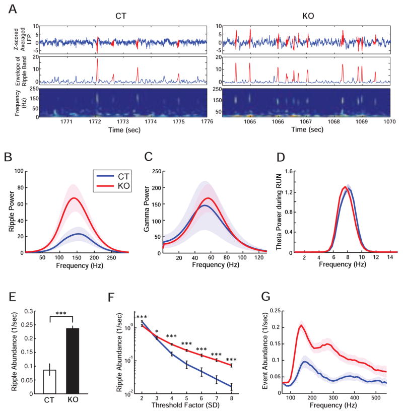
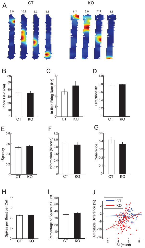
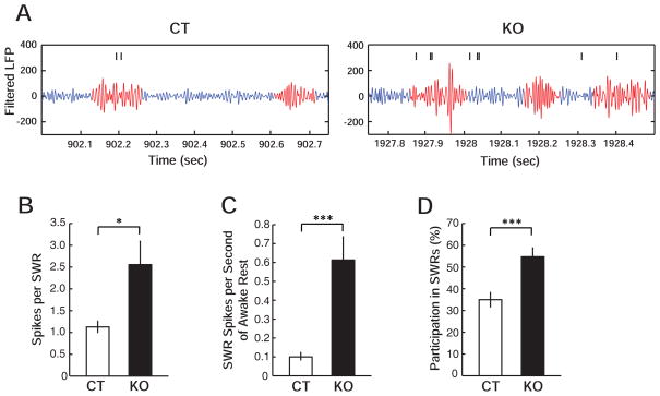
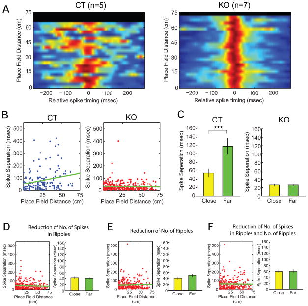
The cognitive symptoms of schizophrenia presumably result from impairments of information processing in neural circuits. We recorded neural activity in the hippocampus of freely behaving mice that had a forebrain-specific knockout of the synaptic plasticity-mediating phosphatase calcineurin and were previously shown to exhibit behavioral and cognitive abnormalities, recapitulating the symptoms of schizophrenia. Calcineurin knockout (KO) mice exhibited a 2.5-fold increase in the abundance of sharp-wave ripple (SWR) events during awake resting periods and single units in KO were overactive during SWR events. Pairwise measures of unit activity, however, revealed that the sequential reactivation of place cells during SWR events was completely abolished in KO. Since this relationship during postexperience awake rest periods has been implicated in learning, working memory, and subsequent memory consolidation, our findings provide a mechanism underlying impaired information processing that may contribute to the cognitive impairments in schizophrenia.


Figures

Figure 1
Figure 1. Increased hippocampal ripple activity in calcineurin KO mice during awake resting periods
(A) Examples of EEG recording from CT (left) and KO (right) mice. Each EEG trace is shown as z-scored raw EEG (top), envelope of smoothed ripple-band-filtered EEG (middle) and wavelet power spectrogram of raw EEG (bottom). Note that sharp waves and their associated ripples are clearly isolated events in this spectrogram. (B–C) Comparison of spectral power of EEG filtered at ripple (B, 100–240 Hz) and gamma (C, 25–80 Hz) frequency bands, in both cases for EEG. (D) Comparison of spectral power of z-scored raw EEG filtered at theta (4–12 Hz) band during run. (E) Comparison of ripple abundance during awake resting period. (F) Quantitative measurement of ripple abundance at different threshold factors (standard deviations of z-scored, smoothed and filtered EEG). (G) The abundance of EEG events measured by a 50 Hz frequency window that filtered raw EEG at different frequency bands. Data are represented as mean ± SEM (shaded area in B, C, D and G).
Figure 2
Figure 2. Similar basic properties of place cells in CT and KO mice in run periods
(A) Examples of color-coded firing rate maps of CA1 place cells during run on a 10 × 76 cm linear track. Peak firing rates in Hz are shown above each rate map. (B–G) Quantitative description of place fields of CT and KO mice: (B) size of place field, (C) mean in-field firing rate, (D) directionality, (E) sparsity, (F) spatial information, and (G) spatial coherence. (H–J) Quantification of spike activity during burst: (H) number spikes per burst per cell, and (I) the proportion of spikes, which were burst spikes, per cell. Data are represented as mean ± SEM. (J) The percentage of attenuation in spike amplitude within bursts as a function of in-burst inter-spike interval (ISI) for each cell (CT: 48 cells; KO: 97 cells).
Figure 3
Figure 3. Increased spike activity of place cells in calcineurin KO mice during ripple events
(A) A representative train of spikes is displayed with simultaneously recorded EEG filtered in ripple frequency range, for CT and KO. Ripple events are highlighted in red. (B) The number of spikes per SWR event, per cell (over all cells that fired at least one spike during at least one SWR event). (C) The number of SWR spikes per second of awake resting period, per cell. (D) The fractional participation in SWRs, ie the fraction of SWR events for which a cell fired at least one spike, averaged across all cells. Data are represented as mean ± SEM.
Figure 4
Figure 4. Impaired reactivation of spatial experience on the linear track during awake resting periods on the linear track in calcineurin KO mice
(A) For each pair of neurons, the pairwise cross-correlogram of the two spike trains around ripple events (± 300 ms) is plotted at a y position given by the linear distance between the corresponding two place field peaks. Wherever more than one pair occupies the same y position (ie has the same inter-peak spatial distance), the cross-correlograms have been averaged. Pairwise data from all sessions are shown together on the left for CT and on the right for KO. (B) Distribution of temporal spike separations during ripples of all pairs of neurons is plotted as a function of the distance between place field peaks on the track. (C) Comparison of the average spike separation for pairs of cells with place field peaks less than 10 cm apart (close cells) and pairs of cells with place field peaks more than 40 cm apart (far cells). (D–F) For KO mice, the reactivation assessment shown in (B) was reanalyzed while only extra spikes (D), only extra ripples (E), or both extra spikes and ripples (F) were randomly decimated. Data are represented as mean ± SEM.

Comment in

References

    1. Arguello PA, Gogos JA. Modeling madness in mice: one piece at a time. Neuron. 2006;52:179–196. - PubMed
    1. Behrens CJ, van den Boom LP, de Hoz L, Friedman A, Heinemann U. Induction of sharp wave-ripple complexes in vitro and reorganization of hippocampal networks. Nat Neurosci. 2005;8:1560–1567. - PubMed
    1. Broyd SJ, Demanuele C, Debener S, Helps SK, James CJ, Sonuga-Barke EJ. Default-mode brain dysfunction in mental disorders: a systematic review. Neurosci Biobehav Rev. 2009;33:279–296. - PubMed
    1. Buckner RL, Andrews-Hanna JR, Schacter DL. The brain’s default network: anatomy, function, and relevance to disease. Ann N Y Acad Sci. 2008;1124:1–38. - PubMed
    1. Buckner RL, Carroll DC. Self-projection and the brain. Trends Cogn Sci. 2007;11:49–57. - PubMed

Publication types

MeSH terms