. 2013 Oct 20:8:170.
doi: 10.1186/1750-1172-8-170.

GPI-anchor and GPI-anchored protein expression in PMM2-CDG patients

Affiliations

GPI-anchor and GPI-anchored protein expression in PMM2-CDG patients

Maria E de la Morena-Barrio et al. Orphanet J Rare Dis. .

Abstract

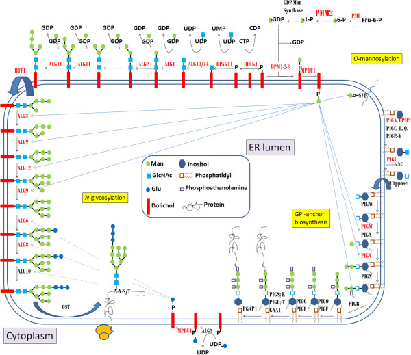
Background: Mutations in PMM2 impair phosphomannomutase-2 activity and cause the most frequent congenital disorder of glycosylation, PMM2-CDG. Mannose-1-phosphate, that is deficient in this disorder, is also implicated in the biosynthesis of glycosylphosphatidyl inositol (GPI) anchors.

Objective: To evaluate whether GPI-anchor and GPI-anchored proteins are defective in PMM2-CDG patients.

Methods: The expression of GPI-anchor and seven GPI-anchored proteins was evaluated by flow cytometry in different cell types from twelve PMM2-CDG patients. Additionally, neutrophil CD16 and plasma hepatic proteins were studied by Western blot. Transferrin glycoforms were evaluated by HPLC.

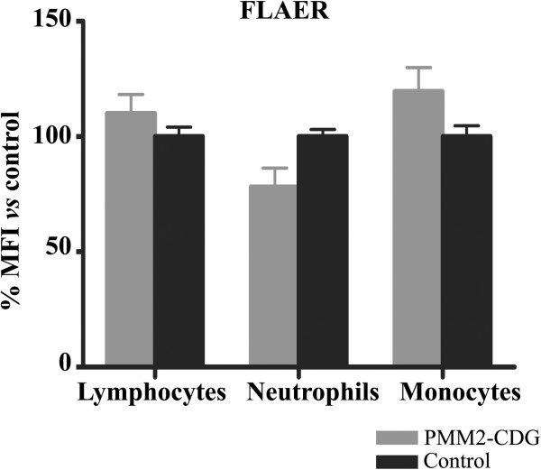
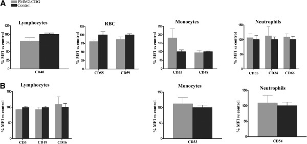
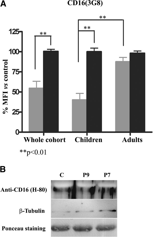
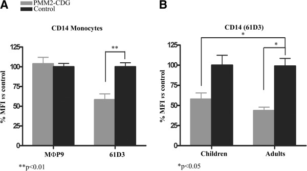
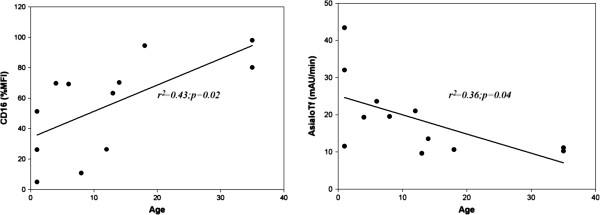
Results: Patients and controls had similar surface expression of GPI-anchor and most GPI-anchored proteins. Nevertheless, patients displayed a significantly diminished binding of two anti-CD16 antibodies (3G8 and KD1) to neutrophils and also of anti-CD14 (61D3) to monocytes. Interestingly, CD16 immunostaining and asialotransferrin levels significantly correlated with patients' age. Analysis by flow cytometry of CD14 with MΦP9, and CD16 expression in neutrophils by Western blot using H-80 ruled out deficiencies of these antigens.

Conclusions: PMM2 mutations do not impair GPI-anchor or GPI-anchored protein expression. However, the glycosylation anomalies caused by PMM2 mutations might affect the immunoreactivity of monoclonal antibodies and lead to incorrect conclusions about the expression of different proteins, including GPI-anchored proteins. Neutrophils and monocytes are sensitive to PMM2 mutations, leading to abnormal glycosylation in immune receptors, which might potentially affect their affinity to their ligands, and contribute to infection. This study also confirms less severe hypoglycosylation defects in older PMM2-CDG patients.


Figures

Figure 1
Figure 1
Biosynthetic pathways depending on mannose-1-phosphate: N-glycosylation, GPI-anchor biosynthesis, and O-mannosylation. Mannose-1-phosphate is the product of the phosphomannomutase activity, encoded by the PMM2 gene. The steps involved in the synthesis of the lipid-linked oligosaccharides required for the N-glycosylation of proteins, and glycosylphosphatidyl inositol required for the anchor of other proteins are detailed. The genes involved in each step are indicated, and those with mutations associated with congenital disorders of glycosylation are marked in red.
Figure 2
Figure 2
Level of expression of glycosylphosphatidyl inositol (GPI) anchored-proteins on different blood cells in PMM2-CDG patients and control subjects. The study was done by flow cytometry using proaerolysin variant (FLAER). Values are expressed as % mean fluorescence intensity (MFI) vs that observed in controls. PMN: Polymorphonuclear cells.
Figure 3
Figure 3
Expression of A) GPI-anchored proteins, and B) non GPI-anchored proteins on different blood cells in PMM2-CDG and control subjects. The study was done by flow cytometry using the monoclonal antibodies indicated in Material and Methods. Values are expressed as % mean fluorescence intensity (MFI) vs that observed in controls. RBC: red blood cells.
Figure 4
Figure 4
Expression of CD16 in neutrophils of PMM2-CDG patients and control subjects. A) Flow cytometry analysis using 3G8 monoclonal antibody in the whole cohort of patients and according to age. Values are expressed as % mean fluorescence intensity (MFI) vs that observed in controls. B) Western blot analysis using the H-80 polyclonal antibody. As loading controls, we stained the membrane with Ponceau Red and evaluated the expression of tubulin.
Figure 5
Figure 5
Expression of CD14 in monocytes of PMM2-CDG patients and control subjects evaluated by flow cytometry. A) Results obtained with MФp9 and 61D3 monoclonal antibodies in the whole cohort of patients. B) Expression of CD14 in monocytes according to age. The study was done with the 61D3 monoclonal antibody. Values are expressed as % mean fluorescence intensity (MFI) vs that observed in controls. **p< 0.01; *p< 0.05.
Figure 6
Figure 6
Glycoforms of antithrombin and α1-antitrypsin in plasma of one control, three adults, and three children with PMM2-CDG. Normal glycoforms are indicated by solid arrows and hypoglycosylated glycoforms by dashed arrows.
Figure 7
Figure 7
Correlations of CD16 binding to neutrophils (%mean fluorescence intensity -MFI- vs controls) and asialotransferrin according to age in PMM2-CDG patients.

Similar articles

Cited by

References

    1. Hennet T. Diseases of glycosylation beyond classical congenital disorders of glycosylation. Biochim Biophys Acta. 1820;8:1306–17. - PubMed
    1. Jaeken J. Congenital disorders of glycosylation (CDG): it’s (nearly) all in it! J Inherit Metab Dis. 2011;8:853–8. doi: 10.1007/s10545-011-9299-3. - DOI - PubMed
    1. Theodore M, Morava E. Congenital disorders of glycosylation: sweet news. Curr Opin Pediatr. 2011;8:581–7. doi: 10.1097/MOP.0b013e32834cd693. - DOI - PubMed
    1. Jaeken J. Congenital disorders of glycosylation. Ann N Y Acad Sci. 2010;8:190–8. doi: 10.1111/j.1749-6632.2010.05840.x. - DOI - PubMed
    1. Van Schaftingen E, Jaeken J. Phosphomannomutase deficiency is a cause of carbohydrate-deficient glycoprotein syndrome type I. FEBS Lett. 1995;8:318–20. doi: 10.1016/0014-5793(95)01357-1. - DOI - PubMed

Publication types

Substances