. 2013 Nov 4;23(21):2140-5.
doi: 10.1016/j.cub.2013.08.054. Epub 2013 Oct 17.

Quantitative imaging of transcription in living Drosophila embryos links polymerase activity to patterning

Affiliations

Quantitative imaging of transcription in living Drosophila embryos links polymerase activity to patterning

Hernan G Garcia et al. Curr Biol. .

Abstract

Spatiotemporal patterns of gene expression are fundamental to every developmental program. The resulting macroscopic domains have been mainly characterized by their levels of gene products. However, the establishment of such patterns results from differences in the dynamics of microscopic events in individual cells such as transcription. It is unclear how these microscopic decisions lead to macroscopic patterns, as measurements in fixed tissue cannot access the underlying transcriptional dynamics. In vivo transcriptional dynamics have long been approached in single-celled organisms, but never in a multicellular developmental context. Here, we directly address how boundaries of gene expression emerge in the Drosophila embryo by measuring the absolute number of actively transcribing polymerases in real time in individual nuclei. Specifically, we show that the formation of a boundary cannot be quantitatively explained by the rate of mRNA production in each cell, but instead requires amplification of the dynamic range of the expression boundary. This amplification is accomplished by nuclei randomly adopting active or inactive states of transcription, leading to a collective effect where the fraction of active nuclei is modulated in space. Thus, developmental patterns are not just the consequence of reproducible transcriptional dynamics in individual nuclei, but are the result of averaging expression over space and time.


Figures

Figure 1
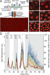
Figure 1. In vivo tracking of transcriptional activity using mRNA stem loops
(A) The hb P2 enhancer controlling the hb P2 promoter transcribes a lacZ gene with 24 MS2 stem loops located at its 5’-end. The MCP-GFP protein that binds to the stem loops is provided maternally. (B) Snapshots (26×26µm2) of the anterior region of an embryo expressing the MS2-MCP system in nuclear cycles 9 through 14, showing MCP-GFP (green) and Histone-RFP (red) fluorescence. Brightness and contrast of each time point adjusted independently. (C) Typical field of view of an embryo between 30–50% egg length (EL), anterior facing left. Scale bar is 10µm. See also Movie S1. (D) Fluorescence traces corresponding to individual spots of transcription (thin lines) color-coded by their nuclear position along the embryo as shown in (B) and corresponding mean fluorescence over position-binned nuclei (thick lines).
Figure 2
Figure 2. Rate of transcript elongation and dynamics of initiation and termination
(A) Comparison of expression dynamics of a single allele of the enhancer-construct in two nuclei (different embryos) with stem loops located at the 5’-end and the 3’-end of the lacZ gene, respectively. Images (7×7µm2) show Histone-RFP (red) and MCP-GFP (green) fluorescence; time 0 min corresponds to anaphase 13. Histogram shows the distribution of times of first spot detection. The difference of the distribution means (i.e. (5.4±0.1) min (red) and (7.6±0.2) min (blue)) is used to measure the rate of transcript elongation relongation = (1.54±0.14) kb/min (difference between 5’ and 3’ stem loop locations is 3.4kb; errors are propagated from the standard error of the distributions; number of nuclei: n5’=34 and n3’=22). (B) Average fluorescence in n.c. 14 as measured by the 5’ and 3’ constructs. The ratio between the maximum 5’ and 3’ fluorescence level is 3.3±0.5, consistent with the predicted ratio of 3.6 based on gene length. Red dashed line is the 5’ signal rescaled by 3.6. Grey bar is the estimated detection limit of 6±3 nascent mRNA molecules per spot (Figure S2H and Movie S2).
Figure 3
Figure 3. Dynamics of boundary formation
(A) Total amount of mRNA produced as a function of AP position for n.c. 12 (blue; n=13), n.c. 13 (black; n=24) and n.c. 14 (red; n=24) (see text and Figure S3A–C). mRNA production is normalized per equivalent n.c. 14 cell (i.e. the production per cell in n.c.12 and n.c. 13 is divided by four and two, respectively); error bars are standard errors over multiple embryos. The dynamic range, defined as the ratio between maximum and minimum expression of the pattern (Figure S1B), is (5.8±0.8) and (26±2) for n.c. 13 and n.c. 14, respectively. (B, C) Model of transcriptional dynamics: transcription is turned on at a time ton after mitosis with a constant rate of polymerase loading, resulting in a linear increase in fluorescence. After a time telongation= (3.4±0.3) min (i.e. the ratio between the length of the gene of 5.4kb and relongation) the first polymerase that was loaded will terminate transcription and leave the transcription site. A steady state of polymerase density (i.e. a stable fluorescence level) between newly loaded and terminating polymerases will persist until the promoter is turned off at toff. Polymerase loading ceases and remaining polymerases terminate transcription at the reverse (negative) rate with which they were loaded. Green curve shows a typical three-parameter fit to the mean fluorescence of nuclei located in a bin of size 2.5%EL centered around 30%EL in n.c. 13 (B) and n.c. 14 (C). In (C) only ton and the rate of polymerase loading are determined by the fit; for determination of toff in (C) see Figures S4A,B. (All errors are standard errors over multiple nuclei.)
Figure 4
Figure 4. Formation of the pattern boundary has three independent dynamical components
(A, B) Mean rate of polymerase loading (A), and mean window of time for transcription (B) as a function of position along the AP axis (for n.c. 12 (blue; n=13 embryos), n.c. 13 (black; n=24), and n.c. 14 (red; n=24), Figure S4A–E). Background values corresponding to a non-functional Bcd fly line are shown as horizontal bars using the same color-coding (Figure S5 and Movie S3). See Figure S4F,G for a summary of transcription dynamics. (C, D) mRNA produced as a function of AP position in n.c. 13 (C) and n.c. 14 (D). Data is normalized to the posterior end of the profiles. The direct measurement corresponds to the data shown in Figure 3A. Predictions (green and cyan) are obtained by multiplying the different values obtained in (A, B and F). (E) Representative fields of view (Histone-RFP) of nuclei in n.c. 14 in the activation (left, 29%EL) and transition regions (right, 61%EL). Nuclei where transcription was detected at any point over the entire n.c. are circled in red (Movies S4). Non-circled nuclei did not display any detectable transcription over the whole n.c. Scale bar is 10 µm. (F) Mean fraction of active nuclei as a function of position along the AP axis (Figure S6B). Color coding is as in (A,B). (A,B,F, error bars are standard errors over multiple embryos; C,D, error bars in the predictions are obtained by propagating the errors from A,B,F).

Comment in

Similar articles

Cited by

References

    1. Poustelnikova E, Pisarev A, Blagov M, Samsonova M, Reinitz J. A database for management of gene expression data in situ. Bioinformatics. 2004;20:2212–2221. - PubMed
    1. Jaeger J, Surkova S, Blagov M, Janssens H, Kosman D, Kozlov KN, Manu, Myasnikova E, Vanario-Alonso CE, Samsonova M, et al. Dynamic control of positional information in the early Drosophila embryo. Nature. 2004;430:368–371. - PubMed
    1. Dubuis JO, Samanta R, Gregor T. Accurate measurements of dynamics and reproducibility in small genetic networks. Molecular systems biology [electronic resource] 2013;9:639. - PMC - PubMed
    1. Pare A, Lemons D, Kosman D, Beaver W, Freund Y, McGinnis W. Visualization of individual Scr mRNAs during Drosophila embryogenesis yields evidence for transcriptional bursting. Curr Biol. 2009;19:2037–2042. - PMC - PubMed
    1. McHale P, Mizutani CM, Kosman D, MacKay DL, Belu M, Hermann A, McGinnis W, Bier E, Hwa T. Gene length may contribute to graded transcriptional responses in the Drosophila embryo. Dev Biol. 2011;360:230–240. - PMC - PubMed

Publication types

MeSH terms

Substances

LinkOut - more resources