. 2013 Nov 4;23(21):2176-80.
doi: 10.1016/j.cub.2013.08.061. Epub 2013 Oct 17.

Evidence for sex and recombination in the choanoflagellate Salpingoeca rosetta

Affiliations

Evidence for sex and recombination in the choanoflagellate Salpingoeca rosetta

Tera C Levin et al. Curr Biol. .

Abstract

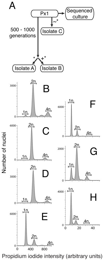
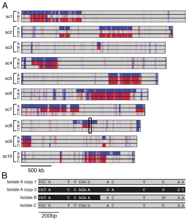
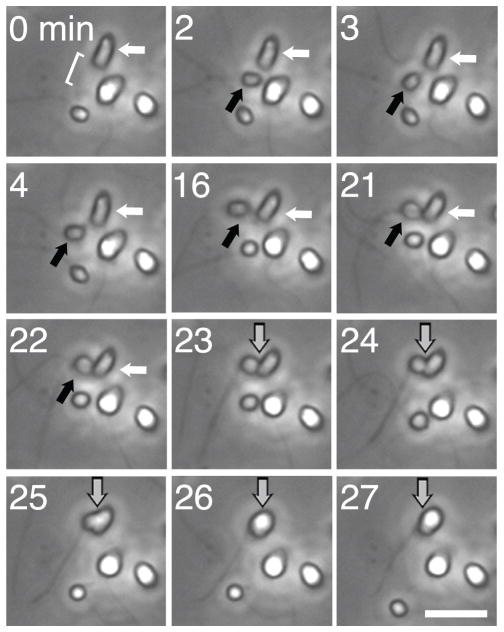
Nearly all animals reproduce sexually through the production and fusion of sperm and egg cells, yet little is known about the ancestry of animal sexual reproduction. Moreover, the sexual cycle of the closest living relatives of animals, the choanoflagellates, remains completely unknown. The choanoflagellate Monosiga brevicollis possesses a "meiotic toolkit" of genes, but the lack of polymorphisms detected during genome sequencing precluded inferences about its ploidy or sexual cycle. Here, we report that a related choanoflagellate, Salpingoeca rosetta, has a sexual life cycle and transitions between haploid and diploid states. Haploid cultures of S. rosetta became diploid in response to nutrient limitation. This ploidy shift coincided with anisogamous mating, during which small flagellated cells fused with larger flagellated cells. Distributions of polymorphisms in laboratory strains of S. rosetta provided independent evidence of historical recombination and mating. The ability of S. rosetta to produce morphologically differentiated gametes and to engage in sexual reproduction has implications for both reconstructing the evolution of sex in the progenitors of animals and establishing classical genetics in choanoflagellates.


Figures

Figure 1
Figure 1. S. rosetta cultures transition between haploid and diploid states in response to changing nutrient availability
(A) The S. rosetta cultures used in this study were all derived from a Px1 culture [6] through clonal isolation (*), during which one cell was isolated and propagated to establish the new culture line. For isolate A, a Px1 culture was propagated for 500–1000 generations before the new culture was established through clonal isolation. For isolate B, clonal isolation occurred after EMS mutagenesis (+). For isolate C, clonal isolation occurred after replacing the feeder bacteria from Px1 with a new species, Echinicola pacifica (~). During preparation for genome sequencing, Px1 was passaged rapidly, which may have resulted in changes in ploidy (see Supplemental Experimental Methods). (B) – (E) S. rosetta laboratory cultures can be haploid, diploid, or have a mix of haploid and diploid cells, as revealed by flow cytometry of propidium iodide-stained nuclei from unsynchronized cultures of Px1 (B), isolate A (C), isolate B (D), and isolate C (E). The expected locations of the 1n, 2n, and 4n peaks were based on an internal standard of S. cerevisiae (Fig. S1A – D). (F) – (H) Nutrient limitation induces haploid cultures to become diploid. Flow cytometry showed that isolate C was haploid after growth in HN medium for six days (F). Following growth of isolate C in unenriched sea water for six days, most cells were diploid (G). Transferring diploid isolate C cells back to HN medium for three days resulted in a return to the haploid state (H). Haploid-to-diploid transitions under nutrient limitation occurred within clonal cultures and did not require mixing of multiple clones. See also Figure S1.
Figure 2
Figure 2. Patterns of polymorphism reveal a history of sex and recombination in S. rosetta
(A) Mapping of SNP positions in isolates A, B, and C on the ten largest supercontigs suggests a history of recombination in S. rosetta. Sites that match the reference genome sequence are indicated in grey, homozygous SNPs in red and heterozygous SNPs in blue. Most SNPs are concentrated in large, contiguous haplotype blocks with sharp cut-offs that mark inferred sites of historical recombination. (B) SNPs in segregating haplotype blocks are shared among isolates. The boxed region of supercontig 8 indicated in (A) was cloned and sequenced from isolates A, B, and C to reveal the physical linkage among SNPs (see Fig. S2A for full alignment). Two distinct sequences were obtained from the heterozygous diploid isolate A; copy 1 (grey) matched isolate C and the reference genome, whereas copy 2 (black) contained many SNPs relative to the reference genome. In isolate B, the 5′ end of the region shares the copy 2 (black) haplotype, while the 3′ end is more similar to the reference sequence (grey). The transition from black to grey in isolate B indicates the inferred region of recombination. Isolate B also has a single SNP (*) that was not detected in any other sequenced sample and likely arose within isolate B. See also Figure S2.
Figure 3
Figure 3. Cell differentiation and cell fusion during haploid-to-diploid transitions
Time-lapse microscopy shows the fusion of morphologically distinct cells in an isolate C culture following 24 hours of nutrient limitation. A small, rounded cell (the ‘male gamete’, black arrow) entered the field of view (minute 2) and adhered to a larger, ovoid cell (‘female gamete’, white arrow; minutes 3–22) before cell fusion (gray arrow; minutes 23–27). The bracket in the first panel indicates the location of the microvillar collar of the female gamete. Cells were imaged once per minute using 40x phase contrast microscopy. Scale bar is 10 μm. See also Figure S3 and Movie S1.

Comment in

Similar articles

Cited by

References

    1. King N, Westbrook MJ, Young SL, Kuo A, Abedin M, Chapman J, Fairclough S, Hellsten U, Isogai Y, Letunic I, et al. The genome of the choanoflagellate Monosiga brevicollis and the origin of metazoans. Nature. 2008;451:783–788. - PMC - PubMed
    1. Carr M, Leadbeater BSC, Hassan R, Nelson M, Baldauf SL. Molecular phylogeny of choanoflagellates, the sister group to Metazoa. Proc Natl Acad Sci USA. 2008;105:16641–16646. - PMC - PubMed
    1. Carr M, Leadbeater BSC, Baldauf SL. Conserved Meiotic Genes Point to Sex in the Choanoflagellates. Journal of Eukaryotic Microbiology. 2010;57:56–62. - PubMed
    1. Fairclough SR, Dayel MJ, King N. Multicellular development in a choanoflagellate. Curr Biol. 2010;20:R875–6. - PMC - PubMed
    1. Dayel MJ, Alegado RA, Fairclough SR, Levin TC, Nichols SA, McDonald K, King N. Cell differentiation and morphogenesis in the colony-forming choanoflagellate Salpingoeca rosetta. Dev Biol. 2011;357:73–82. - PMC - PubMed

Publication types