

Chronic cellular imaging of entire cortical columns in awake mice using microprisms
- PMID: 24139817
- PMCID: PMC3840091
- DOI: 10.1016/j.neuron.2013.07.052
Chronic cellular imaging of entire cortical columns in awake mice using microprisms
Abstract
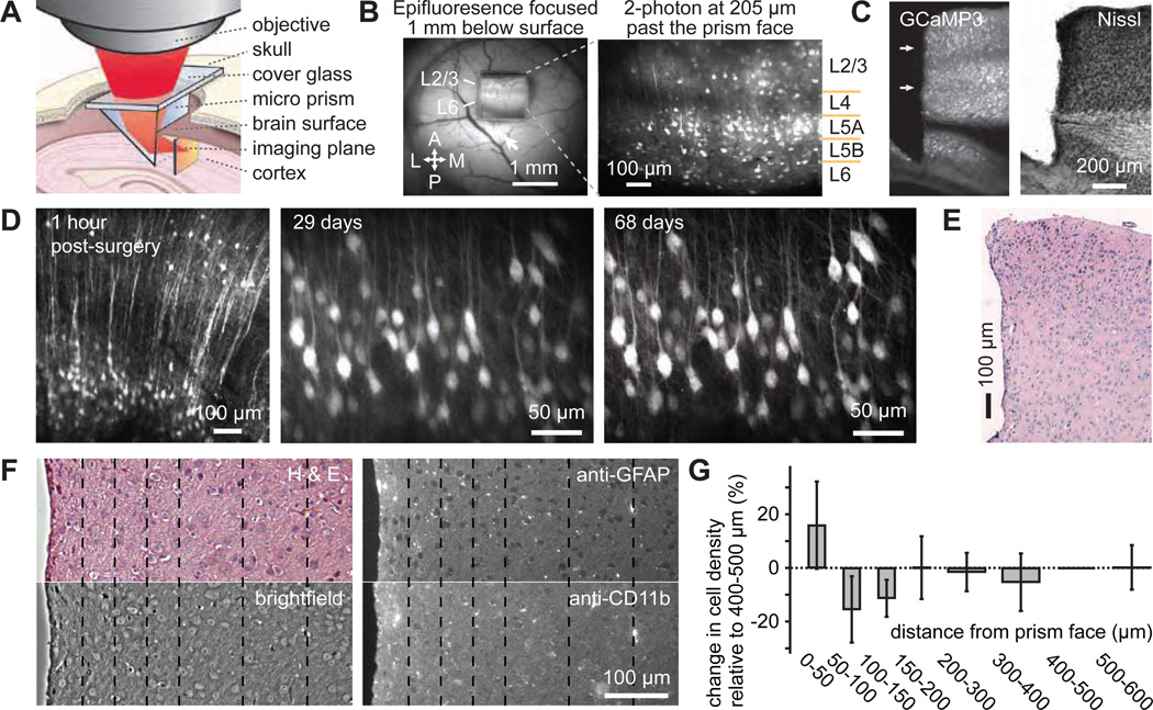
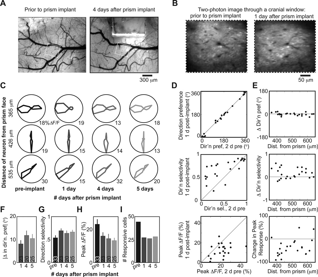
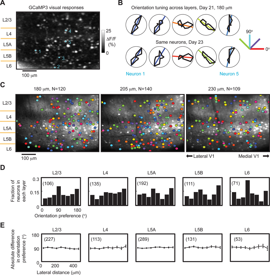
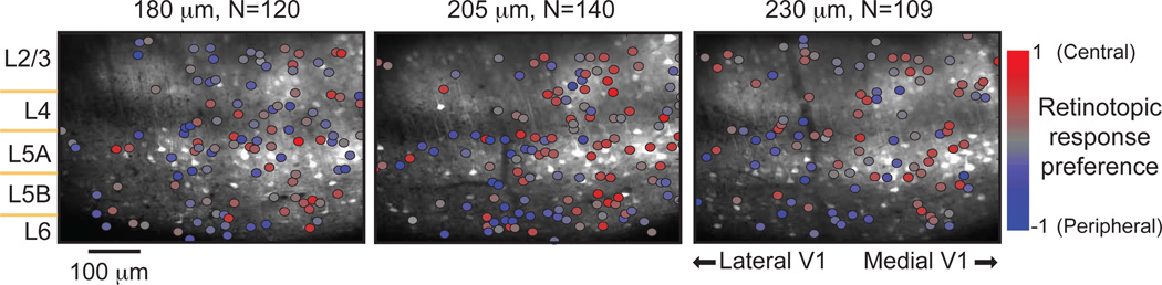
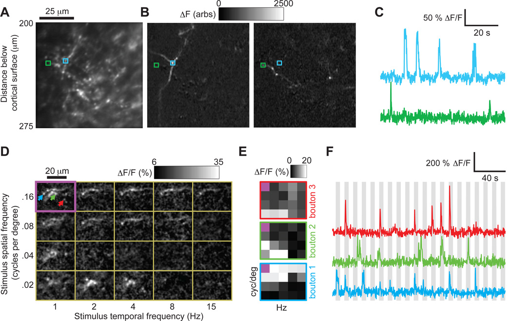
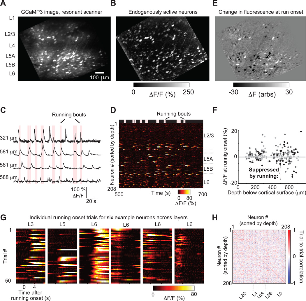
Two-photon imaging of cortical neurons in vivo has provided unique insights into the structure, function, and plasticity of cortical networks, but this method does not currently allow simultaneous imaging of neurons in the superficial and deepest cortical layers. Here, we describe a simple modification that enables simultaneous, long-term imaging of all cortical layers. Using a chronically implanted glass microprism in barrel cortex, we could image the same fluorescently labeled deep-layer pyramidal neurons across their entire somatodendritic axis for several months. We could also image visually evoked and endogenous calcium activity in hundreds of cell bodies or long-range axon terminals, across all six layers in visual cortex of awake mice. Electrophysiology and calcium imaging of evoked and endogenous activity near the prism face were consistent across days and comparable with previous observations. These experiments extend the reach of in vivo two-photon imaging to chronic, simultaneous monitoring of entire cortical columns.
Copyright © 2013 Elsevier Inc. All rights reserved.
Figures
References
-
- Amir W, Carriles R, Hoover EE, Planchon TA, Durfee CG, Squier JA. Simultaneous imaging of multiple focal planes using a two-photon scanning microscope. Opt Lett. 2007;32:1731–1733. - PubMed
Publication types
MeSH terms
Substances
Grants and funding
LinkOut - more resources
Full Text Sources
Other Literature Sources
