

Is cancer a metabolic disease?
- PMID: 24139946
- PMCID: PMC3873478
- DOI: 10.1016/j.ajpath.2013.07.035
Is cancer a metabolic disease?
Abstract
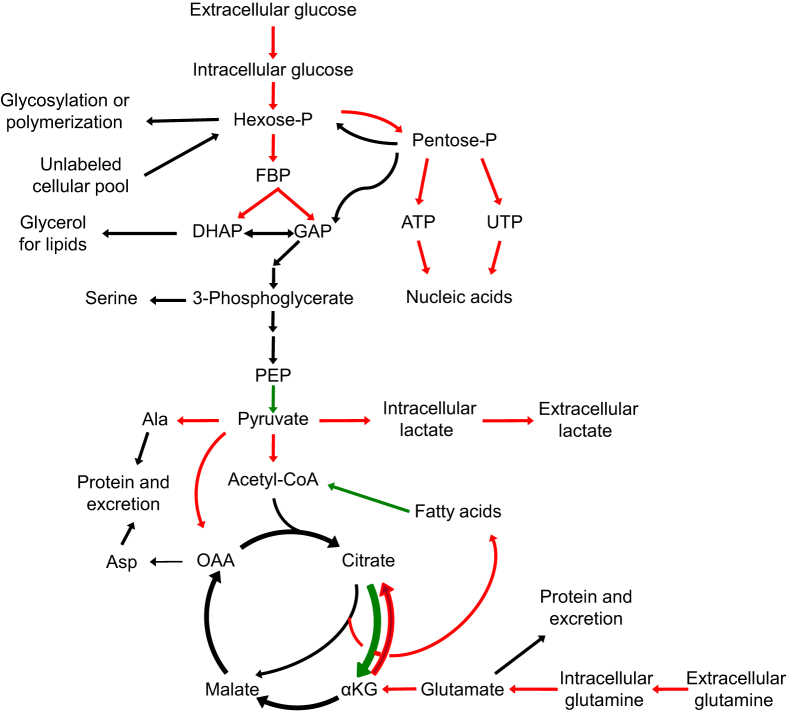
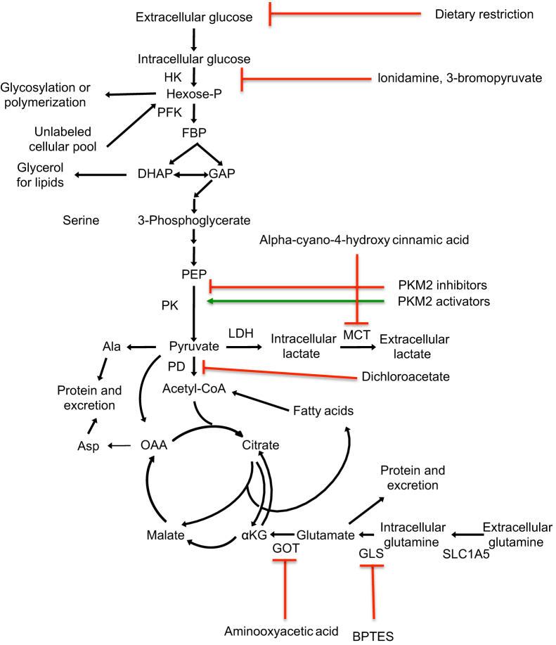
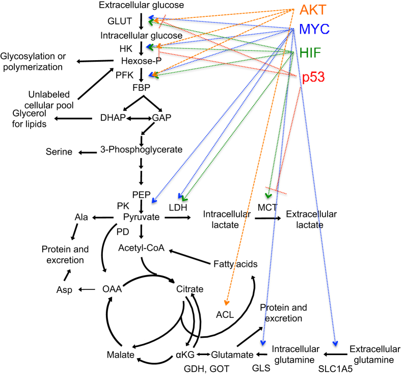
Although cancer has historically been viewed as a disorder of proliferation, recent evidence has suggested that it should also be considered a metabolic disease. Growing tumors rewire their metabolic programs to meet and even exceed the bioenergetic and biosynthetic demands of continuous cell growth. The metabolic profile observed in cancer cells often includes increased consumption of glucose and glutamine, increased glycolysis, changes in the use of metabolic enzyme isoforms, and increased secretion of lactate. Oncogenes and tumor suppressors have been discovered to have roles in cancer-associated changes in metabolism as well. The metabolic profile of tumor cells has been suggested to reflect the rapid proliferative rate. Cancer-associated metabolic changes may also reveal the importance of protection against reactive oxygen species or a role for secreted lactate in the tumor microenvironment. This article reviews recent research in the field of cancer metabolism, raising the following questions: Why do cancer cells shift their metabolism in this way? Are the changes in metabolism in cancer cells a consequence of the changes in proliferation or a driver of cancer progression? Can cancer metabolism be targeted to benefit patients?
Copyright © 2014 American Society for Investigative Pathology. Published by Elsevier Inc. All rights reserved.
Figures
References
-
- Warburg O. On the origin of cancer cells. Science. 1956;123:309–314. - PubMed
-
- Czernin J., Phelps M.E. Positron emission tomography scanning: current and future applications. Ann Rev Med. 2002;53:89–112. - PubMed
-
- Kunkel M., Reichert T.E., Benz P., Lehr H.A., Jeong J.H., Wieand S., Bartenstein P., Wagner W., Whiteside T.L. Overexpression of Glut-1 and increased glucose metabolism in tumors are associated with a poor prognosis in patients with oral squamous cell carcinoma. Cancer. 2003;97:1015–1024. - PubMed
-
- Mochiki E., Kuwano H., Katoh H., Asao T., Oriuchi N., Endo K. Evaluation of 18F-2-deoxy-2-fluoro-D-glucose positron emission tomography for gastric cancer. World J Surg. 2004;28:247–253. - PubMed
-
- Podoloff D.A., Advani R.H., Allred C., Benson A.B., 3rd, Brown E., Burstein H.J., Carlson R.W., Coleman R.E., Czuczman M.S., Delbeke D., Edge S.B., Ettinger D.S., Grannis F.W., Jr., Hillner B.E., Hoffman J.M., Kiel K., Komaki R., Larson S.M., Mankoff D.A., Rosenzweig K.E., Skibber J.M., Yahalom J., Yu J.M., Zelenetz A.D. NCCN task force report: positron emission tomography (PET)/computed tomography (CT) scanning in cancer. J Natl Compr Canc Netw. 2007;5(Suppl 1):S1–S22. - PubMed
Publication types
MeSH terms
Grants and funding
LinkOut - more resources
Full Text Sources
Other Literature Sources
