
Defects in the IFT-B component IFT172 cause Jeune and Mainzer-Saldino syndromes in humans

Jan Halbritter et al. Am J Hum Genet. .

Abstract

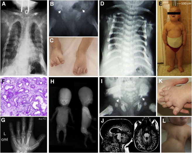
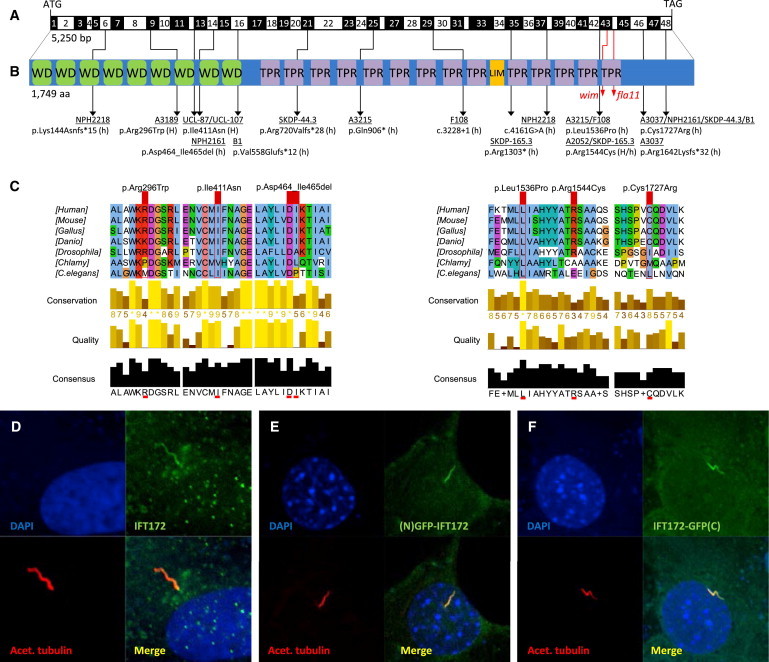
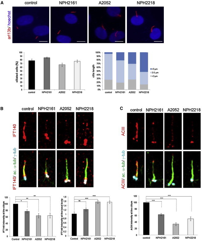
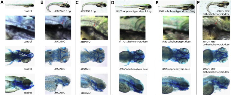
Intraflagellar transport (IFT) depends on two evolutionarily conserved modules, subcomplexes A (IFT-A) and B (IFT-B), to drive ciliary assembly and maintenance. All six IFT-A components and their motor protein, DYNC2H1, have been linked to human skeletal ciliopathies, including asphyxiating thoracic dystrophy (ATD; also known as Jeune syndrome), Sensenbrenner syndrome, and Mainzer-Saldino syndrome (MZSDS). Conversely, the 14 subunits in the IFT-B module, with the exception of IFT80, have unknown roles in human disease. To identify additional IFT-B components defective in ciliopathies, we independently performed different mutation analyses: candidate-based sequencing of all IFT-B-encoding genes in 1,467 individuals with a nephronophthisis-related ciliopathy or whole-exome resequencing in 63 individuals with ATD. We thereby detected biallelic mutations in the IFT-B-encoding gene IFT172 in 12 families. All affected individuals displayed abnormalities of the thorax and/or long bones, as well as renal, hepatic, or retinal involvement, consistent with the diagnosis of ATD or MZSDS. Additionally, cerebellar aplasia or hypoplasia characteristic of Joubert syndrome was present in 2 out of 12 families. Fibroblasts from affected individuals showed disturbed ciliary composition, suggesting alteration of ciliary transport and signaling. Knockdown of ift172 in zebrafish recapitulated the human phenotype and demonstrated a genetic interaction between ift172 and ift80. In summary, we have identified defects in IFT172 as a cause of complex ATD and MZSDS. Our findings link the group of skeletal ciliopathies to an additional IFT-B component, IFT172, similar to what has been shown for IFT-A.


Figures

Figure 1
Figure 1
Clinical Characteristics of Individuals with Recessive Mutations in IFT172 (A) A chest X-ray of individual A3215-21 shows a narrowed, bell-shaped thorax and short ribs. Note the tracheostomy for ventilation. (B) A hip X-ray of individual UCL-87 demonstrates a trident acetabular roof with spurs (white arrowheads). (C) Postaxial polydactyly of the feet in individual UCL-87. (D) A chest X-ray of individual UCL-107 shows a narrowed, bell-shaped thorax. (E) Obesity and short stature of individual NPH2218 at 10 years of age. (F) Renal histology of individual NPH2218 exhibits dilated tubules, disruption of the tubular basement membrane, and extensive interstitial fibrosis. (G) A hand X-ray of individual A3037-21 shows brachydactyly with cone-shaped epiphysis of the middle phalanges. (H) A babygram of individual SKDP-165.3 shows a turricephaly-like skull shape, absent nasal bone, postaxial tetramelic hexadactyly, shortened and curved long bones, short ribs, mild platyspondyly, and spur-like projections of the acetabular roof. (I) A trident acetabular roof with spurs (white arrowheads) in individual UCL-107. (J) Cranial MRI depicts partial agenesis of the cerebellar vermis in individual NPH2218. (K) Brachydactyly of individual NPH2218. (L) Narrow thorax of individual UCL-107.
Figure 2
Figure 2
Biallelic IFT172 Mutations, Deduced Impact at Protein Level, and Subcellular Localization of WT IFT172 (A) Exon structure of human IFT172 cDNA. The positions of the start codon (ATG) and stop codon (TGA) are indicated. (B) Domain structure of IFT172, which contains 9 WD-40 repeats (WD), located N-terminal to 14 tetratricopeptide repeats (TPR) and 1 LIM domain. For the mutations detected, black arrows indicate positions in relation to exons and protein domains. Family numbers are underlined. Abbreviations are as follows: H, homozygous; and h, heterozygous. IFT172 animal mutants wim (mouse, p.Leu1564Pro) and fla11 (C. reinhardtii, p.Leu1615Pro) are indicated by red arrows. Note the proximity of the detected missense changes p.Leu1536Pro and p.Arg1544Cys to the wim locus at position Leu1564. (C) A partial protein alignment of IFT172 shows evolutionary conservation of the identified missense changes (p.Arg296Trp, p.Ile411Asn, p.Leu1536Pro, p.Arg1544Cys, and p.Cys1727Arg). (D) Antibody staining (polyclonal rabbit antibody, Abcam, 1:100) of WT IFT172 in human control fibroblasts shows axonemal and pericentriolar localization in comparison to acetylated tubulin (anti-acetylated alpha tubulin, mouse monoclonal antibody, Abcam, 1:1000). (E and F) Localization of human WT IFT172 constructs, once with an N-terminal GFP tag (E) and once with a C-terminal GFP tag (F), after transfection of a 48 hr serum-starved NIH 3T3 cell line. Immunofluorescence on a confocal microscope (Zeiss, LSM 720) confirmed axonemal localization with enrichment at the ciliary base upon overexpression.
Figure 3
Figure 3
Alteration of Ciliogenesis and Ciliary Composition in Human Mutant Fibroblasts Control and mutant fibroblasts from individuals NPH2161, A2052-21, and NPH2218 were starved for 48 hr for inducing ciliogenesis and were fixed with MetOH. (A) Staining of ARL13B (polyclonal rabbit antibody, Proteintech; 1:400), quantification of ciliated cells, and measurement of cilia length with the use of Lucia G on Nikon DXM 1200 Software. Compared to controls, mutant fibroblasts displayed elongated cilia. The scale bar represents 10 μm. (B) Staining of acetylated-tubulin (mouse monoclonal antibody, Sigma Aldrich; 1:10,000), γ-tubulin (goat polyclonal antibody, Santa Cruz; 1:200), and IFT140 (polyclonal rabbit antibody, Proteintech; 1:100) showed a decrease in ciliary and an increase in basal body IFT140 staining intensity in mutant fibroblasts compared to controls. (C) Staining of adenylyl cyclase III (ACIII, rabbit polyclonal antibody, Santa Cruz; 1:100) showed a decrease in ciliary ACIII-staining intensity in mutant fibroblasts compared to controls. Images in (B) and (C) were recorded with a Leica SP8 confocal microscope and analyzed with ImageJ. All graphs show the mean ± SEM of at least three independent experiments. “ns” stands for not significant. p < 0.05, ∗∗p < 0.01, and ∗∗∗p < 0.001 were calculated via Dunn’s Multiple Comparison Test after the analysis of variance ANOVA test.
Figure 4
Figure 4
Knockdown of ift172 and ift80 and Genetic Epistasis between ift172 and ift80 in Zebrafish (A–C) When compared to the control (A), both ift172 (B) and ift80 (C) morphants displayed similar ciliopathy phenotypes, including ventral body-axis curvature (first row), formation of renal cysts (red arrows, second row), and cartilage defects with hypoplasia of the Meckel’s cartilage (mc) and widening of ceratohyal angle (cha), as shown by Alcian-blue staining (third and fourth row). (D–E) Zebrafish injected with subphenotypic doses of either ift172 (D) or ift80 (E) MO appeared no different than the control (A). (F) Similar to a full dose of each MO alone, combined injection of subphenotypic doses of ift172 MO and subphenotypic doses of ift80 MO resulted in body-axis curvature, formation of renal cysts, and cartilage defects.

References

    1. Baker K., Beales P.L. Making sense of cilia in disease: the human ciliopathies. Am. J. Med. Genet. C. Semin. Med. Genet. 2009;151C:281–295. - PubMed
    1. Hildebrandt F., Benzing T., Katsanis N. Ciliopathies. N. Engl. J. Med. 2011;364:1533–1543. - PMC - PubMed
    1. Badano J.L., Mitsuma N., Beales P.L., Katsanis N. The ciliopathies: an emerging class of human genetic disorders. Annu. Rev. Genomics Hum. Genet. 2006;7:125–148. - PubMed
    1. Huber C., Cormier-Daire V. Ciliary disorder of the skeleton. Am. J. Med. Genet. C. Semin. Med. Genet. 2012;160C:165–174. - PubMed
    1. Czarnecki P.G., Shah J.V. The ciliary transition zone: from morphology and molecules to medicine. Trends Cell Biol. 2012;22:201–210. - PMC - PubMed

Publication types

MeSH terms

Supplementary concepts