. 2013 Nov 7;93(5):812-24.
doi: 10.1016/j.ajhg.2013.09.009. Epub 2013 Oct 17.

Germline mutations in NFKB2 implicate the noncanonical NF-κB pathway in the pathogenesis of common variable immunodeficiency

Affiliations

Germline mutations in NFKB2 implicate the noncanonical NF-κB pathway in the pathogenesis of common variable immunodeficiency

Karin Chen et al. Am J Hum Genet. .

Abstract

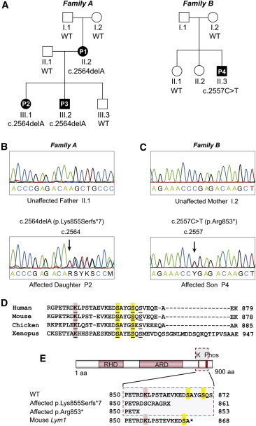
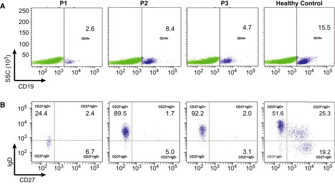
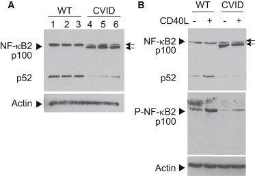
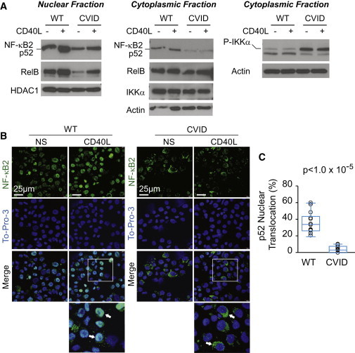
Common variable immunodeficiency (CVID) is a heterogeneous disorder characterized by antibody deficiency, poor humoral response to antigens, and recurrent infections. To investigate the molecular cause of CVID, we carried out exome sequence analysis of a family diagnosed with CVID and identified a heterozygous frameshift mutation, c.2564delA (p.Lys855Serfs(∗)7), in NFKB2 affecting the C terminus of NF-κB2 (also known as p100/p52 or p100/p49). Subsequent screening of NFKB2 in 33 unrelated CVID-affected individuals uncovered a second heterozygous nonsense mutation, c.2557C>T (p.Arg853(∗)), in one simplex case. Affected individuals in both families presented with an unusual combination of childhood-onset hypogammaglobulinemia with recurrent infections, autoimmune features, and adrenal insufficiency. NF-κB2 is the principal protein involved in the noncanonical NF-κB pathway, is evolutionarily conserved, and functions in peripheral lymphoid organ development, B cell development, and antibody production. In addition, Nfkb2 mouse models demonstrate a CVID-like phenotype with hypogammaglobulinemia and poor humoral response to antigens. Immunoblot analysis and immunofluorescence microscopy of transformed B cells from affected individuals show that the NFKB2 mutations affect phosphorylation and proteasomal processing of p100 and, ultimately, p52 nuclear translocation. These findings describe germline mutations in NFKB2 and establish the noncanonical NF-κB signaling pathway as a genetic etiology for this primary immunodeficiency syndrome.


Figures

Figure 1
Figure 1
NFKB2 Mutations in CVID (A) Pedigrees for two families affected by childhood-onset CVID with adrenal insufficiency. Mutation status of NFKB2 c.2564 for family A and NFKB2 c.2557 for family B are indicated beneath symbols for each individual (others were unavailable for testing). WT indicates wild-type. (B) Sanger sequencing of two individuals in family A over the c.2564 position in NFKB2. Upper panel shows wild-type c.2564 position and lower panel shows the c.2564delA variant. Arrow points to c.2564delA variant. (C) Sanger sequencing of two individuals in family B over the c.2557 position in NFKB2. Upper panel shows wild-type c.2557 position and lower panel shows the c.2557C>T variant. Arrow points to the c.2557C>T variant. (D) Multiple sequence alignment of amino acid sequences in the C terminus of NF-κB2. The conserved lysine that acts as an acceptor for ubiquitin is underlined and highlighted in red. The two conserved serines required for phosphorylation are underlined and highlighted in yellow. (E) Schematic of NF-κB2 with the Rel Homology Domain (RHD) and Ankyrin Repeat Domain (ARD) indicated by red boxes. The location of the conserved lysine (K) and two conserved serines (Phos) is indicated at top. The wild-type amino acid sequence is listed below the protein figure with the conserved lysine and serines marked with red and yellow boxes, respectively. The amino acid sequence for affected individuals in family A is shown second. The amino acid sequence of the affected individuals in family B is shown third, and the amino acid sequence for the mouse Lym1 mutation is shown fourth.
Figure 2
Figure 2
B Cell Immunophenotyping (A) Dot plots demonstrate gating for CD19+ B cells. (B) CD19+ B cells were then analyzed for CD27 and IgD expression. Affected individuals (P1, P2, P3) demonstrate reduced absolute numbers and percent of switched-memory B cells (CD19+CD27+IgD) as well as marginal zone/nonswitched memory B cells (CD19+CD27+IgD+) compared to a healthy control. In addition, P1 demonstrates markedly reduced CD19+ B cell numbers.
Figure 3
Figure 3
NF-κB2 Substitutions Lead to Protein Truncation and Phosphorylation Defects (A) Immunoblot of wild-type and truncated mutant NF-κB2/p100 (arrows) from whole-cell lysates of EBV-B cells derived from unaffected (pediatric control, lane 1; A.I.2, lane 2; and A.II.1, lane 3) and affected (P2, lane 4; P3, lane 5; P4, lane 6) individuals are shown. (B) Top panel shows immunoblot of wild-type and mutant NF-κB2 (arrows) from whole-cell lysates of EBV-B cells derived from unaffected (WT, A.II.1) and affected (CVID, P3) individuals. Cells were either treated (+) with CD40 ligand (CD40L) to stimulate the NF-κB2 pathway or left untreated (−) to compare the levels of phosphorylation in wild-type and mutant proteins. Bottom panel shows the same whole-cell lysates probed with an antibody that specifically recognizes the phosphorylated p100 protein (P-NF-κB2). Immunoblotting of actin was used as a loading control. Arrows point to full-length (top) and truncated (bottom) forms of NF-κB2. Arrowheads denote signals for protein being probed.
Figure 4
Figure 4
NF-κB2 Substitutions Affect Nuclear Localization of p52 (A) Nuclear (left) and cytoplasmic (middle and right) fractions of EBV-B cells derived from unaffected (WT, A.II.1) and affected (CVID, P2) individuals were subjected to immunoblotting with NF-κB2, RelB, HDAC1, IKKα, phospho-IKKα, and actin antibodies with (+) and without (−) induction with CD40 ligand (CD40L) for 4 hr. HDAC1 and actin were used as loading controls for the nuclear and cytoplasmic fractions, respectively. (B) EBV-B cells derived from unaffected (WT, A.II.1) and affected (CVID, P3) individuals were stained with a monoclonal antibody against NF-κB2/p52 (green) and To-Pro-3 (blue) nuclear stain. Scale bars represent 25 μM. Cells were either stimulated with CD40 ligand for 4 hr (CD40L) or not stimulated (NS). Inset box in the merged CD40L images is zoomed in the image directly below. Arrows highlight WT cells with NF-κB2 nuclear colocalization, compared to those with reduced NF-κB2 nuclear colocalization in the CVID cells. (C) Percent nuclear translocation of p52 was quantified via immunofluorescence confocal imaging of wild-type and mutant EBV-B cell lines after CD40L stimulation for 4 hr. A minimum of 300 cells per slide, per cell line were counted in the control (WT; pediatric control, A.I.2, and A.II.1) and affected (CVID; P2, P3, and P4) groups. Circles represent one microscopy field for each of three cell lines per group. n = 14 fields per group. Box plot denotes quartiles including median. Whiskers indicate min and max values. p value was calculated by a nonparametric Wilcoxon rank-sum test.

References

    1. Primary immunodeficiency diseases. Report of a WHO scientific group. Clin. Exp. Immunol. 1997;109(Suppl 1):1–28. - PubMed
    1. Oksenhendler E., Gérard L., Fieschi C., Malphettes M., Mouillot G., Jaussaud R., Viallard J.F., Gardembas M., Galicier L., Schleinitz N., DEFI Study Group Infections in 252 patients with common variable immunodeficiency. Clin. Infect. Dis. 2008;46:1547–1554. - PubMed
    1. Geha R.S., Notarangelo L.D., Casanova J.L., Chapel H., Conley M.E., Fischer A., Hammarström L., Nonoyama S., Ochs H.D., Puck J.M., International Union of Immunological Societies Primary Immunodeficiency Diseases Classification Committee Primary immunodeficiency diseases: an update from the International Union of Immunological Societies Primary Immunodeficiency Diseases Classification Committee. J. Allergy Clin. Immunol. 2007;120:776–794. - PMC - PubMed
    1. Cunningham-Rundles C., Bodian C. Common variable immunodeficiency: clinical and immunological features of 248 patients. Clin. Immunol. 1999;92:34–48. - PubMed
    1. Hammarström L., Vorechovsky I., Webster D. Selective IgA deficiency (SIgAD) and common variable immunodeficiency (CVID) Clin. Exp. Immunol. 2000;120:225–231. - PMC - PubMed

Publication types

MeSH terms