. 2013 Nov 7;52(3):459-67.
doi: 10.1016/j.molcel.2013.09.017. Epub 2013 Oct 17.

VprBP has intrinsic kinase activity targeting histone H2A and represses gene transcription

Affiliations

VprBP has intrinsic kinase activity targeting histone H2A and represses gene transcription

Kyunghwan Kim et al. Mol Cell. .

Abstract

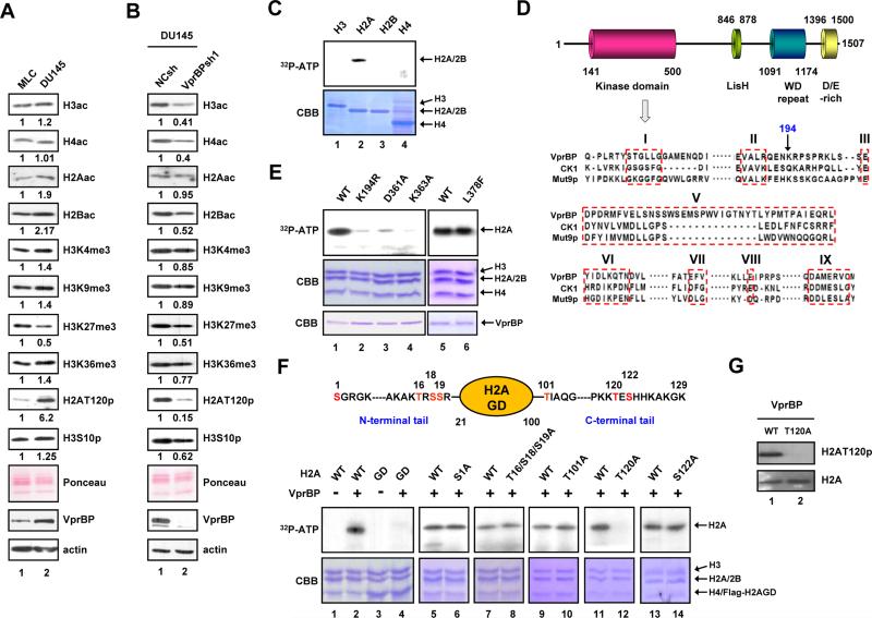
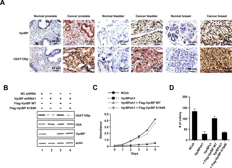
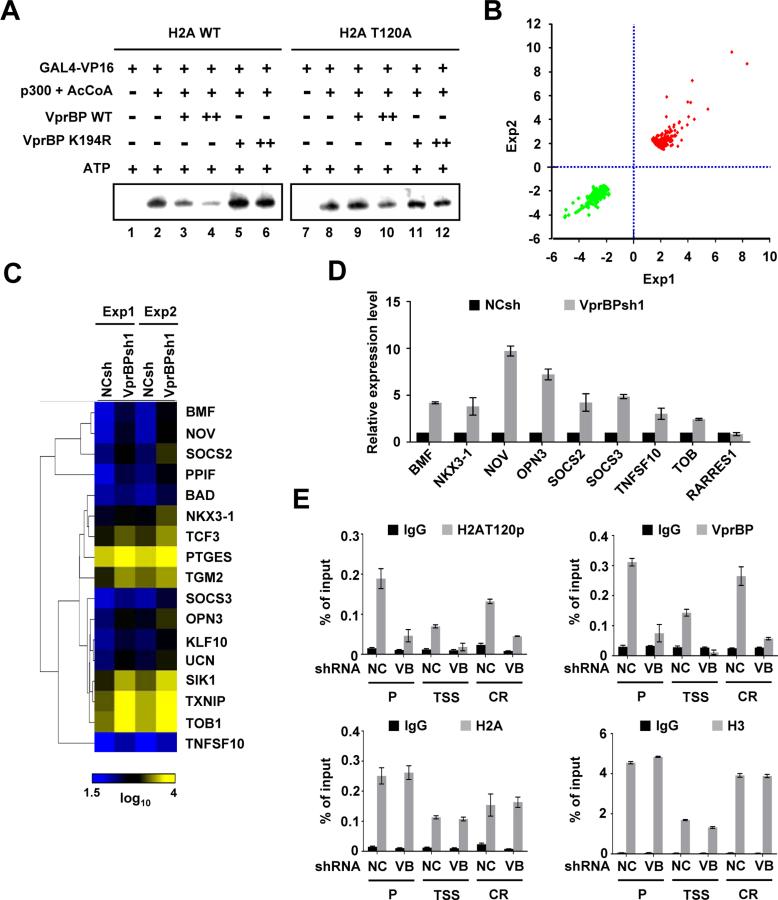
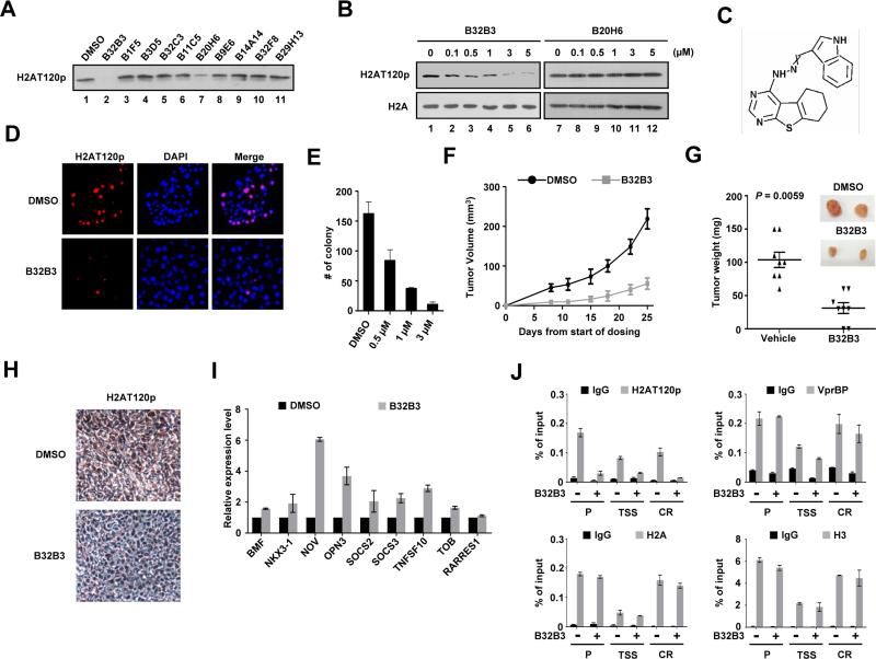
Histone modifications play important roles in the regulation of gene expression and chromatin organization. VprBP has been implicated in transcriptionally silent chromatin formation and cell-cycle regulation, but the molecular basis underlying such effects remains unclear. Here we report that VprBP possesses an intrinsic protein kinase activity and is capable of phosphorylating histone H2A on threonine 120 (H2AT120p) in a nucleosomal context. VprBP is localized to a large set of tumor suppressor genes and blocks their transcription, in a manner that is dependent on its kinase activity toward H2AT120. The functional significance of VprBP-mediated H2AT120p is further underscored by the fact that RNAi knockdown and small-molecule inhibition of VprBP reactivate growth regulatory genes and impede tumor growth. Our findings establish VprBP as a major kinase responsible for H2AT120p in cancer cells and suggest that VprBP inhibition could be a new strategy for the development of anticancer therapeutics.


Figures

Figure 1
Figure 1. VprBP phosphorylates histone H2A at T120
(A) Chromatin was prepared from human prostate cancer (DU145) and normal (MLC) cell lines and subjected to Western blotting with the indicated antibodies. Ponceau S staining and β-actin served as loading controls in all Western blot analyses in this study. Quantifications of the band intensities by densitometry are shown below the Western blots, and similar results were obtained from two additional experiments. ac, acetylation; p, phosphorylation; me3, trimethylation. (B) DU145 cells were infected with lentiviruses expressing VprBP shRNA (lane 2) or non-specific control shRNA (lane 1), and chromatin fractions were analyzed by Western blotting as in (A). (C) Individual core histones were incubated with recombinant VprBP in the presence of [γ-32P] ATP. The reactions were resolved by 15% SDS-PAGE and analyzed by autoradiography (upper panel) and Coomassie blue staining (lower panel). (D) VprBP contains the putative kinase domain in the N-terminal region, the Lis homology motif in the central region, and the WD repeat and D/E-rich motif in the C-terminal region. Numbers denote amino acid positions. Sequence alignment of the putative kinase domain in VprBP with the kinase domains of human CK1 and Mut9p is shown in the lower panel. The boxed regions correspond to the conserved kinase subdomains I-III and V-IX. (E) Nucleosomes were reconstituted on a 207 bp 601 nucleosome positioning sequence using recombinant histones and incubated with wild type VprBP or the indicated mutants. H2A phosphorylation was detected by autoradiography. (F) Kinase assays were performed as in (E), but using nucleosomes containing wild type, tailless or mutant H2A. The residues that were mutated are indicated at the top. The H2A mutations did not affect histone octamer and nucleosome formation during reconstitution (data not shown). (G) Nucleosomes containing H2A wild type or T120A mutant were incubated with VprBP and ATP. H2AT120p was analyzed by Western blotting with anti-H2AT120p antibody. See also Figure S1.
Figure 2
Figure 2. VprBP is overexpressed in tumors and required for cell proliferation
(A) Tissue microarrays containing primary tumor and adjacent normal samples from cancer patients were subjected to immunohistochemistry with VprBP and H2AT120p antibodies. High power magnifications are shown for six representative samples. Scale bars correspond to 50 μm. See also Table S1. (B) DU145 cells were depleted of VprBP and infected with lentiviruses expressing the VprBP wild type (WT) or VprBP kinase-dead mutant K194R (KD). The levels of VprBP and H2AT120p were determined by Western blotting. (C) VprBP-depleted DU145 cells were complemented with VprBP WT or KD, and cell proliferation was measured by MTT assay. Results represent the means ± S.D. of three experiments performed in triplicate. (D) VprBP-depleted DU145 cells were infected with VprBP WT or KD as in (C), and the colonies grown up in soft agar were stained and counted. The Y axis indicates the number of colonies with a diameter of > 0.05 mm per view. Three independent experiments in triplicate wells were performed. Data represent the means ± S.D. of three independent experiments. See also Figure S2.
Figure 3
Figure 3. Functional analysis of VprBP-mediated H2AT120p
(A) Chromatin templates containing wild type or T120-mutated H2A were transcribed in the presence of Gal4-VP16, p300 + AcCoA and/or VprBP as indicated above the panel. VprBP was added to the reaction before p300. The results shown are representative of three independent experiments. (B) Shown are scatter plots of the global gene expression patterns comparing VprBP-depleted DU145 cells with mock-depleted cells. Dots represent expression values for the genes with a change > 1.7-fold in either of two independent experiments. (C) Clustering and heat map representation of the genes upregulated upon VprBP depletion and related to cell death and proliferation. Yellow and blue indicate high and low expression, respectively. See also Tables S2 and S3. (D) RNA was isolated from VprBP-depleted DU145 cells as in (B) and subjected to real-time qRT-PCR using primers specific for the indicated genes and listed in Supplemental Experimatal Procedures. Expression levels were normalized to β-actin level and shown relative to those of mock-depleted cells, and were arbitrarily assigned a value of 1. Data represent the means ± S.D. of three independent experiments. (E) The levels of H2AT120p, H2A, VprBP and H3 at the OPN3 gene were assessed in mock- and VprBP-depleted DU145 cells by ChIP analysis. Precipitation efficiencies were determined for promoter (P), transcription stat site (TSS) and coding region (CR) by quantitative PCR (qPCR) with primers listed in Supplemental Experimental Procedures. Quantitative results were averaged from three separate determinations. Results represent the means ± S.D. of three independent experiments. See also Figure S3.
Figure 4
Figure 4. Discovery and characterization of a small-molecule VprBP inhibitor
(A) In vitro kinase assays were performed with recombinant H2A and VprBP in the presence of the indicated compounds (5 μM). The effects of the compounds were evaluated by Western blotting with H2AT120p antibody. (B) DU145 cells were grown in the presence of the indicated concentrations of either B32B3 or B20H6 for 24 h, and immunoblotted with H2A and H2AT120p antibodies. (C) Molecular structure of B32B3. (D) DU145 cells were treated with DMSO or 0.5 μM B32B3 for 24 h. The cellular levels of H2AT120p were assessed by immunostaining. (E) DU145 cells were treated with increasing concentrations of B32B3 for 24 h, and the colonies were counted 3 weeks after seeding the cells on soft agar. The data are the means of three independent experiments ± S.D. (F) Nude mice were implanted with 1 ×10⁷ DU145 cells on the left flank. Five days after implantation, mice bearing established tumors were randomized into groups and treated with twice-weekly i.p. injections of either DMSO or B32B3 at a dose of 5 mg/kg (n = 8 per group). Tumor volumes (mm3) were measured at the indicated time points and shown as mean tumor volumes ± S.E.M. (G) Mice were killed at day 25 of the tumor growth, and the tumors were dissected and weighed. Mean tumor weights ± S.E.M. are shown, and the P value was calculated by unpaired Student's t test. (H) DU145 xenografts were excised from DMSO-treated and B32B3-treated mice, and were analyzed by immunohistochemistry. Representative view was photographed (I) Relative mRNA levels of the VprBP target genes in DMSO-treated (black bars) and B32B3-treated (1 μM, gray bars) DU145 cells were determined by qRT-PCR. Data represent the means ± S.D. of three independent experiments. (J) DU145 cells exposed to DMSO or 1 μM B32B3 were subject to ChIP analysis using the indicated antibodies. The data are the means of three independent experiments ± S.D. See also Figure S4.

References

    1. Aihara H, Nakagawa T, Yasui K, Ohta T, Hirose S, Dhomae N, Takio K, Kaneko M, Takeshima Y, Muramatsu M, et al. Nucleosomal histone kinase-1 phosphorylates H2A Thr 119 during mitosis in the early Drosophila embryo. Genes Dev. 2004;18:877–888. - PMC - PubMed
    1. Banerjee T, Chakravarti D. A peek into the complex realm of histone phosphorylation. Mol Cell Biol. 2011;31:4858–4873. - PMC - PubMed
    1. Casas-Mollano JA, Jeong BR, Xu J, Moriyama H, Cerutti H. The MUT9p kinase phosphorylates histone H3 threonine 3 and is necessary for heritable epigenetic silencing in Chlamydomonas. Proc Natl Acad Sci U S A. 2008;105:6486–6491. - PMC - PubMed
    1. Chi P, Allis CD, Wang GG. Covalent histone modifications--miswritten, misinterpreted and mis-erased in human cancers. Nat Rev Cancer. 2010;10:457–469. - PMC - PubMed
    1. Choi HS, Choi BY, Cho YY, Mizuno H, Kang BS, Bode AM, Dong Z. Phosphorylation of histone H3 at serine 10 is indispensable for neoplastic cell transformation. Cancer Res. 2005;65:5818–5827. - PMC - PubMed

Publication types

Associated data