RATS: Rapid Automatic Tissue Segmentation in rodent brain MRI
- PMID: 24140478
- PMCID: PMC3914765
- DOI: 10.1016/j.jneumeth.2013.09.021
RATS: Rapid Automatic Tissue Segmentation in rodent brain MRI
Abstract
Background: High-field MRI is a popular technique for the study of rodent brains. These datasets, while similar to human brain MRI in many aspects, present unique image processing challenges. We address a very common preprocessing step, skull-stripping, which refers to the segmentation of the brain tissue from the image for further processing. While several methods exist for addressing this problem, they are computationally expensive and often require interactive post-processing by an expert to clean up poorly segmented areas. This further increases total processing time per subject.
New method: We propose a novel algorithm, based on grayscale mathematical morphology and LOGISMOS-based graph segmentation, which is rapid, robust and highly accurate.
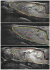
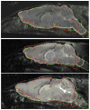
Results: Comparative results obtained on two challenging in vivo datasets, consisting of 22 T1-weighted rat brain images and 10 T2-weighted mouse brain images illustrate the robustness and excellent performance of the proposed algorithm, in a fraction of the computational time needed by existing algorithms.
Comparison with existing methods: In comparison to current state-of-the-art methods, our approach achieved average Dice similarity coefficient of 0.92 ± 0.02 and average Hausdorff distance of 13.6 ± 5.2 voxels (vs. 0.85 ± 0.20, p<0.05 and 42.6 ± 22.9, p << 0.001) for the rat dataset, and 0.96 ± 0.01 and average Hausdorff distance of 21.6 ± 12.7 voxels (vs. 0.93 ± 0.01, p <<0.001 and 33.7 ± 3.5, p <<0.001) for the mouse dataset. The proposed algorithm took approximately 90s per subject, compared to 10-20 min for the neural-network based method and 30-90 min for the atlas-based method.
Conclusions: RATS is a robust and computationally efficient method for accurate rodent brain skull-stripping even in challenging data.
Keywords: Brain segmentation; Rat model; Small animal MR.
Copyright © 2013 Elsevier B.V. All rights reserved.
Figures


Similar articles
-
Automatic macaque brain segmentation based on 7T MRI.Magn Reson Imaging. 2022 Oct;92:232-242. doi: 10.1016/j.mri.2022.07.001. Epub 2022 Jul 13. Magn Reson Imaging. 2022. PMID: 35842194
-
LOGISMOS-B: layered optimal graph image segmentation of multiple objects and surfaces for the brain.IEEE Trans Med Imaging. 2014 Jun;33(6):1220-35. doi: 10.1109/TMI.2014.2304499. Epub 2014 Feb 7. IEEE Trans Med Imaging. 2014. PMID: 24760901 Free PMC article.
-
RU-Net: skull stripping in rat brain MR images after ischemic stroke with rat U-Net.BMC Med Imaging. 2023 Mar 27;23(1):44. doi: 10.1186/s12880-023-00994-8. BMC Med Imaging. 2023. PMID: 36973775 Free PMC article.
-
Robust skull stripping using multiple MR image contrasts insensitive to pathology.Neuroimage. 2017 Feb 1;146:132-147. doi: 10.1016/j.neuroimage.2016.11.017. Epub 2016 Nov 15. Neuroimage. 2017. PMID: 27864083 Free PMC article.
-
An open, multi-vendor, multi-field-strength brain MR dataset and analysis of publicly available skull stripping methods agreement.Neuroimage. 2018 Apr 15;170:482-494. doi: 10.1016/j.neuroimage.2017.08.021. Epub 2017 Aug 12. Neuroimage. 2018. PMID: 28807870 Review.
Cited by
-
Automated identification of piglet brain tissue from MRI images using Region-based Convolutional Neural Networks.PLoS One. 2023 May 11;18(5):e0284951. doi: 10.1371/journal.pone.0284951. eCollection 2023. PLoS One. 2023. PMID: 37167205 Free PMC article.
-
Perturbed neurochemical and microstructural organization in a mouse model of prenatal opioid exposure: A multi-modal magnetic resonance study.PLoS One. 2023 Jul 20;18(7):e0282756. doi: 10.1371/journal.pone.0282756. eCollection 2023. PLoS One. 2023. PMID: 37471385 Free PMC article.
-
Automatic Brain Extraction for Rodent MRI Images.Neuroinformatics. 2020 Jun;18(3):395-406. doi: 10.1007/s12021-020-09453-z. Neuroinformatics. 2020. PMID: 31989442 Free PMC article.
-
A Comparative Study of Automatic Approaches for Preclinical MRI-based Brain Segmentation in the Developing Rat.Annu Int Conf IEEE Eng Med Biol Soc. 2018 Jul;2018:652-655. doi: 10.1109/EMBC.2018.8512402. Annu Int Conf IEEE Eng Med Biol Soc. 2018. PMID: 30440481 Free PMC article.
-
Automated Skull Stripping in Mouse Functional Magnetic Resonance Imaging Analysis Using 3D U-Net.Front Neurosci. 2022 Mar 10;16:801769. doi: 10.3389/fnins.2022.801769. eCollection 2022. Front Neurosci. 2022. PMID: 35368273 Free PMC article.
References
-
- Chou N, Wu J, Bingren JB, Qiu A, Chuang KH. Robust automatic rodent brain extraction using 3D pulse-coupled neural networks (PCNN) IEEE Trans Image Process. 2011;20:2554–64. - PubMed
-
- Firbank MJ, Coulthard A, Harrison RM, Williams ED. A comparison of two methods for measuring the signal to noise ratio on MR images. Phys Med Biol. 1999;44:N261–4. - PubMed
-
- Leemput KV, Maes F, Vandermeulen D, Suetens P. Automated model-based tissue classification of MR images of the brain. IEEE Trans Med Imaging. 1999;18:897–908. - PubMed
Publication types
MeSH terms
Grants and funding
LinkOut - more resources
Full Text Sources
Other Literature Sources
Medical

