

Toll-like receptor 3 plays a role in myocardial infarction and ischemia/reperfusion injury
- PMID: 24140513
- PMCID: PMC3879925
- DOI: 10.1016/j.bbadis.2013.10.006
Toll-like receptor 3 plays a role in myocardial infarction and ischemia/reperfusion injury
Abstract
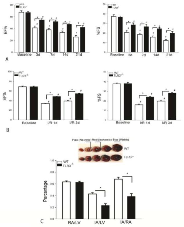
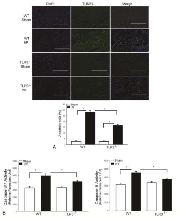
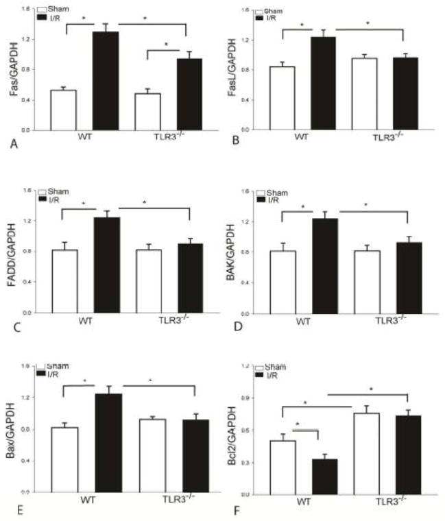
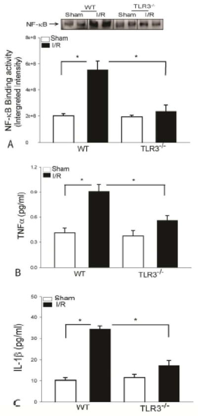
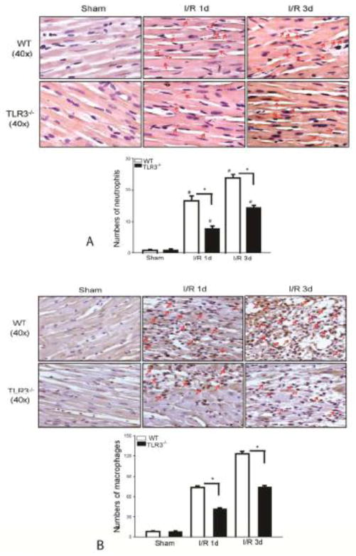
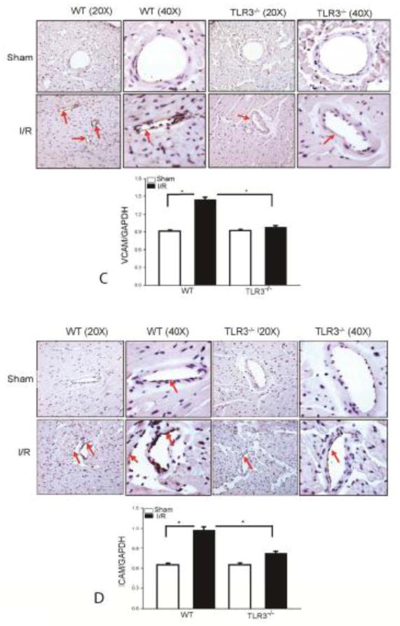
Innate immune and inflammatory responses mediated by Toll like receptors (TLRs) have been implicated in myocardial ischemia/reperfusion (I/R) injury. This study examined the role of TLR3 in myocardial injury induced by two models, namely, myocardial infarction (MI) and I/R. First, we examined the role of TLR3 in MI. TLR3 deficient (TLR3(-/-)) and wild type (WT) mice were subjected to MI induced by permanent ligation of the left anterior descending (LAD) coronary artery for 21days. Cardiac function was measured by echocardiography. Next, we examined whether TLR3 contributes to myocardial I/R injury. TLR3(-/-) and WT mice were subjected to myocardial ischemia (45min) followed by reperfusion for up to 3days. Cardiac function and myocardial infarct size were examined. We also examined the effect of TLR3 deficiency on I/R-induced myocardial apoptosis and inflammatory cytokine production. TLR3(-/-) mice showed significant attenuation of cardiac dysfunction after MI or I/R. Myocardial infarct size and myocardial apoptosis induced by I/R injury were significantly attenuated in TLR3(-/-) mice. TLR3 deficiency increases B-cell lymphoma 2 (BCL2) levels and attenuates I/R-increased Fas, Fas ligand or CD95L (FasL), Fas-Associated protein with Death Domain (FADD), Bax and Bak levels in the myocardium. TLR3 deficiency also attenuates I/R-induced myocardial nuclear factor KappaB (NF-κB) binding activity, Tumor necrosis factor alpha (TNF-α) and Interleukin-1 beta (IL-1β) production as well as I/R-induced infiltration of neutrophils and macrophages into the myocardium. TLR3 plays an important role in myocardial injury induced by MI or I/R. The mechanisms involve activation of apoptotic signaling and NF-κB binding activity. Modulation of TLR3 may be an effective approach for ameliorating heart injury in heart attack patients.
Keywords: Apoptosis; Inflammatory cytokine; Myocardial I/R; NF-κB; TLR.
© 2013.
Conflict of interest statement
There was no conflict of interest for the authors in the present study.
Figures
Similar articles
-
Role of extracellular RNA and TLR3-Trif signaling in myocardial ischemia-reperfusion injury.J Am Heart Assoc. 2014 Jan 3;3(1):e000683. doi: 10.1161/JAHA.113.000683. J Am Heart Assoc. 2014. PMID: 24390148 Free PMC article.
-
Poly (I:C) therapy decreases cerebral ischaemia/reperfusion injury via TLR3-mediated prevention of Fas/FADD interaction.J Cell Mol Med. 2015 Mar;19(3):555-65. doi: 10.1111/jcmm.12456. Epub 2014 Oct 29. J Cell Mol Med. 2015. PMID: 25351293 Free PMC article.
-
κ-Opioid receptor stimulation modulates TLR4/NF-κB signaling in the rat heart subjected to ischemia-reperfusion.Cytokine. 2013 Mar;61(3):842-8. doi: 10.1016/j.cyto.2013.01.002. Epub 2013 Feb 10. Cytokine. 2013. PMID: 23402995
-
The Role of NF-κB in Myocardial Ischemia/Reperfusion Injury.Curr Protein Pept Sci. 2022;23(8):535-547. doi: 10.2174/1389203723666220817085941. Curr Protein Pept Sci. 2022. PMID: 35980051 Review.
-
Tumor Necrosis Factor Family Members and Myocardial Ischemia-Reperfusion Injury: State of the Art and Therapeutic Implications.Int J Mol Sci. 2023 Feb 27;24(5):4606. doi: 10.3390/ijms24054606. Int J Mol Sci. 2023. PMID: 36902036 Free PMC article. Review.
Cited by
-
The Biological Basis for Cardiac Repair After Myocardial Infarction: From Inflammation to Fibrosis.Circ Res. 2016 Jun 24;119(1):91-112. doi: 10.1161/CIRCRESAHA.116.303577. Circ Res. 2016. PMID: 27340270 Free PMC article. Review.
-
Human myocardium releases heat shock protein 27 (HSP27) after global ischemia: the proinflammatory effect of extracellular HSP27 through toll-like receptor (TLR)-2 and TLR4.Mol Med. 2014 Jun 9;20(1):280-9. doi: 10.2119/molmed.2014.00058. Mol Med. 2014. PMID: 24918749 Free PMC article.
-
The innate immune receptor NLRX1 is a novel required modulator for mPTP opening: implications for cardioprotection.Basic Res Cardiol. 2025 Aug;120(4):707-725. doi: 10.1007/s00395-025-01124-x. Epub 2025 Jun 19. Basic Res Cardiol. 2025. PMID: 40536683 Free PMC article.
-
Identification of long non-coding RNAs biomarkers for early diagnosis of myocardial infarction from the dysregulated coding-non-coding co-expression network.Oncotarget. 2016 Nov 8;7(45):73541-73551. doi: 10.18632/oncotarget.11999. Oncotarget. 2016. PMID: 27634901 Free PMC article.
-
TLR3 Mediates Repair and Regeneration of Damaged Neonatal Heart through Glycolysis Dependent YAP1 Regulated miR-152 Expression.Cell Death Differ. 2018 May;25(5):966-982. doi: 10.1038/s41418-017-0036-9. Epub 2018 Jan 22. Cell Death Differ. 2018. PMID: 29358670 Free PMC article.
References
-
- Chong AJ, Shimamoto A, Hampton CR, Takayama H, Spring DJ, Rothnie CL, Yada M, Pohlman TH, Verrier ED. Toll-like receptor 4 mediates ischemia/reperfusion injury of the heart. J Thorac Cardiovasc Surg. 2004;128:170–179. - PubMed
Publication types
MeSH terms
Substances
Grants and funding
LinkOut - more resources
Full Text Sources
Other Literature Sources
Medical
Molecular Biology Databases
Research Materials
Miscellaneous
