. 2014 Feb:67:211-20.
doi: 10.1016/j.freeradbiomed.2013.10.009. Epub 2013 Oct 16.

Contribution of endogenously produced reactive oxygen species to the activation of podocyte NLRP3 inflammasomes in hyperhomocysteinemia

Affiliations

Contribution of endogenously produced reactive oxygen species to the activation of podocyte NLRP3 inflammasomes in hyperhomocysteinemia

Justine M Abais et al. Free Radic Biol Med. 2014 Feb.

Abstract

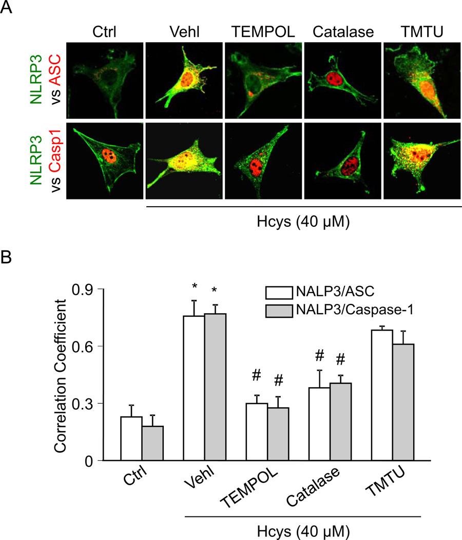
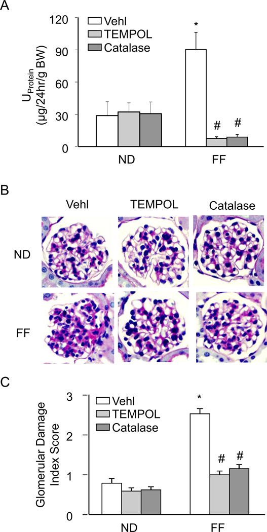
Hyperhomocysteinemia (hHcys) is an important pathogenic factor contributing to the progression of end-stage renal disease. Recent studies have demonstrated the implication of nicotinamide adenine dinucleotide phosphate oxidase-mediated NLRP3 inflammasome activation in the development of podocyte injury and glomerular sclerosis during hHcys. However, it remains unknown which reactive oxygen species (ROS) are responsible for this activation of NLRP3 inflammasomes and how such action of ROS is controlled. This study tested the contribution of common endogenous ROS including superoxide (O2(-)), hydrogen peroxide (H2O2), peroxynitrite (ONOO(-)), and hydroxyl radical (OH) to the activation of NLRP3 inflammasomes in mouse podocytes and glomeruli. In vitro, confocal microscopy and size-exclusion chromatography demonstrated that dismutation of O2(-) by 4-hydroxy-2,2,6,6-tetramethylpiperidine 1-oxyl (Tempol) and decomposition of H2O2 by catalase prevented Hcys-induced aggregation of NLRP3 inflammasome proteins and inhibited Hcys-induced caspase-1 activation and IL-1β production in mouse podocytes. However, scavenging of ONOO(-) or OH had no significant effect on either Hcys-induced NLRP3 inflammasome formation or activation. In vivo, scavenging of O2(-) by Tempol and removal of H2O2 by catalase substantially inhibited NLRP3 inflammasome formation and activation in glomeruli of hHcys mice as shown by reduced colocalization of NLRP3 with ASC or caspase-1 and inhibition of caspase-1 activation and IL-1β production. Furthermore, Tempol and catalase significantly attenuated hHcys-induced glomerular injury. In conclusion, endogenously produced O2(-) and H2O2 primarily contribute to NLRP3 inflammasome formation and activation in mouse glomeruli resulting in glomerular injury or consequent sclerosis during hHcys.

Keywords: Free radicals; Glomerular sclerosis; Homocysteine; NLRP3 inflammasome; Redox signaling.


Figures

Figure 1
Figure 1. TEMPOL and catalase attenuate Hcys-induced inflammasome formation in podocytes
A. Confocal images representing the colocalization of NLRP3 (green) with ASC or caspase-1 (red) in cultured podocytes. B. Summarized data showing the fold change of PCC for the colocalization of NLRP3 with ASC and NLRP3 with caspase-1 (n=4). Ctrl: Control; Vehl: Vehicle; TMTU: tetramethylthiourea, PCC: Pearson coefficient correlation. * P<0.05 vs. Control; # P<0.05 vs. Hcys.
Figure 2
Figure 2. Size-exclusion chromatography demonstrates inhibition of Hcys-induced ASC protein redistribution after O2•− and H2O2 scavenging
A. Representative chromatogram illustrating the elution profile of proteins from both a standard and podocyte sample taken from an absorbance of 280 nm. Elution of a gel filtration standard allowed for the determination of the molecular mass of the samples. B. Selected protein fractions were analyzed by Western blot, probed with an anti-ASC antibody. C. Summarized data determined from the ASC band intensities of the inflammasome complex fractions (3–7) (n=4–6). Ctrl: Control; Vehl: Vehicle; TMTU: tetramethylthiourea. * P<0.05 vs. Control; # P<0.05 vs. Hcys.
Figure 3
Figure 3. Effects of ROS scavenging on caspase-1 activity and IL-1β secretion in the presence of Hcys
A. Caspase-1 activity, shown as folds versus Ctrl, measured in podocytes treated with Hcys in the presence of various ROS scavengers (n=6–8). B. IL-1β production measured in the supernatant of podocytes treated with Hcys in the presence of various ROS scavengers (n=6–8). Ctrl: Control; Vehl: Vehicle; TMTU: tetramethylthiourea. * P<0.05 vs. Control; # P<0.05 vs. Hcys.
Figure 4
Figure 4. In vivo effect of O2•− and H2O2 inhibition on hHcys-induced inflammasome formation in podocytes of hyperhomocysteinemic mice
A. Colocalization of NLRP3 (green) with ASC or caspase-1 (red) in the mouse glomeruli of vehicle, TEMPOL, or catalase-treated mice fed a normal or FF diet. B–C. Summarized data showing the correlation coefficient between NLRP3 with ASC or caspase-1 (n=6). Casp1: caspase-1, Vehl: Vehicle, ND: Normal diet, FF: Folate-free diet. *P<0.05 vs. Vehl on ND; # P<0.05 vs. Vehl on FF Diet.
Figure 5
Figure 5. hHcys-induced inflammasome formation occurs primarily in podocytes of hyperhomocysteinemic mice glomeruli
A. Colocalization of NLRP3 (green) with podocyte markers podocin or desmin (red) in the mouse glomeruli of vehicle, TEMPOL, or catalase-treated mice fed a normal or FF diet. B–C. Summarized data showing the correlation coefficient between NLRP3 with podocin or desmin (n=6). Podo: Podocin; Des: Desmin, Vehl: Vehicle, ND: Normal diet, FF: Folate-free diet. *P<0.05 vs. Vehl on ND; # P<0.05 vs. Vehl on FF Diet.
Figure 6
Figure 6. In vivo administration of TEMPOL and catalase prevents caspase-1 activation, IL-1β production, and O2•− production
A. Caspase-1 activity in Vehl, TEMPOL, or catalase-treated mice with hHcys induced by the FF diet (n=7). B. TEMPOL and catalase administration prevented hHcys-induced IL-1β production (n=6–7). C. O2•− production induced by hHcys was inhibited in mice treated with TEMPOL or catalase (n=6). Vehl: Vehicle, ND: Normal diet, FF: Folate-free diet. *P<0.05 vs. Vehl on ND; # P<0.05 vs. Vehl on FF Diet.
Figure 7
Figure 7. Renal protective effects of O2•− and H2O2 inhibition on hHcys-induced glomerular injury
A. hHcys-induced proteinuria was attenuated in mice receiving TEMPOL or catalase treatment (n=6). B. Microscopic observation of glomeruli in PAS stained kidney sections demonstrated TEMPOL and catalase prevented hHcys-induced changes in glomerular morphological structure by preventing capillary collapse, fibrosis, cellular proliferation and mesangial cell expansion (n=4–6). C. Extent of glomerular sclerosis was assessed semiquantitatively and expressed as the Glomerular damage index (GDI). Vehl: Vehicle, ND: Normal diet, FF: Folate-free diet. *P<0.05 vs. Vehl on ND; # P<0.05 vs. Vehl on FF Diet.

References

    1. Perla-Kajan J, Jakubowski H. Paraoxonase 1 and homocysteine metabolism. Amino Acids. 2012;43:1405–1417. - PubMed
    1. Cavalca V, Cighetti G, Bamonti F, Loaldi A, Bortone L, Novembrino C, De Franceschi M, Belardinelli R, Guazzi MD. Oxidative stress and homocysteine in coronary artery disease. Clin Chem. 2001;47:887–892. - PubMed
    1. Bialecka M, Kurzawski M, Roszmann A, Robowski P, Sitek EJ, Honczarenko K, Gorzkowska A, Budrewicz S, Mak M, Jarosz M, Golab-Janowska M, Koziorowska-Gawron E, Drozdzik M, Slawek J. Association of COMT, MTHFR, and SLC19A1(RFC-1) polymorphisms with homocysteine blood levels and cognitive impairment in Parkinson's disease. Pharmacogenet Genomics. 2012;22:716–724. - PubMed
    1. Schalinske KL, Smazal AL. Homocysteine imbalance: a pathological metabolic marker. Adv Nutr. 2012;3:755–762. - PMC - PubMed
    1. Yu M, Sturgill-Short G, Ganapathy P, Tawfik A, Peachey NS, Smith SB. Age-related changes in visual function in cystathionine-beta-synthase mutant mice, a model of hyperhomocysteinemia. Exp Eye Res. 2012;96:124–131. - PMC - PubMed

Publication types

MeSH terms

LinkOut - more resources