. 2014 Feb:62:286-95.
doi: 10.1016/j.nbd.2013.10.009. Epub 2013 Oct 16.

Evidence that collaboration between HIF-1α and Notch-1 promotes neuronal cell death in ischemic stroke

Affiliations

Evidence that collaboration between HIF-1α and Notch-1 promotes neuronal cell death in ischemic stroke

Yi-Lin Cheng et al. Neurobiol Dis. 2014 Feb.

Abstract

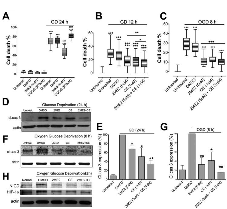
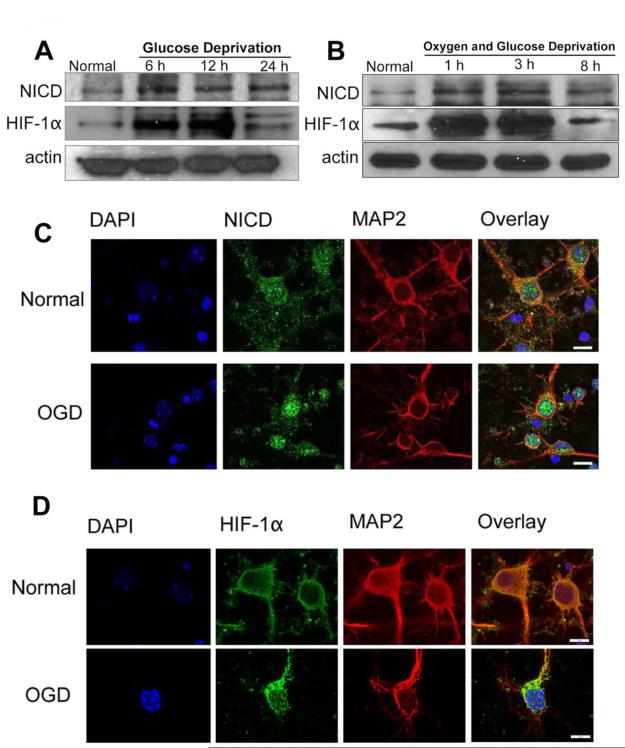
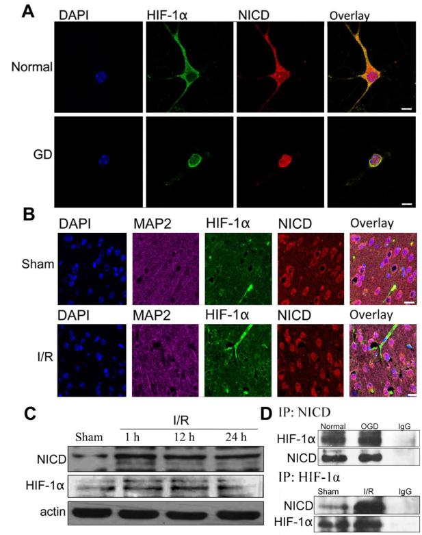
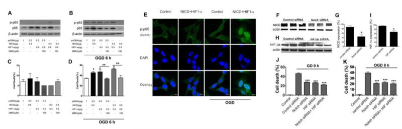
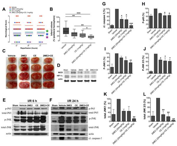
Recent findings suggest that Notch-1 signaling contributes to neuronal death in ischemic stroke, but the underlying mechanisms are unknown. Hypoxia inducible factor-1α (HIF-1α), a global regulator of cellular responses to hypoxia, can interact with Notch and modulate its signaling during hypoxic stress. Here we show that Notch signaling interacts with the HIF-1α pathway in the process of ischemic neuronal death. We found that a chemical inhibitor of the Notch-activating enzyme, γ-secretase, and a HIF-1α inhibitor, protect cultured cortical neurons against ischemic stress, and combined inhibition of Notch-1 and HIF-1α further decreased neuronal death. HIF-1α and Notch intracellular domain (NICD) are co-expressed in the neuronal nucleus, and co-immunoprecipitated in cultured neurons and in brain tissue from mice subjected to focal ischemic stroke. Overexpression of NICD and HIF-1α in cultured human neural cells enhanced cell death under ischemia-like conditions, and a HIF-1α inhibitor rescued the cells. RNA interference-mediated depletion of endogenous NICD and HIF-1α also decreased cell death under ischemia-like conditions. Finally, mice treated with inhibitors of γ-secretase and HIF-1α exhibited improved outcome after focal ischemic stroke, with combined treatment being superior to individual treatments. Additional findings suggest that the NICD and HIF-1α collaborate to engage pro-inflammatory and apoptotic signaling pathways in stroke.

Keywords: Apoptosis; HIF-1α; Ischemic stroke; Neuronal cell death; Notch.


Figures

Figure 1
Figure 1. Inhibition of NICD and HIF-1α protects cultured cortical neurons against death under ischemia-like conditions.
(A) Treatment with 2ME2 significantly reduced cell death, and treatment with DMOG increased cell death, under GD condition. n=6, ***p< 0.005 compared with the vehicle-treated group under normal culture conditions; +++, ###p<0.005 compared with the vehicle treated group under GD condition. (B) Compound E and 2ME2 each significantly reduced GD-induced cell death, and combined treatment further reduced cell death during GD. n=6, ***p<0.005 compared with the untreated group under normal conditions; +++p<0.005 compared with the vehicle treated group under GD conditions; **p<0.01 2ME2 compared with the Compound E+2ME2 treated group; *p<0.05 Compound E compared with the Compound E+2ME2 treated groups. (C) Neuronal cell death was quantified in cultures that had been subjected to OGD. n=6, ***p<0.005 compared with the untreated group under normal condition; +++p<0.005 compared with the vehicle treated group under OGD condition. (D-G) Treatment with either Compound E or 2ME2 reduced the level of cleaved caspase-3, and combined treatment further reduced cleaved caspase-3 levels under GD (D, E) and OGD (F, G) conditions. (H) NICD and HIF-1α expression in 2ME2 and CE treated cultures. n=4 cultures in each group, * p<0.05 compared with vehicle-treated group; **p<0.01 compared with vehicle-treated group.
Figure 2
Figure 2. NICD and HIF-1α levels in primary cortical neurons under normal, GD and OGD conditions.
(A-B) Immunoblots showing NICD and HIF-1α protein levels at different time points during GD (A) and OGD (B) conditions. NICD levels were increased with GD and OGD treatment. HIF-1α levels increased from 6 h to 12 h of GD and from 1 h to 3 h of OGD, and then decreased at later time points. (C, D) Immunofluorescence images showing staining of NICD (C, green), HIF-1α (D, green), the neuronal marker MAP2 (red) and the nuclear marker 4′,6-diamidino- 2- phenylindol (DAPI) (blue). Both NICD and HIF-1α staining showed a slight increase at 8 h of OGD. Scale bar: 10 μm.
Figure 3
Figure 3. NICD and HIF-1α interaction in cortical neurons and mouse brain under ischemic conditions.
(A) Immunofluorescence staining of NICD and HIF-1α in mouse cortical neurons and neurons under GD conditions. NICD (red), HIF-1α (green) and DAPI (blue) staining showing translocation of NICD and HIF-1α from cytoplasm to the nucleus at 24 h GD. Scale bar: 20 μm. (B) Immunofluorescence staining of NICD and HIF-1α in the ipsilateral peri-infarct tissue section showing NICD (red), HIF-1α (green), MAP2 (magenta) and DAPI (blue) staining. The immunoreactivity of NICD and HIF-1α was more robust at 24 h after ischemia/reperfusion (I/R) compared to the sham control. Scale bar: 40 μm. (C) The expression level of NICD and HIF-1α were increased in the ipsilateral hemisphere after 1 h, 12 h and 24 h of I/R compared to sham. (D) Co-immunoprecipitation of NICD and HIF-1α in neurons under OGD 8h and in brain tissue from the ipsilateral cerebral cortex following 1h ischemia and 24 h reperfusion showing increased binding of NICD and HIF-1α compared to controls.
Figure 4
Figure 4. Effect of over-expression and inhibition of NICD and HIF-1α in neurons under ischemia-like conditions.
NICD and HIF-1α were transfected to SH-SY5Y cells to induce NICD and HIF-1α expression; Notch-1, HIF-1α siRNA were transfected to primary cortical neurons to inhibit endogenous NICD and HIF-1α expression. (A) Cells overexpressing NICD or HIF-1α showed an increased level of NF-κB p65/p-p65 when compared to cells transfected with control plasmid cDNA (pcDNA). The level of NF-κB p65/p-p65 was further increased when both NICD, HIF-1α were overexpressed. The NF-κB p65/p-p65 levels were reduced by treatment with 2ME2 under normal conditions (A) and 6 h of OGD (B). (C) Cell death in SH-SY5Y cells overexpressing NICD and HIF-1α. (D) Cell death in SH-SY5Y neurons was increased when NICD and HIF-1α were over-expressed, and was significantly reduced at 6 h OGD when treated with 2ME2. (E) Immunofluorescence staining of phosphorylated p65 in SH-SY5Y cells overexpressing NICD and HIF1-α under OGD (9 h) condition. Scale bar: 10 μm. (F, G) Knockdown of endogenous Notch-1 using Notch-1 siRNA. (H, I) Knockdown of endogenous HIF-1α using HIF-1α siRNA. (J) Cell death in neurons transfected with Notch-1 and HIF-1α siRNA was significantly reduced at 9 h GD (K) and 8 h OGD (J). n=3 cultures. *p< 0.05, **p< 0.01, ***p< 0.005 compared with the vehicle treated group.
Figure 5
Figure 5. Inhibition of NICD and HIF-1α reduces focal ischemic brain injury in vivo.
(A and B) Mice underwent 1 h MCAO and 24-72 h reperfusion. Mice treated with CE (n=11) and 2ME2 (n=10), alone or in combination (n=13), exhibited improved neurological function and reduced brain injury after stroke, compared to vehicle-treated (n=11) mice. Neurological score was measured at 24, 48, and 72 h following reperfusion; infarct volume was measured at 72 h following reperfusion. Mice treated with both 2ME2 and CE treated showed significant reduction in infarct size compared to mice treated with either CE or 2ME2 alone. Data are represented as mean ± SEM. (C) Representative images of brains from each treatment group following 1 h MCAO and 72 h reperfusion. (D) NICD and HIF-1α expression in 2ME2 and CE treated mouse brains after MCAO and 24 h of reperfusion. n=5 in each group. (E) NF-κB p65/p-p65, total JNK/p-JNK expression in the ipsilateral brain hemisphere at 6 h I/R from mice treated with either 2ME2, CE, or both 2ME2 and CE. (F) Cleaved caspase-3, NF-κB p65/p-p65, total JNK/p-JNK expression in the ipsilateral brain hemisphere at 24 h I/R from mice treated with 2ME2, CE, and both 2ME2 and CE. (G, H, I, J, K, L) Quantification of cleaved caspase 3, p-p65 and total JNK/p-JNK expression. n=5 in each group, *p< 0.05, **p< 0.01, ***p< 0.005 compared with the vehicle treated group.

Similar articles

Cited by

References

    1. Antoniou X, Sclip A, Ploia C, Colombo A, Moroy G, Borsello T. JNK contributes to Hif-1alpha regulation in hypoxic neurons. Molecules. 2009;5:114–127. - PMC - PubMed
    1. Arumugam TV, Chan SL, Jo DG, Yilmaz G, Tang SC, Cheng A, Gleichmann M, Okun E, Dixit VD, Chigurupati S, Mughal MR, Ouyang X, Miele L, Magnus T, Poosala S, Granger DN, Mattson MP. Gamma secretase-mediated Notch signaling worsens brain damage and functional outcome in ischemic stroke. Nat Med. 2006;12:621–623. - PubMed
    1. Arumugam TV, Cheng YL, Choi Y, Choi YH, Yang S, Yun YK, Park JS, Yang DK, Thundyil J, Gelderblom M, Karamyan VT, Tang SC, Chan SL, Magnus T, Sobey CG, Jo DG. Evidence that gamma-secretase-mediated Notch signaling induces neuronal cell death via the nuclear factor-kappaB-Bcl-2-interacting mediator of cell death pathway in ischemic stroke. Mol Pharmacol. 2011;80:23–31. - PubMed
    1. Arumugam TV, Salter JW, Chidlow JH, Ballantyne CM, Kevil CG, Granger DN. Contributions of LFA-1 and Mac-1 to brain injury and microvascular dysfunction induced by transient middle cerebral artery occlusion. Am J Physiol Heart Circ Physiol. 2004;287:H2555–2560. - PubMed
    1. Baranova O, Miranda LF, Pichiule P, Dragatsis I, Johnson RS, Chavez JC. Neuron-specific inactivation of the hypoxia inducible factor 1 alpha increases brain injury in a mouse model of transient focal cerebral ischemia. J Neurosci. 2007;27:6320–6332. - PMC - PubMed

Publication types

Substances