. 2014 Feb 1:446:44-52.
doi: 10.1016/j.ab.2013.10.018. Epub 2013 Oct 17.

Crude and purified proteasome activity assays are affected by type of microplate

Affiliations

Crude and purified proteasome activity assays are affected by type of microplate

Ziyou Cui et al. Anal Biochem. .

Abstract

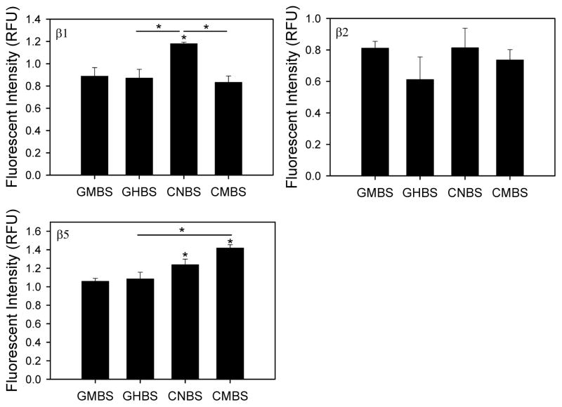
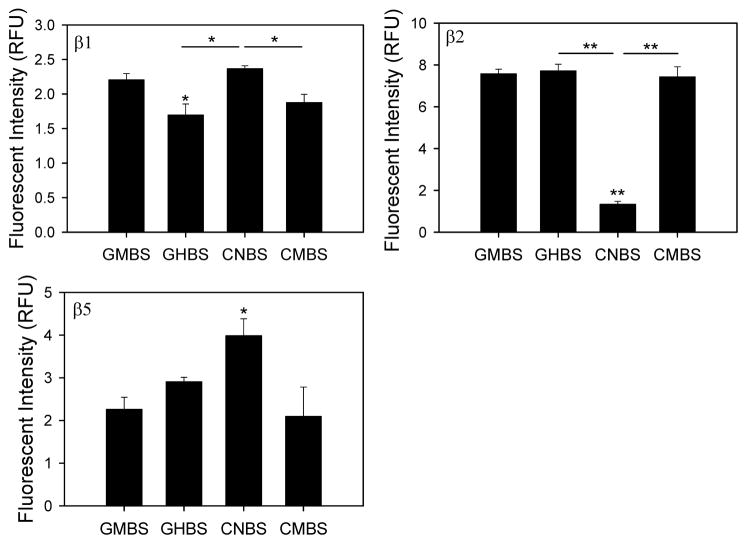
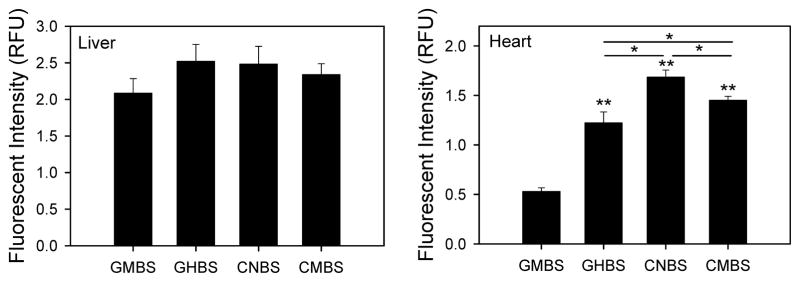
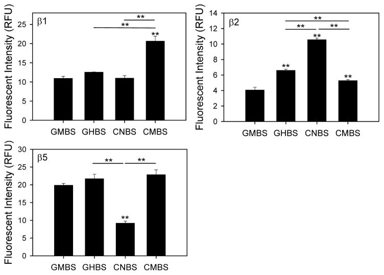
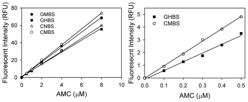
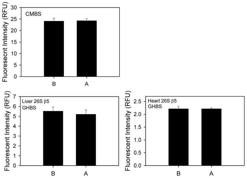
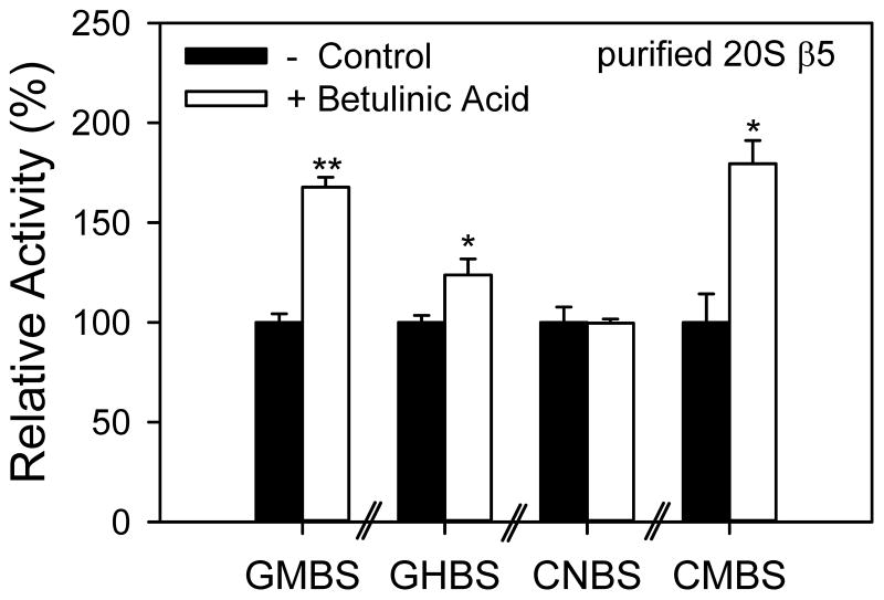
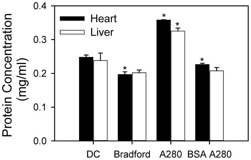
Measurement of proteasome activity is fast becoming a commonly used assay in many laboratories. The most common method to measure proteasome activity involves measuring the release of fluorescent tags from peptide substrates in black microplates. Comparisons of black plates used for measuring fluorescence with different properties show that the microplate properties significantly affect the measured activities of the proteasome. The microplate that gave the highest reading of trypsin-like activity of the purified 20S proteasome gave the lowest reading of chymotrypsin-like activity of the 20S proteasome. Plates with medium binding surfaces from two different companies showed an approximately 2-fold difference in caspase-like activity for purified 20S proteasomes. Even standard curves generated using free 7-amino-4-methylcoumarin (AMC) were affected by the microplate used. As such, significantly different proteasome activities, as measured in nmol AMC released/mg/min, were obtained for purified 20S proteasomes as well as crude heart and liver samples when using different microplates. The naturally occurring molecule betulinic acid activated the chymotrypsin-like proteasome activity in three different plates but did not affect the proteasome activity in the nonbinding surface microplate. These findings suggest that the type of proteasome activity being measured and sample type are important when selecting a microplate.

Keywords: Betulinic acid; Microplate; Proteasome; Proteolytic activity.


Figures

Figure 1
Figure 1. 26S proteasome activities from heart lysates in different black microplates
Caspase- (β1), trypsin- (β2), and chymotrypsin- (β5) like proteasome activities of heart cytosolic lysates were determine in four different microplates. Heart lysates (20 μg) were incubated with different substrates depending on the proteolytic activity of the proteasome that was being measured. Each assay was conducted in the absence and presence of the specific proteasome inhibitor bortezomib. AMC fluorescent tags released from substrates by the specific proteasome activity were measured using a Fluoroskan Ascent fluorometer at an excitation wavelength of 390 nm and an emission wavelength of 460 nm. *, p < 0.05; **, p < 0.001.
Figure 2
Figure 2. 26S proteasome activities from liver lysates in different black microplates
Caspase- (β1), trypsin- (β2), and chymotrypsin- (β5) like proteasome activities of liver cytosolic lysates were determine in four different microplates. Liver lysates (20 μg) were incubated with different substrates depending on the proteolytic activity of the proteasome that was being measured. Each assay was conducted as described in figure 1 legend and in the methods. *, p < 0.05; **, p < 0.001.
Figure 3
Figure 3. 20S chymotrypsin-like proteasome activities in heart and liver lysates using different black microplates
Heart and liver lysates (20 μg) were incubated with LLVY-AMC substrate in the presence of 20S buffer and AMC released from the proteasome substrate was measured using a Fluoroskan Ascent fluorometer as described in the methods. *, p < 0.05; **, p < 0.001.
Figure 4
Figure 4. Purified 20S proteasome activities measured using different microplates
Caspase- (β1), trypsin- (β2), and chymotrypsin- (β5) like proteasome activities of purified mouse 20S proteasomes (0.1 μg) were determined as described in the methods. *, p < 0.05; **, p < 0.001.
Figure 5
Figure 5. Effect of different 96 well black microplates on free AMC standard curves
The fluorescence of free AMC (excitation wavelength of 390 nm and emission wavelength of 460 nm) at different concentrations (0–0.5 μM and 0–8 μM) were measured a Fluoroskan Ascent fluorometer.
Figure 6
Figure 6. Effect of proteasome inhibitor added at different times on purified 20S and heart and liver 26S chymotrypsin-like activity
Proteasome inhibitor was added before (B) or after (A) buffer was to the microplate to determine if adding the inhibitor to the plate increased the binding of the inhibitor to the plate. Upper left panel shows purified 20S in CMBS plates while the lower left and right panels show liver and heart lysates in GHBS plates.
Figure 7
Figure 7. Effect of betulinic acid on purified 20S chymotrypsin-like proteasome activity
Betulinic acid (10 μg/ml final concentration) was added to purified murine 20S proteasomes and incubated for 20 mins before addition of proteasome substrate. AMC released from the substrate was measured using a Fluoroskan Ascent fluorometer. *, p < 0.05; **, p < 0.001.
Figure 8
Figure 8. Measurement of protein concentrations in heart and liver lysates using different protein assays
Protein concentrations of heart and liver lysates (0.2 mg/ml based upon Bradford assay) was determined using DC, Bradford and A280 protein assays. BSA A280 refers to measurement of protein at an absorbance of 280 nm using BSA as a standard curve. BSA was used as a standard for the DC and Bradford assays. *, p < 0.05; **, p < 0.001.

References

    1. Gomes AV, Waddell DS, Siu R, Stein M, Dewey S, Furlow JD, Bodine SC. Upregulation of proteasome activity in muscle RING finger 1-null mice following denervation. FASEB J. 2012;26:2986–99. - PMC - PubMed
    1. Gomes AV, Young GW, Wang Y, Zong C, Eghbali M, Drews O, Lu H, Stefani E, Ping P. Contrasting proteome biology and functional heterogeneity of the 20 S proteasome complexes in mammalian tissues. Mol Cell Proteomics. 2009;8:302–15. - PMC - PubMed
    1. Gomes AV, Zong C, Edmondson RD, Li X, Stefani E, Zhang J, Jones RC, Thyparambil S, Wang GW, Qiao X, Bardag-Gorce F, Ping P. Mapping the murine cardiac 26S proteasome complexes. Circ Res. 2006;99:362–71. - PubMed
    1. Bedford L, Paine S, Sheppard PW, Mayer RJ, Roelofs J. Assembly, structure, and function of the 26S proteasome. Trends Cell Biol. 2010;20:391–401. - PMC - PubMed
    1. Gomes AV, Zong C, Ping P. Protein degradation by the 26S proteasome system in the normal and stressed myocardium. Antioxid Redox Signal. 2006;8:1677–91. - PubMed

Publication types

Substances