. 2013 Dec;9(12):811-7.
doi: 10.1038/nchembio.1364. Epub 2013 Oct 20.

The Fic protein Doc uses an inverted substrate to phosphorylate and inactivate EF-Tu

Affiliations

The Fic protein Doc uses an inverted substrate to phosphorylate and inactivate EF-Tu

Daniel Castro-Roa et al. Nat Chem Biol. 2013 Dec.

Abstract

Fic proteins are ubiquitous in all of the domains of life and have critical roles in multiple cellular processes through AMPylation of (transfer of AMP to) target proteins. Doc from the doc-phd toxin-antitoxin module is a member of the Fic family and inhibits bacterial translation by an unknown mechanism. Here we show that, in contrast to having AMPylating activity, Doc is a new type of kinase that inhibits bacterial translation by phosphorylating the conserved threonine (Thr382) of the translation elongation factor EF-Tu, rendering EF-Tu unable to bind aminoacylated tRNAs. We provide evidence that EF-Tu phosphorylation diverged from AMPylation by antiparallel binding of the NTP relative to the catalytic residues of the conserved Fic catalytic core of Doc. The results bring insights into the mechanism and role of phosphorylation of EF-Tu in bacterial physiology as well as represent an example of the catalytic plasticity of enzymes and a mechanism for the evolution of new enzymatic activities.


Figures

Figure 1
Figure 1. Doc inhibits translation by inactivation of ternary complex formation
(a). Ribbon representation of the structural superposition of Doc (pdbid 3K33) onto Fic adenylylase from N. meningitides (NmFic; pdbid 3S6A). Doc is colored in blue and NmFic in pink, with respective catalytic loops in light and dark green. (b). Superposition of the catalytic loops of Doc (residues H66-R74) and NmFic (in the nucleotide bound conformation; residues H107-R114). The AMPPNP molecule bound to NmFic is shown in black (c). Luciferase synthesis in a commercially available cell-free translation system was performed in the presence of [35S]-methionine in the absence or presence of Doc and revealed by phosphorimaging. Here and after, full images of gels, TLCs and TLEs are presented in Supplementary Fig. 1. (d). Scheme of the assembly of an in vitro translation system using purified components (PK-pyruvate kinase; PEP-phosphoenol pyruvate). Steps at which Doc was added to the reactions in panels (e) and (f) and in Figure 2a, e are depicted in colors. (e) Synthesis of dipeptide MF in the absence or presence of Doc added after (red) or during (blue) ternary complex (TC) formation. (f). Synthesis of tripeptide MFV in the presence or absence of Doc added during initiation (cyan) or ternary (blue) complex formation. Peptides were analyzed by thin layer electrophoresis and autoradiography.
Figure 2
Figure 2. Doc phosphorylates EF-Tu at the conserved Thr382
(a). Radiolabeling of EF-Tu by Doc added during (blue) or after (for 15 min; red) ternary complex formation in the presence of γ[32P]-GTP (here and after 3 pmol unless otherwise specified) was analyzed by SDS-PAGE and autoradiography. (b). Radiolabeling of EF-Tu by Doc in the presence of α[32P]-ATP, γ[32P]-ATP, α[32P]-GTP or γ[32P]-GTP. (c). Products of EF-Tu modification (30 s) by Doc in the presence of α[32P]-ATP or γ[32P]-ATP were separated by thin layer chromatography (PEI-cellulose in 0.5M K2HPO4) and analyzed by autoradiography. ATP and ADP mobility standards were visualized under UV254, and marked with radioactive spots before phosphorimaging. (d). Phosphorylation of EF-Tu by catalytic mutant of Doc, H66Y. (e). Dipeptide MF synthesis with EF-Tu and EF-TuT382V in the absence or presence of Doc added before ternary complex formation. (f). Phosphorylation of wild-type EF-Tu and mutant EF-TuT382V. (g). Doc and γ[32P]-ATP were added to purified EF-Tu, S30 or S100 lysate fractions. A band migrating above the purified EF-Tu corresponds to in vitro aggregated EF-Tu.
Figure 3
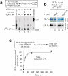
Figure 3. Inhibition of phosphorylation of EF-Tu and its dephosphorylation
(a). A scheme of the experiment is shown above the radiogram (see also Supplementary Fig. 6). EF-Tu 32P-phosphorylated by Doc (ensuring full usage of γ[32P]-ATP) was then incubated with or without Phd and/or ADP. The products were analyzed by TLC. For ATP mobility standard the reaction of EF-Tu phosphorylation in the presence of γ[32P]-ATP was spotted on the TLC plate immediately after start of the reaction. For ADP mobility standard α[32P]-ATP was used in the reaction of EF-Tu phosphorylation, which resulted in formation of α[32P]-ADP. Nonradioactive standards, visualized under UV254 are not displayed. Not all EF-Tu can be dephosphorylated even after prolonged incubation either due to aggregation or to the competition from phosphorylation with the γ[32P]-ATP produced in the reaction. The identity of the of 32P-phosphorylated His-tagged EF-Tu on TLC was verified by addition of Ni2+-NTA-agarose to the reaction. (b). Phosphorylation of EF-Tu by Doc was performed in the absence and presence of equimolar amounts of Phd or its toxin-binding domain Phd52-73. (c). Kinetics of dephosphorylation of immobilized 32P-phosphorylated EF-Tu (see Online Methods) in the presence of GDP and Doc. Data are the mean of three independent experiments and error bars show standard deviations. Data were fitted into a single-exponential equation and normalized to the predicted maximum, which was taken as 100. ± sign represents standard error of the fit.
Figure 4
Figure 4. Solution structure of the Doc:EF-Tu:GDP complex by SAXS
Experimental SAXS scattering curves (grey) for Doc (a), EF-Tu:GDP (b) and Doc:EF-Tu:GDP (c) overlaid with computed scattering curves obtained from rigid body modeling (black) of the particles (see Online Methods for details). Bars are experimental errors of the measurements. The rigid body reconstructed models of the EF-Tu:GDP complex (d) and the Doc:EF-Tu:GDP ternary complex (e) are superimposed onto the ab initio envelopes of the complexes reconstructed from the experimental SAXS data using the program DAMMIF from the ATSAS suite (see also Supplementary Fig. 8). All the ab initio models of the complex were very reproducible in independent runs with average normalized spatial discrepancy (NSD) values below 0.9. (f). 1H/15N HSQC spectra of the titration of Doc (0 μM of EF-Tu in blue) with EF-Tu (34.0 μM (in purple), 58.0 μM (in red), 123.3 μM (in yellow) and 197.3 μM (in green) (see also Supplementary Fig. 7a, b). (g) and (h). I/Io changes above 2σ (green line) are colored blue on the surface of Doc (pdbid 3K33) (see also Figure 5c). (i) Doc (red ribbons) binds to “open” EF-Tu (blue surface; pdbid 1EFC) in a central cavity (in purple) of ~ 16 Å formed between the G-domain and the two β-barrels domains. (j). Phe-tRNAPhe from the ternary complex (pdbid 1TTT) superimposed on the model of the Doc:Tu:GDP complex. Notice that CCA tail of tRNA (in yellow) overlaps with the binding site of Doc.
Figure 5
Figure 5. Chemical shift mapping of the nucleotide and EF-Tu binding sites of Doc
(a) 1H/15N HSQC spectra of Doc (in blue) titrated with AMPPNP (1.4 mM (purple), 2.7 mM (red), 9.0 mM (brown), 15.0 mM (orange), 25.8 mM (yellow) and 40.0 mM (green)). (b) Residue specific AMPPNP-induced chemical shift changes of Doc. (c) Mapping on the surface of Doc of residues (colored raspberry) with chemical shift perturbations (Δδ) > 2σ (green line in panel b) and residues that disappear upon AMPPNP binding (colored salmon). (d) Chemical shift based docking model of the Doc:AMPPNP complex. Doc active site loop is highlighted in red and AMPPNP is shown as spheres (see also Supplementary Fig. 7d and 9).
Figure 6
Figure 6. Proposed catalytic mechanism of the Doc-type Fic kinases
(a). The active site architecture and transposed binding of the NTP (shown in orange) led us to propose a catalytic mechanism used by Doc that, as in the case of other Fic proteins, involves the highly conserved histidine as the general base. The α- and β-phosphates of the NTP are most likely stabilized by the side chains of the conserved Lys73 and Arg74 residues of the catalytic loop. His66 hydrogen bonds (represented as a dotted line in both panels) the attacking hydroxyl group from Thr382 (in blue) and activates the oxygen atom for the nucleophilic attack. The catalytic residues from Doc are shown in raspberry. (b). Cartoon representation of the general reaction mechanism for Doc (left) and Fic (right) proteins. The reorientation of the NTP in the active site enables transferring the γ-phosphate instead of the NMP moiety while maintaining the active site chemistry. Note, that the targets are shown schematically only for representation purposes.

Comment in

  • Enzyme mechanisms: What's up 'Doc'?
    Peti W, Page R. Peti W, et al. Nat Chem Biol. 2013 Dec;9(12):756-7. doi: 10.1038/nchembio.1379. Epub 2013 Oct 20. Nat Chem Biol. 2013. PMID: 24141194 No abstract available.

References

    1. Engel P, et al. Adenylylation control by intra- or intermolecular active-site obstruction in Fic proteins. Nature. 2012;482:107–10. - PubMed
    1. Woolery AR, Luong P, Broberg CA, Orth K. AMPylation: Something Old is New Again. Front Microbiol. 2010;1:113. - PMC - PubMed
    1. Yarbrough ML, Orth K. AMPylation is a new post-translational modiFICation. Nat Chem Biol. 2009;5:378–9. - PubMed
    1. Anantharaman V, Aravind L. New connections in the prokaryotic toxin-antitoxin network: relationship with the eukaryotic nonsense-mediated RNA decay system. Genome Biol. 2003;4:R81. - PMC - PubMed
    1. Garcia-Pino A, et al. Doc of prophage P1 is inhibited by its antitoxin partner Phd through fold complementation. J Biol Chem. 2008;283:30821–7. - PMC - PubMed

Publication types

MeSH terms