. 2013 Dec;16(12):1888-1895.
doi: 10.1038/nn.3549. Epub 2013 Oct 20.

Common medial frontal mechanisms of adaptive control in humans and rodents

Affiliations

Common medial frontal mechanisms of adaptive control in humans and rodents

Nandakumar S Narayanan et al. Nat Neurosci. 2013 Dec.

Abstract

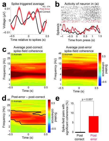
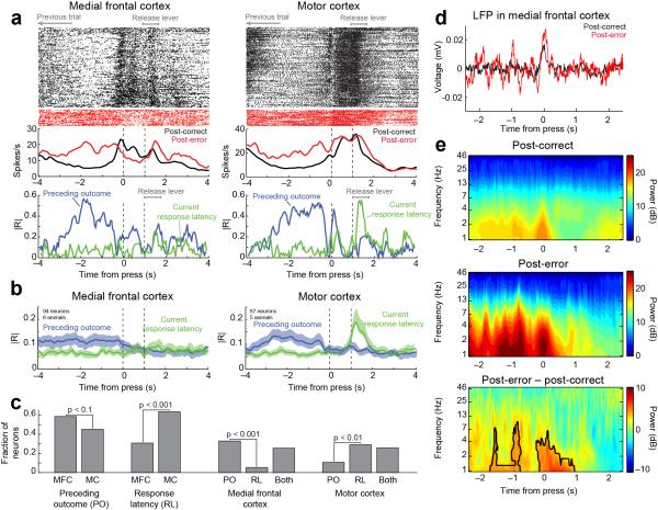
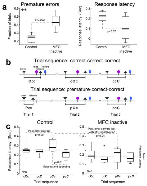
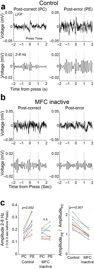
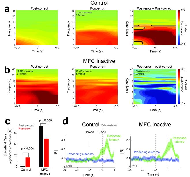
In this report we describe how common brain networks within the medial frontal cortex (MFC) facilitate adaptive behavioral control in rodents and humans. We demonstrate that after errors, low-frequency oscillations below 12 Hz are modulated over the midfrontal cortex in humans and within the prelimbic and anterior cingulate regions of the MFC in rats. These oscillations were phase locked between the MFC and motor areas in both rats and humans. In rats, single neurons that encoded prior behavioral outcomes were phase coherent with low-frequency field oscillations, particularly after errors. Inactivating the medial frontal regions in rats led to impaired behavioral adjustments after errors, eliminated the differential expression of low-frequency oscillations after errors and increased low-frequency spike-field coupling within the motor cortex. Our results describe a new mechanism for behavioral adaptation through low-frequency oscillations and elucidate how medial frontal networks synchronize brain activity to guide performance.


Figures

Figure 1
Figure 1
Common mechanisms of medial frontal cortical oscillations during adaptive control in rats and humans. a) Sequences of events in the time-estimation task on post-correct vs post-error trials (black). All analyses here are restricted to correct trials as a function of prior outcome. ▼ - press; formula image - 
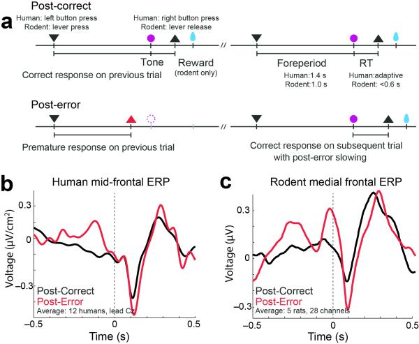
tone; ▲ - release, and formula image – reward. Imperative tones occurred at the target time on 50% of trials. b) Average event-related potentials over mid-frontal cortex (electrode Cz) in humans aligned to the target time. Amplitudes were significantly increased on post-error (red) vs. post-correct (black) trials. c) Rodent medial frontal field potentials were also significantly increased on post-error (red) vs. post-correct (black) trials, and highly similar to humans. Data is from 28 medial frontal channels in 5 rats and is aligned to the target time.
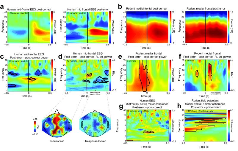
Figure 2
Figure 2
Time-frequency analysis reveals enhanced low-frequency power after errors. a) Humans: on post-correct trials, there was less low-frequency power than on post-error trials. b) Rodents: on post-correct trials, there was less low-frequency power than on post-error trials. c) Humans: direct comparison of post-error and post-correct trials revealed stronger theta modulation to the imperative tone on post-error trials. d) Humans: trial-to-trial variation in low frequency EEG signals was significantly correlated with subsequent response latency (electrode Cz). Current source density of scalp topographies (shown below) revealed that these effects were prominent over medial frontal regions. b) Rodents: similar patterns were seen in rodents, although broader bands of low-frequency modulation were observed. e) Rodents: direct comparison of post-error and post-correct trials revealed dramatically stronger low-frequency modulation to the imperative tone on post-error trials. f) Rodents: trial-to-trial variation in 4-25 Hz frequencies were strongly correlated with subsequent response latency. Time aligned to the target time; black contours indicate significant differences via a t-test between post-error and post-error trials (p<0.05) or Spearman’s (non-parametric) correlations (p <0.05). g) Humans: midfrontal and motor sites had significantly more low-frequency coherence on post-error compared to post-correct trials. h) Rodents: A similar pattern was observed in rodents between 12 medial frontal and 12 motor cortex channels in three rats. Time for g-h aligned to trial initiation; black contours indicate significant differences via a t-test between post-error and post-error trials (p<0.05).
Figure 3
Figure 3
Medial frontal single neuron spiking is coupled with low-frequency oscillations. a) Spike-triggered average of a medial frontal neuron where local field potentials are averaged across all spikes within 2 seconds of lever press for a single neuron. For this neuron, the spike-triggered average revealed robust low-frequency oscillations on post-error trials compared to post-correct trials. b) Peri-event raster of the neuron in a) revealed different patterns of activity after errors as well as low-frequency oscillations in firing rates. c) Spike-field coherence to post-correct and post-error trials. d) Post-error trials were characterized by enhanced low-frequency spike-field coupling. e) 9% of medial frontal neurons had significant low-frequency (below 12 Hz) spike-field coherence on post-error trials, compared to no significant spike-field pairs on post-correct trials. Time aligned to the target time (▲ - release); black contours indicate significant differences via a t-test between post-error and post-error trials (p<0.05).
Figure 4
Figure 4
Encoding of previous outcomes in the medial frontal and motor cortices. a) Examples of spike activity and correlation coefficients from the partial correlation analysis are shown for neurons in the medial frontal and motor cortices. b) Group summary for the sliding-window partial correlation analysis revealed that neurons in both cortical areas were sensitive to the previous outcome (in blue) and that only neurons in the motor cortex were sensitive to variations in response latency (in green). Error bars represent SEM. c) Fractions of neurons that were selective to the previous outcome and current response latency and that were sensitive to either or both of these behavioral factors are summarized in the lower plot. Significance was assessed over all data windows (±2 sec around the press event). d) Spiking correlates of previous outcomes were accompanied by increased low-frequency oscillations in the field potential, as was apparent in the trial-averaged ERP and event-related spectral power. e) Medial frontal cortex local field potentials had prominent low-frequency modulation around the time of the response; plots aligned to trial initiation; black contours indicate significant differences via a t-test between post-error and post-error trials (p<0.05)
Figure 5
Figure 5
Loss of adaptive control following inactivation of the medial frontal cortex. a) Reversible inactivation of the medial frontal cortex in 6 rats increased the fraction of trials with premature responses and reduced the overall response time.b) Given erratic performance in the inactivation sessions with runs of premature errors, it was essential to confirm that effects on response latency adjustments would be found in controlled sequences of trials in which rats made two consecutive correct responses after making either a correct response or premature error response. c) Analysis of the trial sequences revealed clear evidence for slowing of response latencies after premature errors and a subsequent speeding in the control session (saline infused into the medial frontal cortex). This was not observed in sessions with medial frontal cortex inactivated. Inactivation of medial frontal cortex also led to an overall speeding of responses and eliminated the post-error slowing and subsequent speeding after the corrected response. Boxes – IQR; Whiskers 1.5x IQR
Figure 6
Figure 6
Inactivation of medial frontal cortex eliminated post-error increases in low-frequency oscillations in motor cortex. a) Peri-event averages of wideband field potentials (top row) and bandpass filtered signals (2-8 Hz; lower row) are shown from the motor cortex of one rat. In the control session, low frequency oscillations were elevated on post-error trials. b) Sessions with medial frontal cortex inactivated. C) Z-transformed amplitude in the range between 2 and 8 Hz was measured using the Hilbert transform. Medial inactivation caused low frequency oscillations to become equivalent on the post-correct and post-error trials. This effect was found every field potential examined from three rats.
Figure 7
Figure 7
Rat medial frontal cortex directly influences post-error low-frequency oscillations in motor cortex in the service of adaptive control. a) In control sessions, low-frequency spike-field coherence in motor cortex on post-correct trials was less prominent than on post-error trials, as was apparent in comparisons of spike-field coherence between post-error and post-correct trials (right column). b) Medial frontal inactivation increase post-correct spike-field coherence and abolished differences between post-error and post-correct trials (right column). Black contours indicate significant differences via a t-test between post-error and post-error trials (p<0.05); see Fig S5 for comparison between control and medial frontal inactivation sessions. c) Medial frontal inactivation increased the numbers of neurons with post-significant spike-field coherence on post-correct trials. These data suggest that with medial frontal inactivation, low-frequency coherence is no longer specific to post-error trials. Error bars represent SEM. d) Changes to spike-field coupling occurred in the absence of any effects of medial frontal inactivation on the sensitivity of the motor cortical neurons to the prior behavioral outcome or response latency; note that this is a subset of data in Fig 4 with slightly less predictive power for previous outcome.

Comment in

Similar articles

Cited by

References

    1. Bellman R, Kalaba R. A MATHEMATICAL THEORY OF ADAPTIVE CONTROL PROCESSES. Proc. Natl. Acad. Sci. U.S.A. 1959;45:1288–1290. - PMC - PubMed
    1. Ridderinkhof KR, Ullsperger M, Crone EA, Nieuwenhuis S. The Role of the Medial Frontal Cortex in Cognitive Control. Science. 2004;306:443–447. - PubMed
    1. Holroyd CB, Coles MGH. The neural basis of human error processing: reinforcement learning, dopamine, and the error-related negativity. Psychol Rev. 2002;109:679–709. - PubMed
    1. Rushworth MFS, Behrens TEJ. Choice, uncertainty and value in prefrontal and cingulate cortex. Nature Neuroscience. 2008;11:389–397. - PubMed
    1. vanMeel CS, Heslenfeld DJ, Oosterlaan J, Sergeant JA. Adaptive control deficits in attention-deficit/hyperactivity disorder (ADHD): the role of error processing. Psychiatry Res. 2007;151:211–220. - PubMed

Publication types