. 2013 Dec;14(12):1266-76.
doi: 10.1038/ni.2741. Epub 2013 Oct 20.

The kinase mTOR modulates the antibody response to provide cross-protective immunity to lethal infection with influenza virus

Affiliations

The kinase mTOR modulates the antibody response to provide cross-protective immunity to lethal infection with influenza virus

Rachael Keating et al. Nat Immunol. 2013 Dec.

Abstract

Highly pathogenic avian influenza viruses pose a continuing global threat. Current vaccines will not protect against newly evolved pandemic viruses. The creation of 'universal' vaccines has been unsuccessful because the immunological mechanisms that promote heterosubtypic immunity are incompletely defined. We found here that rapamycin, an immunosuppressive drug that inhibits the kinase mTOR, promoted cross-strain protection against lethal infection with influenza virus of various subtypes when administered during immunization with influenza virus subtype H3N2. Rapamycin reduced the formation of germinal centers and inhibited class switching in B cells, which yielded a unique repertoire of antibodies that mediated heterosubtypic protection. Our data established a requirement for the mTORC1 complex in B cell class switching and demonstrated that rapamycin skewed the antibody response away from high-affinity variant epitopes and targeted more conserved elements of hemagglutinin. Our findings have implications for the design of a vaccine against influenza virus.


Figures
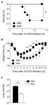
Figure 1
Figure 1
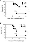
Rapamycin treatment during primary infection decreases mortality following a lethal, heterosubtypic H5N1 secondary infection. (a–c) C57BL/6 mice received 75 μg/kg rapamycin or PBS, i.p. beginning 1 day prior to and daily for 28 days after primary infection with 108 EID50 of the H3N2 HKx31 i.p. On day 28, mice were challenged with 4.5 x 10⁵ EID50 of the H5N1 ΔVn1203 i.n. and (a) monitored for survival (**P = 0.003, Mantel-Cox test, n = 9 per group), and (b) weight loss. *P < 0.05 and **P < 0.01, (unpaired t test). Data are representative of more than 10 separate experiments. (c) ΔVn1203 viral titers in the lung were determined by plaque assay 5 days after secondary infection. Data represent average viral titer ± s.e.m. (n = 5 per group; *P = 0.040, Mann-Whitney U test). Data are representative of 6 independent experiments.
Figure 2
Figure 2
Rapamycin protects mice from lethal H7N9 and H1N1 infections. Mice were infected with 108 EID50 of the H3N2 HKx31 i.p., treated with rapamycin daily, challenged i.n. on day 28 with (a) 4.5 x 10⁵ EID50 of A/Anhui/1/2013 or (b) 4.5 x 10⁵ EID50 of PR8 and monitored for survival (*P < 0.05 and ***P < 0.001, Mantel-Cox test, n = 16 and n = 18 per group, respectively). Data are a combination of 2 statistically significant independent experiments.
Figure 3
Figure 3
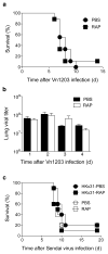
Rapamycin-enhanced protection against lethal ΔVn1203 infection is influenza-specific and requires primary HKx31 infection. (a) Mice were treated with rapamycin or PBS for 28 days, infected with ΔVn1203 i.n., and monitored for survival (n = 9 per group; P > 0.05, Mantel-Cox test). (b) Mice were treated with rapamycin or PBS daily beginning 1 day prior to i.n. infection with 4 x 10⁵ EID50 of ΔVn1203. Lungs were homogenized and virus titer was measured in a plaque assay. Results are expressed as the mean ± s.e.m. (n = 5 per group; P > 0.05, Mann-Whitney U test). (c) Mice were infected with HKx31 virus i.p., treated with rapamycin or PBS daily for 28 days and infected with 2 x 10⁴ pfu of Sendai virus i.n. Control groups received PBS or rapamycin daily with no HKx31 primary infection (n = 10 per group; P > 0.05, Mantel-Cox test). Data represent 2 independent experiments.
Figure 4
Figure 4
Memory CD8+ T cells are not required for rapamycin-mediated protection. (ab) Mice were injected with rapamycin or PBS and infected with HKx31 and ΔVn1203 as described in Fig. 1a. (a) On day 27, prior to secondary infection, a sample of blood from each mouse was analyzed for H-2Db NP366 and H-2Db PA224 tetramer binding. Data represent averages ± s.e.m. of 15 mice per group and are representative of 4 separate experiments (**P < 0.01, Mann-Whitney U test). (b) Five days after secondary challenge with ΔVn1203, the spleen, MLN, and BAL were harvested and analyzed for DbNP366 tetramer binding. Data represents the averages ± s.e.m. of 4 mice per group, which are representative of 4 separate experiments. (*P < 0.05, Mann-Whitney U test). (c) Mice were infected with ΔCD8-HKx31 virus i.p., treated daily with rapamycin or PBS, and challenged with ΔVn1203 after 28 days and monitored for survival (n = 9 for PBS-treated mice, n = 15 for RAP-treated mice; ***P < 0.001, Mantel-Cox test). Data are representative of 1 experiment. (d) CD8+ T cells were depleted with anti-CD8 antibody (Clone 53-6.7) on days −3, −1, 1, 3, 5, and 17. Mice were infected with HKx31 virus, treated daily with rapamycin or PBS daily, and challenged with ΔVn1203 after 28 days. (n > 23 for each group; **P < 0.01 comparing PBS and RAP-treated wildtype mice and comparing PBS and RAP-treated, CD8-depleted mice, Mantel-Cox test). These data are a combination of 3 independent experiments.
Figure 5
Figure 5
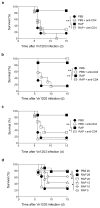
CD4+ T cells are required during primary HKx31 infection for rapamycin-mediated protection. (a–c) Mice were infected with HKx31, received rapamycin or PBS daily, and were challenged with ΔVn1203 as described in Fig. 1. CD4+ T cells were depleted during the (a) primary and secondary infection by injection of anti-CD4 antibody (Clone GK1.5) −3, −1, 2, 5, 22, 24 days after HKx31 infection (n > 6 per group,) (b) during the secondary infection by injecting antibody 22 and 24 days after HK31 infection (n > 6 per group), and (c) during the primary infection by injecting antibody −3, −1, 2, 5 days after HKx31 infection (n > 6 per group). These data are representative of three independent experiments. (d) Mice were infected with HKx31 virus, received rapamycin or PBS daily for 5,10, 15, 20 or 28 days, and were then challenged with Δ Vn1203 virus 28 days following the primary HKx31 infection (n > 8 per group). *P < 0.05 and **P < 0.01, Mantel-Cox test. The 28 day data are representative of more than 10 independent experiments. The 20, 15, 10 and 5 day data are representative of at least 2 independent experiments.
Figure 6
Figure 6
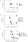
B cells are required for rapamycin-mediated protection. (a) C57BL/6 mice were infected with HKx31 and received rapamycin or PBS daily for 28 days. Six weeks later, the mice were challenged with ΔVn1203 and monitored for survival. (n > 13 per group) (b) C57BL/6 and μMT mice were infected with HKx31, received rapamycin or PBS daily, and were challenged with ΔVn1203 (n ≥ 4 per group). Data are representative of 2 independent experiments (c) One day prior to infection with 4 x 10⁴ EID50 of ΔVn1203, μMT mice received 450 μl of serum i.p. from HKx31-infected mice treated with PBS (n = 14) or rapamycin (n = 13). Control mice received normal mouse serum (n = 10) or serum from ΔVn1203-infected mice (n = 8). The data are combined from 2 independent experiments *P < 0.05 and **P < 0.01, Mantel-Cox test.
Figure 7
Figure 7
Rapamycin-treated mice show elevated influenza-specific IgM antibodies. Mice were infected with HKx31 and received rapamycin or PBS daily as described in Fig. 1. On day 27, serum isolated from each mouse was analyzed by ELISA for (a) IgM specific for HKx31 (n = 8; **P < 0.01 and ***P < 0.001, Mann-Whitney U test) (b) IgG specific for HKx31 (n = 3), (c) IgG specific for ΔVn1203 (n = 4). Each absorbance curve represents 5 independent experiments.
Figure 8
Figure 8
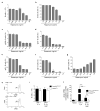
Rapamycin decreases germinal center formation and reduces B cell class-switching. (a) Sera from mice infected with HKx31 and treated with rapamycin or PBS for 25 days were analyzed for Ig subtypes with a Milliplex Immunologlobulin Isotyping Panel (n=10). Data are representative of two independent experiments (b) MLN cells recovered 15 or 20 days after HKx31 infection and daily rapamycin or PBS injections were stained with antibodies to B220 and GL7. Each bar represents the average percent GL7+ of B220+ cells ± s.e.m. (n = 5,). Data are representative of 2 independent experiments. (c) Sections of MLN recovered 15 or 20 days after HKx31 infection and daily rapamycin or PBS injections were stained with Bcl-6 and PNA (data not shown) to identify germinal centers. The average number of germinal centers per section was calculated (n = 9 for day 15 and n = 3 for day 20). (d) Representative pictures of day 15 lymph nodes with arrows indicating germinal centers. (e) Fifteen and 20 days after primary infection, mice received a BrdU pulse 4 hours prior to harvest, and a second pulse 2 hours prior to harvest. The average percent ± s.e.m. of B220+ cells in the MLN incorporating BrdU is shown (n = 5). *P > 0.05 and ***P > 0.001, Mann-Whitney U test.
Figure 9
Figure 9
mTORC1 is required for Aicda transcription and B cell class-switching. (a–g) IgM+ B cells were enriched from the spleen, CFSE labeled, and left unstimulated (US) or stimulated in vitro for 4 days with (a–d) LPS or (e–g) LPS and IL-4 in the presence of increasing doses of rapamycin. (a) Shown is the average number ± s.d. and (b) the percent ± s.d. of live divided B cells. The proportion of live B cells that are positive for (c) IgG2b, (d) IgG3, (e) IgG1 (f) IgE, and (g) IgM is shown. These data are representative of 5 separate experiments. (h–j) Control Rosa26ERcreT and RaptorB/ mice received injections of tamoxifen, i.p. daily for 4 days. Eight days after the last injection, IgM+ B cells were harvested from spleens, stimulated for 24 hours with LPS and IL-4, with or without 0.5 ng/ml of rapamycin. (h) CFSE profile of control cells and (i) cell counts 24 hours after stimulation. (j) RNA was extracted and analyzed by qPCR for the level of Aicda transcripts relative to Cd79b transcripts. Data is a combination of 6 technical replicates and is representative of three biological replicates. (**P = 0.01, Mann-Whitney U test).
Figure 10
Figure 10
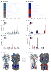
Rapamycin modifies the antibody repertoire. Antigen microarrays were spotted with overlapping peptides that span the HA protein of both HKx31 and ΔVn1203. These were used to probe the IgM and IgG repertoire simultaneously using sera from animals sampled at 20 days following primary infection. Responses of 10 mice from each group were analyzed. (a–b) Samples were clustered using complete-linkage and the similarity between response patterns was defined as one minus the Spearman correlation coefficient between these vectors. Clustering dendrograms of response patterns of (a) IgM and (b) IgG to ΔVn1203 are depicted. (c–f) Specific antigens in which the responses between the rapamycin and PBS-treated mice had a p < 0.05 (Fisher’s exact test) are shown. (c) IgM specific for HKx31 (d) IgG specific for HKx31 (e) IgM specific for ΔVn1203 (f) IgM specific for ΔVn1203. (g–h) The positions of the antigens described in (c–f) are imposed on the structure of (g) HKx31 HA and (h) Δ Vn1203 HA.

Comment in

References

    1. Osterholm MT, Kelley NS. Mammalian-Transmissible H5N1 Influenza: Facts and Perspective. mBio. 2012;3 - PMC - PubMed
    1. Herfst S, et al. Airborne transmission of influenza A/H5N1 virus between ferrets. Science. 2012;336:1534–1541. - PMC - PubMed
    1. Imai M, et al. Experimental adaptation of an influenza H5 HA confers respiratory droplet transmission to a reassortant H5 HA/H1N1 virus in ferrets. Nature. 2012;486:420–428. - PMC - PubMed
    1. Pica N, Palese P. Toward a universal influenza virus vaccine: prospects and challenges. Annu Rev Med. 2013;64:189–202. - PubMed
    1. Thomas PG, Keating R, Hulse-Post DJ, Doherty PC. Cell-mediated protection in influenza infection. Emerging Infect Dis. 2006;12:48–54. - PMC - PubMed

Publication types

MeSH terms