

Extracellular vesicles: structure, function, and potential clinical uses in renal diseases
- PMID: 24141609
- PMCID: PMC3854311
- DOI: 10.1590/1414-431X20132964
Extracellular vesicles: structure, function, and potential clinical uses in renal diseases
Abstract
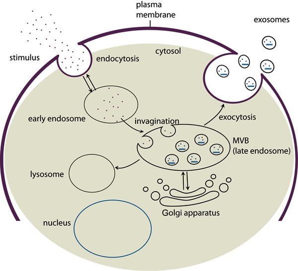
Interest in the role of extracellular vesicles in various diseases including cancer has been increasing. Extracellular vesicles include microvesicles, exosomes, apoptotic bodies, and argosomes, and are classified by size, content, synthesis, and function. Currently, the best characterized are exosomes and microvesicles. Exosomes are small vesicles (40-100 nm) involved in intercellular communication regardless of the distance between them. They are found in various biological fluids such as plasma, serum, and breast milk, and are formed from multivesicular bodies through the inward budding of the endosome membrane. Microvesicles are 100-1000 nm vesicles released from the cell by the outward budding of the plasma membrane. The therapeutic potential of extracellular vesicles is very broad, with applications including a route of drug delivery and as biomarkers for diagnosis. Extracellular vesicles extracted from stem cells may be used for treatment of many diseases including kidney diseases. This review highlights mechanisms of synthesis and function, and the potential uses of well-characterized extracellular vesicles, mainly exosomes, with a special focus on renal functions and diseases.
Figures
References
Publication types
MeSH terms
LinkOut - more resources
Full Text Sources
Other Literature Sources
Medical
