. 2014 Jan;21(1):161-71.
doi: 10.1038/cdd.2013.135. Epub 2013 Oct 18.

Remodelling of a polypyrimidine tract-binding protein complex during apoptosis activates cellular IRESs

Affiliations

Remodelling of a polypyrimidine tract-binding protein complex during apoptosis activates cellular IRESs

H A King et al. Cell Death Differ. 2014 Jan.

Abstract

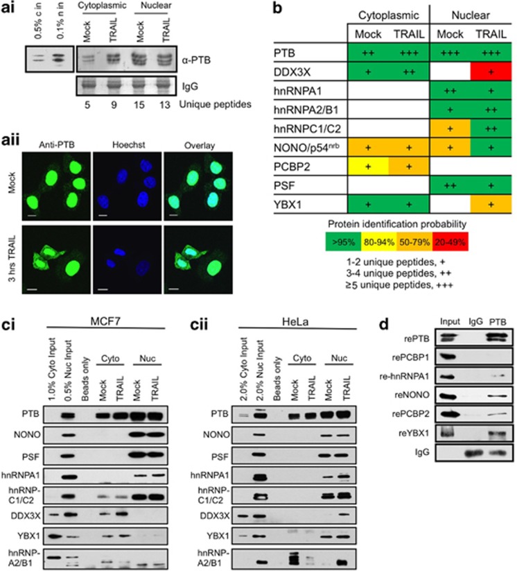
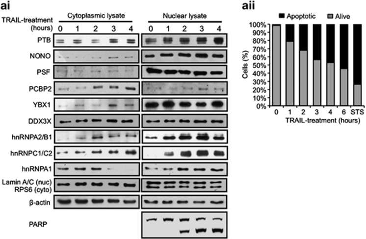
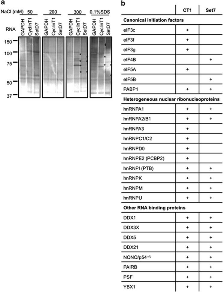
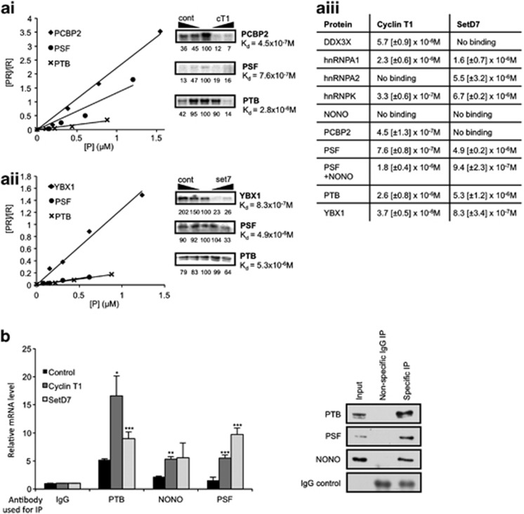
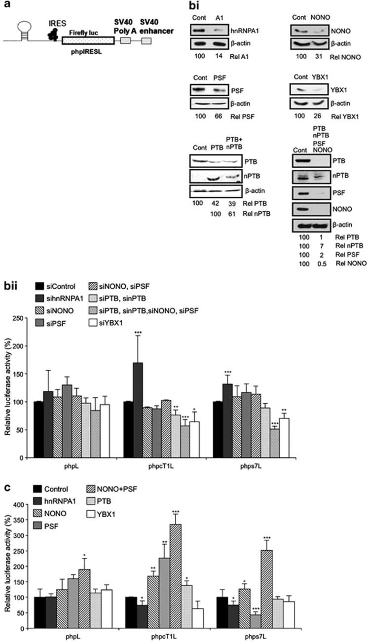
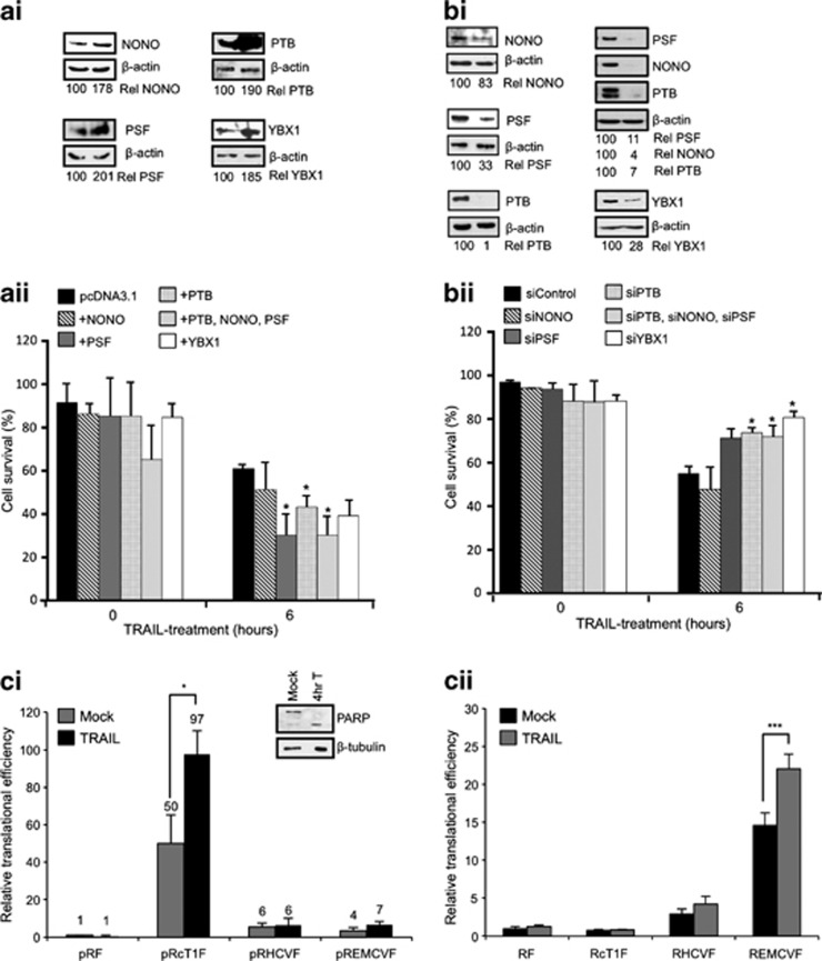
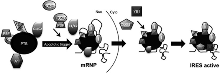
Post-transcriptional control of gene expression is mediated by the interaction of RNA-binding proteins with their cognate mRNAs that specifically regulate their stability, localization and translation. mRNA-binding proteins are multifunctional and it has been proposed therefore that a combinatorial RNA-binding protein code exists that allows specific protein sub-complexes to control cytoplasmic gene expression under a range of pathophysiological conditions. We show that polypyrimidine tract-binding protein (PTB) is central to one such complex that forms in apoptotic cells. Thus, during apoptosis initiated by TNF-related apoptosis inducing ligand there is a change in the repertoire of RNA-binding proteins with which PTB interacts. We show that altering the cellular levels of PTB and its binding partners, either singly or in combination, is sufficient to directly change the rates of apoptosis with increased expression of PTB, YBX1, PSF and NONO/p54(nrb) accelerating this process. Mechanistically, we show that these proteins post-transcriptionally regulate gene expression, and therefore apoptotic rates, by interacting with and stimulating the activity of RNA elements (internal ribosome entry segments) found in mRNAs that are translated during apoptosis. Taken together, our data show that PTB function is controlled by a set of co-recruited proteins and importantly provide further evidence that it is possible to dictate cell fate by modulating cytoplasmic gene expression pathways alone.


Figures

Figure 1
Figure 1
PTB-containing complex that forms during TRAIL-mediated apoptosis. MCF7 cell were induced to undergo apoptosis via treatment with 0.5 μg/μl TRAIL for 3 h, then harvested and fractionated to produce nuclear and cytoplasmic lysates. Mock and TRAIL-treated cytoplasmic/nuclear lysates were used in an anti-PTB immunoprecipitation, which were then analyzed via immunoblotting and LC-MS/MS mass spectrometry. (ai) Western blot with α-PTB antibody. IgG was used as a loading control from the IP reaction. Number of unique peptides identified for PTB is shown. (aii) Immunofluorescence was used to confirm the relocalization of PTB following TRAIL-treatment. MCF cells±TRAIL for 3 h were stained with α-PTB antibody and observed by confocal microscopy. White bar represents 10 μm. (b) Table showing PTB-binding partners identified by LC-MS/MS mass spectrometry. A color scale is used to illustrate the number of unique peptides. Number of plus (e.g., +/++/+++) are used to represent the relative abundance of each species detected. (ci) Western blotting of the control and TRAIL treated MCF7 cell lysates was performed using antibodies to the indicated proteins to investigate PTB-interacting partners. (cii) The immunoprecipitations of PTB and its interacting partners were then repeated in HeLa cells. Western blotting of control and TRAIL treated HeLa cell lysates was performed using antibodies to the indicated proteins to investigate PTB-interacting partners in this cell type. (d) RNA-independent association of the different PTB partners were confirmed by carrying out anti-PTB immunoprecipitations in vitro following incubation of recombinant-PTB (rePTB) with rePCBP1, re-hnRNPA1, reNONO, rePCBP2 and reYBX1. Western blot using antibodies to the indicated proteins is shown; IgG was used as a loading control
Figure 2
Figure 2
Relocalization of members of the PTB complex during apoptosis. (ai) Western blotting of nuclear and cytoplasmic fractionated lysates of MCF7 cells treated with TRAIL over a 4 h time course using antibodies against indicated proteins. RPS6 and Lamin A/C antibodies were used as cytoplasmic and nuclear markers, respectively. PARP cleavage was used to indicate apoptosis and β-actin was used as a loading control. (aii) Control and TRAIL-treated MCF7 cells were stained with Annexin V-FITC and propidium iodide at the indicated time points and subjected to FACS analysis. Staurosporin-treated MCF7 cells (STS) served as a positive control at 6 h
Figure 3
Figure 3
IRES RNA affinity analysis. (a) In all, 50 μg of biotin-tagged cyclin T1 and SETD7 5' UTRs or 300 nt RNA of GAPDH coding region was used as bait to purify IRES-binding proteins from HeLa cell lysate in a binding buffer (10 mM Tris pH 7.5, 25 mM KCl, 2 mM MgCl2, 0.02% Tween-20, 1 mM ATP, 1 μg/ml yeast tRNA, 1 μg/ml heparin, RNase inhibitor, 1 × protease inhibitor) for 60 min at 4 °C. Streptavidin-conjugated magnetic beads (Invitrogen) were added and incubated for a further 30 min. The bound complexes were washed with buffers that contained increasing salt concentration and eluted samples were applied to SDS-PAGE and stained with colloidal Coomassie blue and silver. Proteins that were specifically eluted from the cyclin T1 or SETD7IRESs, but not the GAPDH are marked (*). (b) Proteins bound to each RNA were identified firstly via LC-MS/MS mass spectrometry (Supplementary Figures S2A and B) and then via immunoblotting analysis (Supplementary Figure S2C). The table shows proteins, which were identified as binding to either or both of the cyclin T1 and SETD7 IRESs, whereby a plus indicates binding, in either the mass spectrometry data, the immunoblotting data or both. The proteins included in the table are those found to be both IRES binding and PTB associated
Figure 4
Figure 4
Members of the PTB-containing complex bind apoptotic IRES RNA in vitro and in vivo. Binding of the identified complex members to the apoptotic IRES RNA was validated by a number of methods. (a) UV-crosslinking (right panels) and filter-binding assays (left panels) using 32P-labelled cyclin T1 or SETD7 5' UTR. (ai) 32P-CTP-labelled cyclin T1 and rePTB, rePSF and rePCBP2 or (aii) 32P-CTP-labelled SETD7 RNA with rePTB, rePSF and reYBX1 were used in UV-crosslinking and filter-binding assays. Specificity of the binding was demonstrated by increasing the amount of cold competitor IRES RNA (right panel), or nonspecific control RNA (left panel). The UV-crosslinked signal was quantified relative to the central no competitor RNA lane, which was set to a 100. (aiii) Dissociation constants for each protein/RNA combination were calculated in the filter-binding assay. A constant level of the 32P-labelled probe was used over a range of molarity of each recombinant protein. S.D. is shown in square brackets, and the data are the average of three repeats. (b) Binding of endogenous PTB, PSF or NONO/p54nrb to the IRES RNA was shown using RNA-IP with antibodies against the proteins indicated, followed by RT-qPCR using primers specific for cyclin T1, SETD7 or control RNA. 15 cm confluent plates of HeLa cells were lysed in lysis buffer (20 mM HEPES pH7.2, 100 mM KCl, 5 mM MgCl2, 1 mM DTT, RNAsin 400 U/ml, 0.5% Triton X-100 and protease inhibitors), incubated for 5 min at 4 °C and centrifuged to pellet the nuclei. The post-nuclear extract was incubated with PTB, PSF or NONO/p54nrb antibodies or IgG (control) coated protein A beads for 1 h at 4 °C. Beads were washed 4 × 30 min with buffer A (20 mM Hepes pH 7.2, 200 mM NaCl, 5 mM MgCl2, 0.5% Triton X-100, 1 mM DTT) and 2 × with buffer B (50 mM Tris pH 7.4, 150 mM NaCl, 1 mM MgCl2 and 0.05% NP40). Beads containing protein-RNA complexes were isolated and re-suspended in 200 μl buffer B and proteinase K treated for 45 min at 55 °C. The RNA was isolated by phenol/chloroform extraction and quantified by RT-qPCR using primers specific for cyclin T1, SETD7 or control RNA. Results are shown relative to a nonspecific IgG immunoprecipitation. Western blot of the IPs using antibodies against proteins indicated is shown on the right. Significance (*P<0.05, **P<0.01 or ***P<0.005) was calculated using an unpaired two-tailed Student's t-test (n=3), error bars represent S.D.
Figure 5
Figure 5
Altering the levels of PTB-interacting proteins effects IRES activity. (a) Schematic representation of the monocistronic constructs used in figures bii and c, which contains a stable hairpin loop downstream of the 5′ m7G cap in order to inhibit cap-dependent translation of the Firefly luciferase reporter. (bi) HeLa cells were co-transfected with siRNA against the indicated proteins together with the monocistronic reporter plasmid, incubated for 48 h then harvested and western blot analysis was carried out to confirm RNAi success. Immunoblots are quantified relative to β-actin levels, and expressed as a percent of levels in the control siRNA treated lysate. (bii) Luciferase assays were carried out on the RNAi lysates. Data are shown relative to a control experiment using control siRNA. Significance (*P<0.05, **P< 0.01 or ***P<0.005) was calculated using an unpaired two-tailed Student's t-test (n=3), error bars represent S.D. (c) Reticulocyte lysates were primed with 100 ng recombinant protein and 100 ng of in vitro transcribed m7G capped and polyadenylated reporter RNA, incubated at 30 °C for 90 min, then assayed for luciferase activity. Data are shown relative to a control experiment with no recombinant protein added. Significance (*P<0.05, **P<0.01 or ***P<0.005) was calculated using an unpaired two-tailed Student's t-test (n=3), error bars represent S.D.
Figure 6
Figure 6
Altered expression of PTB-binding proteins controls apoptotic rates. (a) Overexpression of PTB1, NONO/p54nrb, PSF either individually or together enhances TRAIL-induced apoptosis. (ai) MCF7 cells were transfected with pcDNA3.1, pcDNA3.1-PTB1, pcDNA3.1-NONO/p54nrb, pcDNA3.1-PSF, pcDNA3.1-YBX1 plasmids and subjected to 6-h TRAIL treatment. The overexpression was assessed by western blot using antibodies against the proteins indicated. Immunoblots were quantified relative to β-actin levels and expressed as a percent of levels in the control cells. (aii) FACS analysis of MCF7 cells overexpressing the proteins above. After TRAIL treatment, cells were stained with Annexin V-FITC and propidium iodide and subjected to FACS analysis. Significance (*P<0.05) was calculated using an unpaired two-tailed Student's t-test (n=3) relative to the control. (b) Depletion of endogenous YBX1, PTB, NONO/p54nrb and PSF individually or in combination protected cells against TRAIL-mediated apoptosis. (bi) Western blot of siRNA treated MCF7 cells using antibodies indicated. Immunoblots were quantified relative to β-actin levels and expressed as a percent of levels in the control cells. (bii) FACS analysis of siRNA treated MCF7 cells treated as in aii. Significance (*P<0.05) was calculated using a paired two-tailed Student's t-test (n=3) relative to the control. (ci) MCF7 cells were transfected via nucleofection with dicistronic pRIRESF plasmid, incubated for 48 h before mock or TRAIL treatment for 4 h. Both protein and RNA were harvested. Firefly luciferase counts were normalized to total protein, and luciferase RNA levels were normalized to GAPDH RNA. The translation efficiency is expressed as Firefly luciferase activity/mRNA level. Data are presented relative to this value for pRF in the mock condition. Western blot with αPARP was performed to monitor the degree of apoptosis. Significance (*P<0.05) was calculated using an unpaired two-tailed Student's t-test (n=3), error bars represent S.D. (cii) In all, 1 μg of capped and polyadenylated mRNA was transfected into MCF7 cells, incubated for 2 h before mock or TRAIL-treatment for a further 4 h. Cells were harvested and assayed for both Renilla and Firefly luciferase. Firefly luciferase counts were normalized to Renilla luciferase counts and are presented relative to RF in the mock condition. Significance (***P<0.01) was calculated using an unpaired two-tailed Student's t-test (n=5), error bars represent S.D.
Figure 7
Figure 7
A model to illustrate how formation of a nuclear mRNP containing the PTB complex could be required for cellular IRES activity. Our data suggest remodelling of a PTB-containing complex occurs following treatment with the apoptosis-inducing ligand TRAIL. Thus, the IRES-inhibitory protein hnRNPA1 decreases in association with PTB, whereas the IRES-stimulatory proteins NONO/p54nrb, PSF and hnRNPA2/B1 increase in association. This complex then binds to the IRES RNA, and shuttles to the cytoplasm as an mRNP. Here, further IRES-stimulatory proteins including DDX3X and YBX1 are recruited to the complex, which is then competent to stimulate apoptotic IRES-mediated translation

References

    1. Spriggs KA, Bushell M, Willis AE. Translational regulation of gene expression during conditions of cell stress. Mol Cell. 2010;40:228–237. - PubMed
    1. Bushell M, Stoneley M, Kong YW, Hamilton TL, Spriggs KA, Dobbyn HC, et al. Polypyrimidine tract binding protein regulates IRES-mediated gene expression during apoptosis. Mol Cell. 2006;23:401–412. - PubMed
    1. Pichon X, Wilson LA, Stoneley M, Bastide A, King HA, Somers J, et al. RNA binding protein/RNA element interactions and the control of translation. Curr Protein Pept Sci. 2012;13:294–304. - PMC - PubMed
    1. Powley I, Kondrashov A, Young LA, Dobbyn HC, Hill K, Cannell IG, et al. Translational reprogramming following UVB irradiation is mediated by DNA-PKcs and allows selective recruitment to the polysomes of mRNAs encoding DNA repair enzymes. Genes Dev. 2009;23:1207–1220. - PMC - PubMed
    1. Brown V, Jin P, Ceman S, Darnell JC, O'Donnell WT, Tenenbaum SA, et al. Microarray identification of FMRP-associated brain mRNAs and altered mRNA translational profiles in fragile X syndrome. Cell. 2001;107:477–487. - PubMed

Publication types

MeSH terms

LinkOut - more resources